Current evaluation of causes and treatment for amenorrhea
1. Current evaluation of amenorrhea
The Practice Committee of the American Society for Reproductive Medicine
Birmingham, Alabama
Amenorrhea is the absence or abnormal cessation of the menses. Primary and secondary amenorrhea describe the
occurrence of amenorrhea before and after menarche, respectively. (Fertil SterilÒ 2008;90:S219–25. Ó2008 by
American Society for Reproductive Medicine.)
Amenorrhea is the absence or abnormal cessation of the men-
ses (1). Primary and secondary amenorrhea describe the
occurrence of amenorrhea before and after menarche, respec-
tively. The majority of the causes of primary and secondary
amenorrhea are similar. Timing of the evaluation of primary
amenorrhea recognizes the trend to earlier age at menarche
and is therefore indicated when there has been a failure to
menstruate by age 15 in the presence of normal secondary sex-
ual development (two standard deviations above the mean of
13 years), or within five years after breast development if
that occurs before age 10 (2). Failure to initiate breast develop-
ment by age 13 (two standard deviations above the mean of 10
years) also requires investigation (2). In women with regular
menstrual cycles, a delay of menses for as little as one week
may require the exclusion of pregnancy; secondary amenor-
rhea lasting three months and oligomenorrhea involving less
than nine cycles a year require investigation.
The prevalence of amenorrhea not due to pregnancy, lacta-
tion or menopause is approximately 3% to 4% (3, 4). Al-
though the list of potential causes of amenorrhea is long
(Table 1), the majority of cases are accounted for by four con-
ditions: polycystic ovary syndrome, hypothalamic amenor-
rhea, hyperprolactinemia, and ovarian failure. Other causes
are seldom encountered in a typical reproductive medicine
practice. In highly specialized referral centres, only 10 to
15 patients per annum were seen with primary amenorrhea,
and a similar number with secondary amenorrhea (5–7).
The World Health Organization (WHO) has summarized
the causes: in WHO group I there is no evidence of endoge-
nous estrogen production, normal or low FSH levels, normal
prolactin levels, and no evidence of a lesion in the hypotha-
lamic-pituitary region; WHO group II is associated with ev-
idence of estrogen production and normal levels of prolactin
and FSH; and WHO group III involves elevated serum FSH
levels indicating gonadal failure (8).
Amenorrhea may occur with sexual ambiguity or viriliza-
tion, but usually in these cases amenorrhea is not the primary
complaint. The sexual ambiguity or virilization should be
evaluated as separate disorders, mindful that amenorrhea is
an important component of their presentation (9).
EVALUATION OF THE PATIENT
History, physical examination, and estimation of follicle
stimulating hormone (FSH), thyroid stimulating hormone
(TSH), and prolactin will identify the most common causes
of amenorrhea (Fig. 1). The presence of breast development
means there has been previous estrogen action. Excessive tes-
tosterone secretion is suggested most often by hirsutism and
rarely by increased muscle mass or other signs of virilization.
The history and physical examination should include a thor-
ough assessment of the external and internal genitalia.
The genital examination is abnormal in approximately
15% of women with primary amenorrhea. A blind or absent
vagina with breast development usually indicates Mullerian
agenesis, transverse vaginal septum, or androgen insensitiv-
ity syndrome. If a genital examination is not feasible, an
abdominal ultrasound may be useful to confirm the presence
or absence of the uterus.
When the physical examination is normal (the majority of
cases), the initial investigations should exclude pregnancy
and estimate FSH and prolactin concentrations. Estimation
of TSH is useful to rule out subclinical hypothyroidism,
even in the absence of thyroid-related symptoms. If there is
gonadal failure, a karyotype should be done if the woman
is less than 30 years of age to identify chromosomal abnor-
malities, including the presence of a Y chromosome as may
be seen in mosaic Turner syndrome or Swyer syndrome. If
the serum prolactin is persistently elevated, and there is no
history of medication or drug use that may elevate prolactin,
magnetic resonance imaging (MRI) is preferred to identify
a pituitary tumor. When FSH values are normal or low, the
problem is most often polycystic ovary syndrome or hypotha-
lamic amenorrhea. Tables 2 and 3 show the distribution of the
common causes of primary and secondary amenorrhea,
respectively, in clinical practice (5–7).
CAUSES OF AMENORRHEA
Anatomical Defects
When all or part of the uterus and vagina are absent in the
presence of otherwise normal female sexual characteristics,
Educational Bulletin
Reviewed June 2008.
Received February 20, 2004; revised and accepted February 20, 2004.
No reprints will be available.
Correspondence to: Practice Committee, American Society for Reproduc-
tive Medicine, 1209 Montgomery Highway, Birmingham, Alabama
35216 .
0015-0282/08/$34.00 Fertility and Sterilityâ Vol. 90, Suppl 3, November 2008 S219
doi:10.1016/j.fertnstert.2008.08.038 Copyright ª2008 American Society for Reproductive Medicine, Published by Elsevier Inc.
2. TABLE 1
Classification of amenorrhea (not including
disorders of congenital sexual ambiguity).
I. Anatomic defects (outflow tract)
A. M€ullerian agenesis (Mayer-Rokitansky-
Kuster-Hauser syndrome)
B. Complete androgen resistance (testicular
feminization)
C. Intrauterine synechiae (Asherman
syndrome)
D. Imperforate hymen
E. Transverse vaginal septum
F. Cervical agenesis—isolated
G. Cervical stenosis—iatrogenic
H. Vaginal agenesis—isolated
I. Endometrial hypoplasia or
aplasia—congenital
II. Primary hypogonadism
A. Gonadal dysgenesis
1. Abnormal karyotype
a. Turner syndrome 45,X
b. Mosaicism
2. Normal karyotype
a. Pure gonadal dysgenesis
i. 46,XX
ii. 46,XY (Swyer syndrome)
B. Gonadal agenesis
C. Enzymatic deficiency
1. 17a-Hydroxylase deficiency
2. 17,20-Lyase deficiency
3. Aromatase deficiency
D. Premature ovarian failure
1. Idiopathic
2. Injury
a. Chemotherapy
b. Radiation
c. Mumps oophoritis
3. Resistant ovary
a. Idiopathic
III. Hypothalamic causes
A. Dysfunctional
1. Stress
2. Exercise
3. Nutrition-related
a. Weight loss, diet, malnutrition
b. Eating disorders (anorexia nervosa,
bulimia)
4. Pseudocyesis
B. Other disorders
1. Isolated gonadotropin deficiency
a. Kallmann syndrome
b. Idiopathic hypogonadotropic
hypogonadism
2. Infection
a. Tuberculosis
ASRM Practice Committee. Amenorrhea. Fertil Steril 2008.
TABLE 1
Continued.
b. Syphilis
c. Encephalitis/meningitis
d. Sarcoidosis
3. Chronic debilitating disease
4. Tumors
a. Craniopharyngioma
b. Germinoma
c. Hamartoma
d. Langerhans cell histiocytosis
e. Teratoma
f. Endodermal sinus tumor
g. Metastatic carcinoma
IV. Pituitary causes
A. Tumors
1. Prolactinomas
2. Other hormone-secreting pituitary tu-
mor (ACTH, thyrotropin-stimulating hor-
mone, growth hormone, gonadotropin)
b. Mutations of FSH receptor
c. Mutations of LH receptor
d. Fragile X syndrome
4. Autoimmune disease
5. Galactosemia
V. Other endocrine gland disorders
A. Adrenal disease
1. Adult-onset adrenal hyperplasia
2. Cushing syndrome
B. Thyroid disease
1. Hypothyroidism
2. Hyperthyroidism
C. Ovarian tumors
1. Granulosa-theca cell tumors
2. Brenner tumors
3. Cystic teratomas
4. Mucinous/serous cystadenomas
5. Krukenberg tumors
3. Nonfunctional tumors
(craniopharyngioma)
4. Metastatic carcinoma
B. Space-occupying lesions
1. Empty sella
2. Arterial aneurysm
C. Necrosis
1. Sheehan syndrome
2. Panhypopituitarism
D. Inflammatory/infiltrative
1. Sarcoidosis
2. Hemochromatosis
3. Lymphocytic hypophysitis
E. Gonadotropin mutations (FSH)
VI. Multifactorial causes
A. Polycystic ovary syndrome
ASRM Practice Committee. Amenorrhea. Fertil Steril 2008.
S220 ASRM Practice Committee Amenorrhea Vol. 90, Suppl 3, November 2008
3. the diagnosis is usually Mullerian agenesis, which accounts
for approximately 10% of cases of primary amenorrhea. Mul-
lerian agenesis is associated with urogenital malformations
such as unilateral renal agenesis, pelvic kidney, horseshoe
kidney, hydronephrosis, and ureteral duplication. Mullerian
agenesis must be differentiated from complete androgen in-
sensitivity because the vagina may be absent or short in
both disorders. Complete androgen insensitivity is rare, hav-
ing an incidence as low as 1 in 60,000 (10), but it comprises
approximately 5% of cases of primary amenorrhea (Table 2).
The simplest means of distinguishing between Mullerian
agenesis and complete androgen insensitivity is by measuring
serum testosterone, which is in the normal male range or
higher in the latter condition (11). Complete androgen insen-
sitivity is suggested by family history, the absence of pubic
hair, and the occasional presence of inguinal masses. The di-
agnosis can be confirmed by a 46, XY karyotype. The inci-
dence of gonadal malignancy is 22%, but it rarely occurs
before age 20 (12). A plan should be established for the
timely removal of the gonads following breast development
and the attainment of adult stature.
Other anatomic defects include imperforate hymen (1 in
1,000 women), transverse vaginal septum (1 in 80,000
women), and isolated absence of the vagina or cervix (13).
These conditions are more likely to present with cyclic
pain and an accumulation of blood behind the obstruction
which can lead to endometriosis and pelvic adhesions. Amen-
orrhea after an episode of postpartum endometritis or an op-
erative procedure involving the uterus, particularly curettage
for postpartum hemorrhage, elective abortion, or a missed
abortion, is usually due to intrauterine synechiae. If the vag-
inal opening is patent and the cervix is visualized with a spec-
ulum, a sound or probe can confirm the presence or the
absence of cervical stenosis or scarring (9). To evaluate intra-
uterine synechiae, an imaging procedure (hysterosalpingo-
gram, sonohysterogram, or hysteroscopy) is indicated.
Elevated FSH Levels
Lack of gonadal function is marked by high FSH levels. Go-
nadal failure can occur at any age, even in utero, when it is
usually the result of gonadal agenesis or gonadal dysgenesis.
Gonadal failure in genetically XX individuals is ovarian fail-
ure; when this occurs at any time before onset of sexual mat-
uration, there will be primary amenorrhea and incomplete
breast development. Genetically XY individuals with
gonadal failure will have female genitalia because Mullerian
inhibiting factor and testosterone will not be produced.
Gonadal tumors occur in up to 25% of women with a Y chro-
mosome; unlike complete androgen insensitivity, these
gonads do not secrete hormones and should be removed at
the time of diagnosis (14).
Gonadal dysgenesis (streak gonads) can occur with normal
XX and XY karyotypes and a variety of abnormal karyo-
types, most commonly 45,X (Turner syndrome), in which oo-
cyte loss is accelerated after 18 weeks in utero (15, 16).
Turner syndrome is often diagnosed in early childhood be-
cause of the well-known phenotypic characteristics (short
stature, webbed neck and low hairline), and therefore many
patients do not present for assessment of primary
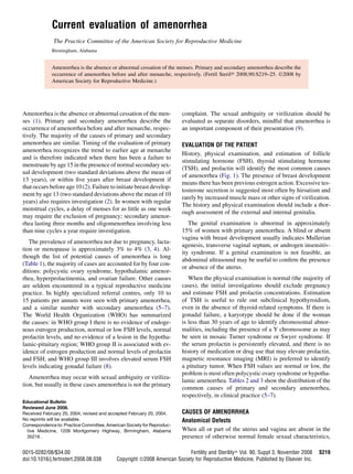
FIGURE 1
Suggested flow diagram aiding in the evaluation of women with amenorrhea.
ASRM Practice Committee. Amenorrhea. Fertil Steril 2008.
Fertility and Sterilityâ S221
4. amenorrhea. Uncommon causes of ovarian failure include
FSH or LH receptor mutations (17, 18), galactosemia, 17
a-hydroxylase or 17,20-lyase deficiency, and aromatase defi-
ciency (19–21).
In premature ovarian failure (POF), amenorrhea, persistent
estrogen deficiency, and elevated FSH levels occur prior to
the age of 40, and this condition affects 1% to 5% of women
(22, 23). Iatrogenic causes of POF, such as chemotherapy and
radiation therapy for malignancy, have a potential for recov-
ery. Ovarian function may fluctuate, with increasingly irreg-
ular menstrual cycles before the final depletion of oocytes
and permanent ovarian failure. The resulting fluctuation in
gonadotropin levels accounts for the lack of accuracy associ-
ated with a single FSH value (24).
Ovarian failure is confirmed by documenting an FSH level
persistently in the menopausal range. In women under 30 with
POF, a karyotype should be obtained to rule out sex chromo-
some translocation, short arm deletion, or the presence of an
occult Y chromosome, which is associated with an increased
risk of gonadal tumors. About 16% of women who are carriers
of the premutation of Fragile X syndrome experience prema-
ture menopause (19). A thorough family history should be
obtained because several autosomal disorders have been
associated with ovarian failure, including mutations of the
phosphomannomutase 2 (PMM2) gene, the galactose-1-phos-
phate uridyltransferase (GALT) gene, the FSH receptor
(FSHR) gene, chromosome 3q containing the Blepharophi-
mosis gene, and the autoimmune regulator (AIRE) gene, re-
sponsible for polyendocrinopathy-candidiasis-ectodermal
dystrophy (25). A further indication for karyotype and genetic
investigation is that some patients with POF have children for
whom the genetic information may be useful.
Up to 40% of women with POF may have autoimmune ab-
normalities, most commonly autoimmune thyroiditis (26,
27). POF is slightly more common in women with insulin-de-
pendent diabetes mellitus, myasthenia gravis, and parathy-
roid disease than in healthy women (28). Autoimmune
lymphocytic oophoritis may be seen in Addison’s disease,
in which 10% to 60% of cases may have ovarian failure,
but this condition is extremely rare (1 per million women).
Ovarian biopsy is not indicated in clinical practice, but be-
cause autoimmune POF could be a component of a polygland-
ular syndrome, patients can be screened for other
abnormalities by means of TSH, thyroid autoantibodies, fast-
ing glucose, and electrolytes (29). Thyroid autoantibodies
may increase the ability to identify individuals likely to de-
velop subsequent primary hypothyroidism. No currently
available validated serum antibody marker can confirm a clin-
ical diagnosis of autoimmune premature ovarian failure.
Also, at this time, no therapy for infertile patients with
autoimmune ovarian failure has been proven effective in
a prospective controlled study.
Patients with ovarian failure should be offered estrogen
and progestin treatment to promote and maintain secondary
sexual characteristics and reduce the risk of developing oste-
oporosis. In adolescents with gonadal failure, the aim is to
TABLE 2
Common causes of primary amenorrhea (4, 6).
Category
Approximate
frequency (%)
Breast development 30
Mullerian agenesis 10
Androgen insensitivity 9
Vaginal septum 2
Imperforate hymen 1
Constitutional delay 8
No breast development: high FSH 40
46 XX 15
46 XY 5
Abnormal 20
No breast development: low FSH 30
Constitutional delay 10
Prolactinomas 5
Kallman syndrome 2
Other CNS 3
Stress, weight loss, anorexia 3
PCOS 3
Congenital adrenal hyperplasia 3
Other 1
ASRM Practice Committee. Amenorrhea. Fertil Steril 2008.
TABLE 3
Common causes of secondary amenorrhea (5).
Category
Approximate
frequency (%)
Low or normal FSH 66
Weight loss/anorexia
Non-specific hypothalamic
Chronic anovulation
including PCOS
Hypothyroidism
Cushing’s syndrome
Pituitary tumor, empty sella,
Sheehan syndrome
Gonadal failure: high FSH 12
46 XX
Abnormal karyotype
High prolactin 13
Anatomic 7
Asherman syndrome
Hyperandrogenic states 2
Ovarian tumor
Non-classic CAH
Undiagnosed
ASRM Practice Committee. Amenorrhea. Fertil Steril 2008.
S222 ASRM Practice Committee Amenorrhea Vol. 90, Suppl 3, November 2008
5. mimic pubertal development with low-dose estrogens, in-
creasing gradually to augment breast development, avoiding
progestin until the breast mound and areola have developed.
Rarely, some ovarian follicles remain in women with ovarian
failure so that spontaneous ovulation and conception are pos-
sible, even in women taking exogenous estrogen with or
without a progestogen (29).
Elevated Prolactin Levels
Hyperprolactinemia is associated with decreased estradiol
concentrations and amenorrhea or oligomenorrhea. Prolactin
concentrations are higher in women with amenorrhea than in
those with oligomenorrhea (30). With persistent hyperprolac-
tinemia, after ruling out primary hypothyroidism, MRI of the
pituitary is indicated. Mildly elevated prolactin levels may be
a sign of another organic central nervous system lesion, such
as congenital aqueductal stenosis, non-functioning adeno-
mas, or any condition which causes pituitary stalk irritability.
In women with hyperprolactinemia, the prevalence of a pitu-
itary tumor is 50% to 60% (31). The likelihood of a pituitary
tumor was unrelated to the level of prolactin (31), and only
16% of the variability in tumor size was associated with
prolactin level (r¼0.40, p<.001) (32).
Usually, however, patients with amenorrhea have larger tu-
mors than patients with oligomenorrhea. The poor correlation
between tumor presence and prolactin level indicates that
MRI should be performed whenever prolactin levels are per-
sistently elevated. In most amenorrheic women with hyper-
prolactinemia, prolactin levels do not decline without
treatment, and the amenorrhea does not resolve as long as
the prolactin levels remain elevated (30, 32). In the absence
of another organic condition, dopamine agonists are the pre-
ferred treatment of hyperprolactinemia with or without a pitu-
itary tumor.
Normal or Low FSH Levels
Amenorrhea associated with normal or low FSH values and
chronic anovulation is frequently unexplained. The most
common diagnostic categories are hypothalamic amenorrhea
and polycystic ovary syndrome, and in each case similar but
less common conditions must be excluded. Hypothalamic
amenorrhea is characterized by inconsistent GnRH drive,
while in polycystic ovary syndrome GnRH pulses are persis-
tently rapid or increased, leading to excessive LH synthesis,
hyperandrogenism, and impaired follicular maturation (33).
Differentiating hypothalamic amenorrhea from polycystic
ovary syndrome depends on clinical judgment aided by the
presence of obesity and androgenization, which are typical
features of polycystic ovary syndrome. This judgment also
is relevant to the prognosis because obesity and androgeniza-
tion tend to reduce the likelihood of conception (34).
Estradiol concentration does not effectively distinguish be-
tween hypothalamic amenorrhea and polycystic ovary syn-
drome. Although hypothalamic amenorrhea implies that
levels of estradiol should be low, while normal levels of estra-
diol are expected with polycystic ovary syndrome, estradiol
concentrations tend to fluctuate and each condition is associ-
ated with both normal and low estrogen production. As an in-
dication of endogenous estrogen levels, the duration of the
amenorrhea and clinical features are more important than
measurement of estradiol, the progesterone challenge test,
or presence of cervical mucus. Although the progesterone
challenge test might seem to characterize estrogen produc-
tion, withdrawal bleeding correlates poorly with estrogen sta-
tus and the test imposes a delay on the diagnostic process. The
false positive rate is high: up to 20% of women with oligome-
norrhea or amenorrhea in whom estrogen is present have no
withdrawal bleeding (35). The false negative rate is also
high; withdrawal bleeding occurs in up to 40% of women
with amenorrhea due to stress, weight loss, exercise, or hyper-
prolactinemia where estrogen production is usually reduced
(36) and in up to 50% of women with ovarian failure (29).
Hypothalamic Amenorrhea
Functional disorders of the hypothalamus or higher centers
are the most common reason for chronic anovulation. Psy-
chogenic stress, weight changes, undernutrition, and exces-
sive exercise are frequently associated with functional
hypothalamic amenorrhea, but the pathophysiologic mecha-
nisms are unclear. More cases of amenorrhea are associated
with weight loss than with anorexia, which is rare (15 cases
per 100,000 women per year), but amenorrhea with anorexia
nervosa is more severe (37, 38). Women involved in compet-
itive sports activities have a three-fold higher risk of primary
or secondary amenorrhea than others, and the highest preva-
lence is among long-distance runners (39). Infrequently, hy-
pothalamic dysfunction occurs before menarche and presents
as primary amenorrhea in approximately 3% of adolescents;
usually secondary sexual characteristics will develop and
menstrual cycles will evolve without therapy (40).
Chronic debilitating diseases, such as uncontrolled juve-
nile diabetes, end-stage kidney disease, malignancy, acquired
immune deficiency syndrome, or malabsorption, which are
uncommon in women of reproductive age, may lead to anov-
ulation and amenorrhea through a central mechanism.
Other rare causes of hypothalamic amenorrhea include iso-
lated gonadotropin deficiency. This is most often due to Kall-
mann syndrome, which is associated with defects in olfactory
bulb development. Thus, these women may have anosmia as
well as amenorrhea and low gonadotropins due to gonadotro-
pin-releasing hormone (GnRH) deficiency (41). Mutations in
gonadotropin-releasing hormone receptor genes also may
be associated with hypogonadotropic hypogonadism (42). Pi-
tuitary disorders that cause anovulation include Sheehan syn-
drome, necrosis of the pituitary gland, and empty sella
syndrome (43). When amenorrhea persists and stress, exces-
sive exercise, or weight loss can be confidently excluded as
causes, MRI may be indicated to rule out organic disease in
the central nervous system, hypothalamus, or pituitary gland.
Women with hypothalamic amenorrhea are susceptible to
the development of osteoporosis (44). Unless the primary
cause can be easily treated, cyclic estrogen-progestin therapy
Fertility and Sterilityâ S223
6. or oral contraceptive pills should be initiated to prevent ex-
cessive bone loss. If pregnancy is desired, good nutrition
and optimal body weight are important objectives but may
be difficult to achieve. Ovulation induction with clomiphene
citrate, exogenous gonadotropins, or pulsatile GnRH therapy
should be offered (45).
Polycystic Ovary Syndrome
When amenorrhea is associated with evidence of androgen
excess, the most common disorder is polycystic ovary syn-
drome (PCOS). Less commonly, amenorrhea with hyperan-
drogenism arises from adrenal diseases, such as non-
classical adrenal hyperplasia and Cushing syndrome or
from androgen-producing tumors (46). Other disorders that
may cause chronic anovulation (Table 1) are much less com-
mon than PCOS, and in each case special characteristics are
likely to direct the investigation toward a specific diagnosis.
PCOS is characterized by menstrual disturbances ranging
from dysfunctional uterine bleeding to oligomenorrhea and
amenorrhea, hyperandrogenism, and infertility. Seventy-
five percent of North American women with PCOS are obese
(47). PCOS patients are more likely to present with oligome-
norrhea (76%) than amenorrhea (24%) (34, 48). The symp-
toms often occur first at menarche, but the signs of
androgen excess may not become evident until several years
later and these signs increase over time.
The 1990 National Institutes of Health/National Institute
of Child Health and Human Development (NIH/NICHHD)
criteria for PCOS, although not a consensus, were as follows:
(1) ovulatory dysfunction, (2) clinical evidence of hyperan-
drogenism (hirsutism, acne, androgenic alopecia) and/or hy-
perandrogenemia, and (3) exclusion of other related disorders
such as hyperprolactinemia, thyroid abnormalities, and non-
classical adrenal hyperplasia (49). An international consen-
sus conference held in 2003 concluded that the syndrome
‘‘encompasses a broader spectrum of signs and symptoms
of ovarian dysfunction than those defined by the original di-
agnostic criteria’’ (50). Therefore, participants concluded
that individuals must have two out of three of the following
features to be classified as having PCOS:
1 Oligo- and/or anovulation.
2 Clinical and/or biochemical signs of hyperandrogenism.
3 Ultrasound evidence of polycystic ovaries.
In addition, other etiologies such as congenital adrenal hy-
perplasia, androgen secreting tumors, and Cushing syndrome
must have been excluded. This definition is open to challenge
because at least one study has noted that ultrasonographic cri-
teria for diagnosis of PCOS are not useful because one-fifth
of women with regular cycles have ovaries that appear poly-
cystic (51). Many of the participants of the conference argued
that hyperandrogenic women with PCOS may have regular
menstrual cycles. No doubt the definition will continue to
evolve with new research findings.
Although several endocrine abnormalities are associated
with PCOS, there is no universally accepted definition based
on hormonal criteria. Serum androgen levels usually range
from upper normal to two-fold higher than normal in women
with PCOS. Prolactin levels are mildly elevated in 10% to
25% of women with PCOS. The LH/FSH ratio may be greater
than 2, but gonadotropin values are not useful to confirm the
diagnosis (47, 49–51).
Women with PCOS are frequently insulin resistant; insulin
sensitivity is reduced by 30% to 40%, leading to hyperinsuli-
nemia, but the insulin response may be inadequate because of
beta-cell dysfunction. Thus, PCOS patients are pre-disposed
to glucose intolerance (52). Impaired glucose tolerance oc-
curs in 31% of PCOS patients, but fasting glucose is elevated
in only 7.5%. It can be argued that women with PCOS should
be screened for type 2 diabetes (52, 53). Obesity further ex-
acerbates insulin resistance, and higher insulin concentra-
tions are associated with higher androgen levels (34).
CONCLUSIONS
Amenorrhea is an uncommon presentation in reproduc-
tive medicine.
The four most common causes are polycystic ovary syn-
drome, hypothalamic amenorrhea, ovarian failure, and
hyperprolactinemia.
The initial useful laboratory tests are FSH, TSH, and
prolactin.
Differentiating hypothalamic amenorrhea from poly-
cystic ovary syndrome depends on clinical judgment,
aided by the presence or absence of androgenization.
Acknowledgment: This report was developed under the direction of the Prac-
tice Committee of the American Society for Reproductive Medicine as a ser-
vice to its members and other practicing clinicians. While this document
reflects appropriate management of a problem encountered in the practice
of reproductive medicine, it is not intended to be the only approved standard
of practice or to dictate an exclusive course of treatment. Other plans of man-
agement may be appropriate, taking into account the needs of the individual
patient, available resources, and institutional or clinical practice limitations.
This report was approved by the Board of Directors of the American Society
for Reproductive Medicine in January 2004.
REFERENCES
1. Stedman’s Medical Dictionary. 27th ed. Philadelphia: Lippincott Wil-
liams Wilkins, 2000:56.
2. Herman-Giddens ME, Slora EJ, Wasserman RC, Bourdony CJ,
Bhapkar MV, Koch GG, et al. Secondary sexual characteristics and men-
ses in young girls seen in office practice: a study from the Pediatric
Research in Office Settings network. Pediatrics 1997;99:505–12.
3. Pettersson F, Fries H, Nillius SJ. Epidemiology of secondary amenor-
rhea. I. Incidence and prevalence rates. Am J Obstet Gynecol
1973;117:80–6.
4. Bachmann G, Kemmann E. Prevalence of oligomenorrhea and amenor-
rhea in a college population. Am J Obstet Gynecol 1982;144:98–102.
5. Reindollar RM, Byrd JR, McDonough PG. Delayed sexual development:
a study of 252 patients. Am J Obstet Gynecol 1981;140:371–80.
6. Reindollar RH, Novak M, Tho SP, McDonough PG. Adult-onset amen-
orrhea: a study of 262 patients. Am J Obstet Gynecol 1986;155:531–43.
7. Mashchak CA, Kletzky OA, Davajan V, Mishell DR. Clinical and labo-
ratory evaluation of patients with primary amenorrhea. Obstet Gynecol
1981;57:715–21.
8. Insler V. Gonadotophin therapy: new trends and insights. Int J Fertil
1988;33:85–97.
S224 ASRM Practice Committee Amenorrhea Vol. 90, Suppl 3, November 2008
7. 9. Doody KM, Carr BR. Amenorrhea. Obstet Gynecol Clin North Am
1990;17:361–87.
10. Jagiello G. Prevalence of testicular feminization. Lancet 1962;1:329.
11. Wilson JD. Syndromes of androgen resistance. Biol Reprod 1992;46:
168–73.
12. Lobo RA. Primary and secondary amenorrhea. In: Fraser IS, Jansen R,
Lobo RA, Whitehead M, eds. Estrogens and progestogens in clinical
practice. London: Churchill Livingstone, 1998.
13. Reid RL. Amenorrhea. In: Copeland LJ, ed. Textbook of gynecology.
2nd ed. Philadelphia: WB Saunders, 1996.
14. Manuel M, Katayama PK, Jones HW Jr. The age of occurrence of
gonadal tumors in intersex patients with a Y chromosome. Am J Obstet
Gynecol 1976;124:293–300.
15. Turner HH. A syndrome of infantilism, congenital webbed neck, and
cubitus valgus. Endocrinology 1938;23:566.
16. Turner’s syndrome. West J Med 1982;137:32–44.
17. Toledo SP, Brunner HG, Kraaij R, Post M, Dahia PL, Hayashida CY,
et al. An inactivating mutation of the luteinizing hormone receptor
causes amenorrhea in a 46,XX female. J Clin Endocrinol Metab
1996;81:3850–4.
18. Aittomaki K, Lucena JL, Pakarinen P, Sistonen P, Tapanainen J,
Gromoll J, et al. Mutation in the follicle-stimulating hormone receptor
gene causes hereditary hypergonadotropic ovarian failure. Cell
1995;82:959–68.
19. Allingham-Hawkins DJ, Babul-Hirji R, Chitayat D, Holden JJ, Yang KT,
Lee C, et al. Fragile X premutation is a significant risk factor for prema-
ture ovarian failure: the International Collaborative POF in Fragile X
study—preliminary data. Am J Med Genet 1999;83:322–5.
20. Laml T, Preyer O, Umek W, Hengstschlager M, Hanzal H. Genetic dis-
orders in premature ovarian failure. Hum Reprod Update 2002;8:483–91.
21. Morishima A, Grumbach MM, Simpson ER, Fisher C, Qin K. Aromatase
deficiency in male and female siblings caused by a novel mutation and
the physiological role of estrogens. J Clin Endocrinol Metab 1995;80:
3689–98.
22. Jones GS, De Moraes-Ruehsen M. A new syndrome of amenorrhea in
association with hypergonadotropism and apparently normal ovarian
follicular apparatus. Am J Obstet Gynecol 1969;104:597–600.
23. Van Campenhout J, Vauclair R, Maraghi K. Gonadotropin-resistant ova-
ries in primary amenorrhea. Obstet Gynecol 1972;40:6–12.
24. Conway GS, Kaltsas G, Patel A, Davies MC, Jacobs HS. Characteriza-
tion of idiopathic premature ovarian failure. Fertil Steril 1996;65:
337–41.
25. Laml T, Preyer J, Umek W, Hengstschlager M, Hanzal E. Genetic disor-
ders in premature ovarian failure. Hum Reprod Update 2002;8:483–91.
26. LaBarbera AR, Miller MM, Ober C, Rebar RW. Autoimmune etiology in
premature ovarian failure. Am J Reprod Immunol Microbiol 1988;16:
115–22.
27. Hoek A, Schoemaker J, Drexhage HA. Premature ovarian failure and
ovarian autoimmunity. Endocr Rev 1997;18:107–34.
28. Nelson LM, Anasti JN, Flack MR. Premature ovarian failure. In:
Adashi EY, RockJA,Rosenwaks Z,eds.Reproductiveendocrinology,sur-
gery, and technology. Philadelphia: Lippincott-Raven, 1996:1393–410.
29. Rebar RW, Connolly HV. Clinical features of young women with hyper-
gonadotropic amenorrhea. Fertil Steril 1990;53:804–10.
30. Touraine P, Plu-Bureau G, Beji C, Mauvais-Jarvis P, Kuttenn F. Long-
term follow-up of 246 hyperprolactinemic patients. Acta Obstet Gynecol
Scand 2001;80:162–8.
31. Brenner SH, Lessing JB, Quagliarello J, Weiss G. Hyperprolactinemia
and associated pituitary prolactinomas. Obstet Gynecol 1985;65:661–4.
32. Schlechte J, Dolan K, Sherman B, Chapler F, Luciano A. The natural his-
tory of untreated hyperprolactinemia: a prospective analysis. J Clin En-
docrinol Metab 1989;68:412–8.
33. Marshall JC, Eagleson CA, McCartney CR. Hypothalamic dysfunction.
Mol Cell Endocinol 2002;183:29–32.
34. Imani B, Eijkemans MJ, te Velde ER, Habbema JD, Fauser BC. A nomo-
gram to predict the probability of live birth after clomiphene citrate
induction of ovulation in normogonadotropic oligoamenorrheic infertil-
ity. Fertil Steril 2002;77:91–7.
35. Rarick LD, Shangold MM, Ahmed SW. Cervical mucus and serum estra-
diol as predictors of response to progestin challenge. Fertil Steril
1990;54:353–5.
36. Nakamura S, Douchi T, Oki T, Ijuin H, Yamamoto S, Nagata Y. Relation-
ship between sonographic endometrial thickness and progestin-induced
withdrawal bleeding. Obstet Gynecol 1996;87:722–5.
37. Frisch RE, McArthur JW. Menstrual cycles: fatness as a determinant of
minimum weight for height necessary for their maintenance or onset.
Science 1974;185:949–51.
38. Lucas AR, Crowson CS, O’Fallon WM, Melton LJ 3rd. The ups and
downs of anorexia nervosa. Int J Eat Disord 1999;26:397–405.
39. Warren MP, Goodman LR. Exercise-induced endocrine pathologies.
J Endocrinol Invest 2003;26:873–8.
40. Rosenfield RL. Clinical review 6: diagnosis and management of delayed
puberty. J Clin Endocrinol Metab 1990;70:559–62.
41. Hall JE. Physiologic and genetic insights into the pathophysiology and
management of hypogonadotropic hypogonadism. Ann Endocrinol
1999;60:93–101.
42. Layman LC, McDonough PG, Cohen DP, Maddox M, Tho SP,
Reindollar RH. Familial gonadotropin-releasing hormone resistance
and hypogonadotropic hypogonadism in a family with multiple affected
individuals. Fertil Steril 2001;75:1148–55.
43. Sheehan HL. Simmond’s disease due to post-partum necrosis of the
anterior pituitary. Q J Med 1939;8:277.
44. Davies MC, Hall ML, Jacobs HS. Bone mineral loss in young women
with amenorrhoea. BMJ 1990;301:790–3.
45. Martin KA, Hall JE, Adams JM, Crowley WF Jr. Comparison of exoge-
nous gonadotropins and pulsatile gonadotropin-releasing hormone for
induction of ovulation in hypogonadotropic amenorrhea. J Clin Endocri-
nol Metab 1993;77:125–9.
46. Moran C, Azziz R, Carmina E, Dewailly D, Fruzzetti F, Ibanez L, et al.
21-Hydroxylase-deficient nonclassic adrenal hyperplasia is a progressive
disorder: a multicenter study. Am J Obstet Gynecol 2000;183:1468–74.
47. Legro RS. Polycystic ovary syndrome: the new millennium. Mol Cell
Endocrinol 2001;184:87–93.
48. Bili H, Laven J, Imani B, Eijkemans MJ, Fauser BC. Age-related differ-
ences in features associated with polycystic ovary syndrome in normogo-
nadotrophic oligo-amenorrhoeic infertile women of reproductive years.
Eur J Endocrinol 2001;145:749–55.
49. Zawadzki JK, Dunaif A. Diagnostic criteria for polycystic ovary syn-
drome: towards a rational approach. In: Dunaif A, Givens JR, Haselti-
ne FP, Merriam GM, eds. Polycystic ovary syndrome. Boston:
Blackwell, 1992:377–84.
50. Rotterdam ESHRE/ASRM-Sponsored PCOS Consensus Workshop
Group. Revised 2003 consensus on diagnostic criteria and long-term
health risks related to polycystic ovary syndrome. Fertil Steril
2004;81:19–25.
51. Dunaif A, Thomas A. Current concepts in the polycystic ovary syn-
drome. Annu Rev Med 2001;52:401–19.
52. Legro RS, Kunselman AR, Dodson WC, Dunaif A. Prevalence and pre-
dictors of risk for type 2 diabetes mellitus and impaired glucose tolerance
in polycystic ovary syndrome: a prospective, controlled study in 254
affected women. J Clin Endocrinol Metab 1999;84:165–9.
53. Dunaif A, Finegood DT. Beta-cell dysfunction independent of obesity
and glucose intolerance in the polycystic ovary syndrome. J Clin Endo-
crinol Metab 1996;81:942–7.
Fertility and Sterilityâ S225