2. April 15, 2006 Volume 73, Number 8 www.aafp.org/afp American Family Physician 1375
Amenorrhea
SORT: KEY RECOMMENDATIONS FOR PRACTICE
Clinical recommendation
Evidence
rating References
A female patient with primary amenorrhea and sexual development, including pubic hair,
should be evaluated for the presence of a uterus and vagina.
C 1, 18
Women with secondary amenorrhea should receive pregnancy tests. C 1-3, 6
Women with polycystic ovary syndrome should be tested for glucose intolerance. C 21
A = consistent, good-quality patient-oriented evidence; B = inconsistent or limited-quality patient-oriented evidence; C = consensus, disease-
oriented evidence, usual practice, expert opinion, or case series. For information about the SORT evidence rating system, see page 1313 or
http://www.aafp.org/afpsort.xml.
TABLE 1
Normal Female Pubertal Development
Developmental stage
(age in years) Anatomic drawing
Tanner stage
Breast
development
Pubic hair
development
Initial growth acceleration (8 to 10) Elevation of papilla only; no pubic hair 1 1
Thelarche (9 to 11) See adrenarche for stage 2 development 2 1
Adrenarche (9 to 11) 2 2
Peak growth (11 to 13) 3 3
Menarche (12 to 14) 4 4
Adult characteristics (13 to 16) 5 5
Illustrations by Renee Cannon.
Information from references 4 and 5.
3. 1376 American Family Physician www.aafp.org/afp Volume 73, Number 8 April 15, 2006
Amenorrhea
7.5 percent of participants had abnormal prolactin levels
and 4.2 percent had abnormal TSH levels.
If TSH and prolactin levels are normal, a progestogen
challenge test (Table 33,14
) can help evaluate for a patent
outflow tract and detect endogenous estrogen that is
affecting the endometrium. A withdrawal bleed usually
occurs two to seven days after the challenge test.3
A nega-
tive progestogen challenge test signifies an outflow tract
abnormality or inadequate estrogenization. An estrogen/
progestogen challenge test (Table 33,14
) can differentiate
the two diagnoses. A negative estrogen/progestogen chal-
lenge test typically indicates an outflow tract obstruc-
tion. A positive test indicates an abnormality within the
hypothalamic-pituitary axis or the ovaries.
TABLE 2
History and Physical Examination Findings Associated with Amenorrhea
Findings Associations
Patient history
Exercise, weight loss, current or previous chronic illness,
illicit drug use
Hypothalamic amenorrhea
Menarche and menstrual history Primary versus secondary amenorrhea
Prescription drug use Multiple, depending on medication
Previous central nervous system chemotherapy or radiation Hypothalamic amenorrhea
Previous pelvic radiation Premature ovarian failure
Psychosocial stressors; nutritional and exercise history Anorexia or bulimia nervosa
Sexual activity Pregnancy
Family history
Genetic defects Multiple causes of primary amenorrhea
Pubic hair pattern Androgen insensitivity syndrome
Infertility Multiple
Menarche and menstrual history (mother and sisters) Constitutional delay of growth and puberty
Pubertal history (e.g., growth delay) Constitutional delay of growth and puberty
Physical examination
Anthropomorphic measurements; growth chart Constitutional delay of growth and puberty
Body mass index Polycystic ovary syndrome
Dysmorphic features (e.g., webbed neck, short stature,
widely spaced nipples)
Turner’s syndrome
Rudimentary or absent uterus; pubic hair Müllerian agenesis
Striae, buffalo hump, significant central obesity, easy bruising,
hypertension, or proximal muscle weakness
Cushing’s disease
Tanner staging (Table 1) Primary versus secondary amenorrhea
Thyroid examination Thyroid disease
Transverse vaginal septum; imperforate hymen Outflow tract obstruction
Undescended testes; external genital appearance; pubic hair Androgen insensitivity syndrome
Virilization; clitoral hypertrophy Androgen-secreting tumor
Review of systems
Anosmia Kallmann syndrome
Cyclic abdominal pain; breast changes Outflow tract obstruction or müllerian agenesis
Galactorrhea; headache and visual disturbances Pituitary tumor
Hirsutism or acne Polycystic ovary syndrome
Signs and symptoms of hypothyroidism or hyperthyroidism Thyroid disease
Vasomotor symptoms Premature ovarian failure
Information from references 2 and 6 through 8.
4. April 15, 2006 Volume 73, Number 8 www.aafp.org/afp American Family Physician 1377
Amenorrhea
Gonadotropin levels can further help determine the
source of the abnormality. Elevated follicle-stimulat-
ing hormone (FSH) or luteinizing hormone (LH) levels
suggest an ovarian abnormality (hypergonadotropic
hypogonadism). Normal or low FSH or LH levels suggest
a pituitary or hypothalamic abnormality (hypogonado-
tropic hypogonadism). Magnetic resonance imaging
(MRI) of the sella turcica can rule out a pituitary
tumor. Normal MRI indicates a hypothalamic cause of
amenorrhea.3
Differential Diagnosis of Primary Amenorrhea
Causes of primary amenorrhea should be evaluated in the
context of the presence or absence of secondary sexual
characteristics. Table 43,6,15
includes the differential diag-
nosis of primary amenorrhea.
PRESENCE OF SECONDARY SEXUAL CHARACTERISTICS
If a patient with amenorrhea has breast development and
minimal or no pubic hair, the usual diagnosis is androgen
insensitivity syndrome (i.e., patient is phenotypically female
but genetically male with undescended testes). A karyotype
analysis is needed to determine proper treatment. If testes
are present, they should be removed because of the high
risk of malignant transformation after puberty.1
If a patient has normal secondary sexual characteris-
tics, including pubic hair, the physician should perform
MRI or ultrasonography to determine if a uterus is
present. Müllerian agenesis (the congenital absence of
a vagina and abnormal uterine development [usually
rudimentary]) causes approximately 15 percent of pri-
mary amenorrhea.16
The etiology is thought to involve
embryonic activation of the antimüllerian hormone,
causing malformation of the female genital tract.7,17
Patients may have cyclic abdominal pain if there is endo-
metrial tissue in the rudimentary uterus, mittelschmerz,
or breast tenderness. An absent or truncated vagina and
an abnormal adult uterus confirm müllerian agenesis.
Karyotype analysis should be performed to determine if
the patient is genetically female.8
If the patient has a normal uterus, outflow tract
obstructionshouldbeconsidered.Animperforatehymen
or a transverse vaginal septum can cause congenital out-
flow tract obstruction, which typically is associated with
cyclic abdominal pain from blood accumulation in the
uterus and vagina.1
If the outflow tract is patent, the
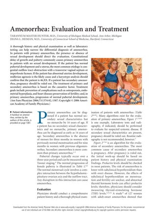
Evaluation of Primary Amenorrhea
Figure 1. Algorithm for the evaluation of primary amenorrhea. (FSH = follicle-stimulating hormone; LH = luteinizing
hormone.)
Information from references 1, 7, 9, and 10.
History and physical examination completed
for a patient with primary amenorrhea
Secondary sexual characteristics present
Perform ultrasonography of uterus.Measure FSH and LH levels.
Hypogonadotropic
hypogonadism (Table 4)
No Yes
Uterus present
or normal
Uterus absent
or abnormal
FSH >20 IU per L and
LH >40 IU per L
FSH and LH <5 IU per L
Hypergonadotropic
hypogonadism
Karyotype analysis
45,XO
Turner’s
syndrome
46,XX
Premature
ovarian failure
Müllerian
agenesis
Androgen
insensitivity
syndrome
Karyotype analysis
46,XX46,XY
Outflow obstruction
Imperforate hymen
or transverse
vaginal septum
Evaluate for secondary
amenorrhea (Figure 2).
No Yes
5. 1378 American Family Physician www.aafp.org/afp Volume 73, Number 8 April 15, 2006
Amenorrhea
physician should continue an evaluation similar to that
for secondary amenorrhea (Figure 21-3,6
).1
ABSENCE OF SECONDARY SEXUAL CHARACTERISTICS
Diagnosis of patients with amenorrhea and no second-
ary sexual characteristics is based on laboratory test
results and karyotype analysis. The most common cause
of hypogonadotropic hypogonadism (low FSH and LH
levels) in primary amenorrhea is constitutional delay of
growth and puberty.16,17
A detailed family history also
may help detect this etiology, because it often is familial.
Hypogonadotropic hypogonadism associated with con-
stitutional delay of growth and puberty is indistinguish-
able from that associated with hypothalamic or pituitary
failure.10
Watchful waiting is appropriate for constitu-
tional delay of growth and puberty. Kallmann syndrome,
which is associated with anosmia, also can cause hypogo-
nadotropic hypogonadism.18
Hypergonadotropic hypogonadism (elevated FSH and
LH levels) in patients with primary amenorrhea is caused
by gonadal dysgenesis or premature ovarian failure.
Turner’s syndrome (45,XO karyotype) is the most com-
mon form of female gonadal dysgenesis. Characteristic
physical findings include webbing of the neck, widely
Evaluation of Secondary Amenorrhea
Figure 2. Algorithm for the evaluation of secondary amenorrhea. (TSH = thyroid-stimulating hormone; MRI = magnetic
resonance imaging; FSH = follicle-stimulating hormone; LH = luteinizing hormone.)
Information from references 1 through 3 and 6.
Patient presenting with secondary
amenorrhea; negative pregnancy test
Check TSH and prolactin levels.
Normal TSH level,
abnormal prolactin level
Progestogen
challenge test
(Table 3)
Withdrawal bleed
Normogonadotropic
hypogonadism (Table 4)
No withdrawal bleed
Estrogen/progestogen
challenge test (Table 3)
Withdrawal bleed
Check FSH and LH levels.
Hypergonadotropic
hypogonadism (Table 4)
Perform MRI to evaluate
for pituitary tumor.
Normal MRI: hypogonadotropic
hypogonadism (Table 4)
No withdrawal bleed
Outflow obstruction
Normal prolactin level,
abnormal TSH level
Thyroid disease
FSH >20 IU per L and
LH >40 IU per L
Prolactin 100 ng per mL
(100 mcg per L)
Prolactin >100 ng per mL
Perform MRI to evaluate
for prolactinoma.Consider other
causes (Table 4).
Both normal
FSH and LH <5 IU per L
Negative MRI
Consider other
causes (Table 4).
6. April 15, 2006 Volume 73, Number 8 www.aafp.org/afp American Family Physician 1379
Amenorrhea
spaced nipples, and short stature. Mosaicism occurs
in approximately 25 percent of patients with Turner’s
syndrome.19
These patients often have a more normal
phenotype with spontaneous onset of puberty and men-
arche. Other rare causes of pure gonadal dysgenesis can
occur with a 46,XY or XX karyotype.7
Differential Diagnosis of Secondary Amenorrhea
After pregnancy, thyroid disease, and hyperprolactinemia
are eliminated as potential diagnoses, the remaining
causes of secondary amenorrhea are classified as nor-
mogonadotropic amenorrhea, hypogonadotropic hypo-
gonadism, and hypergonadotropic hypogonadism; each
is associated with specific etiologies (Table 43,6,15
).
HYPOTHYROIDISM
Other clinical signs of thyroid disease are usually noted
before amenorrhea presents. Mild hypothyroidism is
more often associated with hypermenorrhea or oligo-
menorrhea than with amenorrhea. Treatment of hypo-
thyroidism should restore menses, but this may take
several months.12
HYPERPROLACTINEMIA
A patient with markedly elevated prolactin levels, galac-
torrhea, headaches, or visual disturbances should receive
imaging tests to rule out a pituitary tumor. Adenomas
are the most common cause of anterior pituitary dys-
function.15
A prolactin level more than 100 ng per mL
(100 mcg per L) suggests a prolactinoma, and MRI
should be performed. If tumor is excluded as the cause,
medications (e.g., oral contraceptive pills, antipsychotics,
antidepressants, antihypertensives, histamine H2 block-
ers, opiates) are the next most common cause of hyper-
prolactinemia. Medications usually raise prolactin levels
to less than 100 ng per mL.15
When hyperprolactinemia
is not related to tumor, physicians should identify and
treat or eliminate the underlying cause. Table 43,6,15
lists
common etiologies of hyperprolactinemia.
If asymptomatic microadenomas (smaller than 10
mm) are found on MRI, repeat prolactin measurements
and imaging should be performed to monitor for pro-
gression. Microadenomas are slow growing and rarely
malignant. Treatment of microadenomas should focus
on management of infertility, galactorrhea, and breast
discomfort. A dopamine agonist can help improve
symptoms and fertility. Bromocriptine (Parlodel) is
effective, but cabergoline (Dostinex) has been shown
to be superior in effectiveness and tolerability.20
Mac-
roadenomas may be treated with dopamine agonists or
removed with transsphenoidal resection or craniotomy,
if necessary.
NORMOGONADOTROPIC AMENORRHEA
Two common causes of normogonadotropic amenor-
rhea are outflow tract obstruction and hyperandrogenic
chronic anovulation. The most common cause of out-
flow obstruction in secondary amenorrhea is Asherman’s
syndrome (intrauterine synechiae and scarring, usually
from curettage or infection).3
Hysterosalpingography,
hysteroscopy, or sonohysterography can help diagnose
Asherman’s syndrome. Other causes of outflow tract
obstruction include cervical stenosis and obstructive
fibroids or polyps.
Polycystic ovary syndrome (PCOS) is the most com-
mon cause of hyperandrogenic chronic anovulation.
The National Institutes of Health diagnostic criterion for
PCOS21
is chronic anovulation and hyperandrogenism
TABLE 3
Guidelines for Progestogen and Estrogen/Progestogen Challenge Tests
Drug Dosing Duration
Progestogen challenge test
Medroxyprogesterone acetate (Provera) 10 mg orally once per day Seven to 10 days
Norethindrone (Aygestin) 5 mg orally once per day Seven to 10 days
Progesterone 200 mg parenterally once per day Single dose
Progesterone micronized 400 mg orally once per day Seven to 10 days
Progesterone micronized gel (4 or 8%) Intravaginally every other day Six applications
Estrogen/progestogen challenge test
Conjugated equine estrogen (Premarin)
or
Estradiol (Estrace)
followed by
Progestational agent
1.25 mg orally once per day
2 mg orally once per day
As noted above
21 days
21 days
As noted above
Information from references 3 and 14.
7. 1380 American Family Physician www.aafp.org/afp Volume 73, Number 8 April 15, 2006
Amenorrhea
with no other identified secondary cause. The primary
etiology of PCOS is unknown, but resistance to insulin
is thought to be a fundamental component.21
The diagnosis of PCOS is primarily clinical, although
laboratory studies may be needed to rule out other causes
of hyperandrogenism (Table 56,21
). Significantly elevated
testosterone or dehydroepiandrosterone sulfate levels
indicate a possible androgen-secreting tumor (ovarian
or adrenal). Levels of 17-hydroxyprogesterone can help
diagnose adult-onset congenital adrenal hyperplasia.
Cushing’s disease is rare; therefore, patients should only
be screened when characteristic signs and symptoms
(e.g., striae, buffalo hump, significant central obesity,
easy bruising, hypertension, proximal muscle weakness)
are present.21,22
Patients with PCOS have excess unopposed circulat-
ing estrogen, increasing their risk of endometrial cancer
threefold.21
The insulin resistance associated with PCOS
increases a patient’s risk of diabetes mellitus two- to
fivefold; therefore, testing for glucose intolerance should
be considered.21-24
The primary treatment for PCOS is weight loss
through diet and exercise. Modest weight loss can lower
androgen levels, improve hirsutism, normalize menses,
and decrease insulin resistance. It may take months to
see these results, however.21
Use of oral contraceptive
pills or cyclic progestational agents can help maintain
a normal endometrium. The optimal cyclic progestin
regimen to prevent endometrial cancer is unknown,
but a monthly 10- to 14-day regimen is recommended.21
Insulin sensitizing agents such as metformin (Gluco-
phage) can reduce insulin resistance and improve ovula-
tory function.21,25,26
HYPERGONADOTROPIC HYPOGONADISM
Ovarian failure can cause menopause or can occur pre-
maturely. On average, menopause occurs at 50 years of
age and is caused by ovarian follicle depletion. Premature
TABLE 4
Causes of Amenorrhea
Hyperprolactinemia
Prolactin 100 ng per mL
(100 mcg per L)
Altered metabolism
Liver failure
Renal failure
Ectopic production
Bronchogenic
(e.g., carcinoma)
Gonadoblastoma
Hypopharynx
Ovarian dermoid cyst
Renal cell carcinoma
Teratoma
Breastfeeding
Breast stimulation
Hypothyroidism
Medications
Oral contraceptive pills
Antipsychotics
Antidepressants
Antihypertensives
Histamine H2 receptor
blockers
Opiates, cocaine
Prolactin >100 ng per mL
Empty sella syndrome
Pituitary adenoma
*—Causes of primary amenorrhea only.
Information from references 3, 6, and 15.
Hypogonadotropic hypogonadism
(continued)
Excessive exercise
Excessive weight loss or malnutrition
Hypothalamic or pituitary destruction
Kallmann syndrome*
Sheehan’s syndrome
Normogonadotropic
Congenital
Androgen insensitivity syndrome*
Müllerian agenesis*
Hyperandrogenic anovulation
Acromegaly
Androgen-secreting tumor (ovarian or
adrenal)
Cushing’s disease
Exogenous androgens
Nonclassic congenital adrenal hyperplasia
Polycystic ovary syndrome
Thyroid disease
Outflow tract obstruction
Asherman’s syndrome
Cervical stenosis
Imperforate hymen*
Transverse vaginal septum*
Other
Pregnancy
Thyroid disease
Hypergonadotropic hypogonadism
Gonadal dysgenesis
Turner’s syndrome*
Other*
Postmenopausal ovarian failure
Premature ovarian failure
Autoimmune
Chemotherapy
Galactosemia
Genetic
17-hydroxylase deficiency syndrome
Idiopathic
Mumps
Pelvic radiation
Hypogonadotropic hypogonadism
Anorexia or bulimia nervosa
Central nervous system tumor
Constitutional delay of growth and puberty*
Chronic illness
Chronic liver disease
Chronic renal insufficiency
Diabetes
Immunodeficiency
Inflammatory bowel disease
Thyroid disease
Severe depression or psychosocial stressors
Cranial radiation
8. April 15, 2006 Volume 73, Number 8 www.aafp.org/afp American Family Physician 1381
Amenorrhea
ovarian failure is characterized by amenorrhea, hypoes-
trogenism, and increased gonadotropin levels occurring
before 40 years of age and is not always irreversible27
(0.1 percent of women are affected by 30 years of age
and one percent by 40 years of age).28
Approximately
50 percent of women with premature ovarian failure have
intermittent ovarian functioning29
with a 5 to 10 percent
chance of achieving natural conception.
Women with premature ovarian failure have an
increased risk of osteoporosis and heart disease.29-31
The condition also can be associated with autoim-
mune endocrine disorders such as hypothyroidism,
Addison’s disease, and diabetes mellitus.27,29
Therefore,
fasting glucose, thyroid-stimulating hormone (TSH),
and, if clinically appropriate, morning cortisol levels
should be measured. Other laboratory testing should be
determined based on the individual patient.32
Approxi-
mately 20 to 40 percent of women with premature ovar-
ian failure will develop another autoimmune disorder;
therefore, if initial laboratory tests are normal, periodic
screening should be considered. Patients younger than
30 years should receive a karyotype analysis to rule
out the presence of a Y chromosome and the need for
removal of gonadal tissue.29
Ovarian biopsy and anti-
ovarian antibody testing have not been shown to have
clinical benefit.27,29
HYPOGONADOTROPIC HYPOGONADISM
Hypothalamic amenorrhea is associated with abnor-
malities in gonadotropin-releasing hormone (GnRH)
secretion and disruption of the hypothalamic-pituitary-
ovarian axis. The condition often is caused by excessive
weight loss, exercise, or stress. Other causes are listed
in Table 4.3,6,15
The mechanism of how stress or weight
loss affects GnRH secretion is unknown.33-35
Treatment
of hypothalamic amenorrhea depends on the etiology.
Women with excessive weight loss should be screened
for eating disorders and treated if anorexia nervosa or
bulimia nervosa is diagnosed. Menses usually will return
after a healthy body weight is acheived.35
Young athletes may develop a combination of health
conditions called the female athlete triad that includes
an eating disorder, amenorrhea, and osteoporosis. Men-
ses may return after a modest increase in caloric intake
or a decrease in athletic training. Similar to patients
with eating disorders, athletes with continued amenor-
rhea are at risk of bone loss. In adolescent athletes, the
bone loss occurs during peak bone mass development
and may not be reversible.36,37
Weight-bearing exercise
may partially protect against bone loss.38
In patients with amenorrhea caused by eating disor-
ders or excessive exercise, the use of oral contraceptive
pills or menopausal hormone therapy may decrease
bone turnover and partially reverse bone loss; however,
neither therapy has been shown to significantly increase
bone mass.38
Bisphosphonates, traditionally used to
treat postmenopausal osteoporosis, are possible terato-
gens and have not been studied as a therapy in women
of reproductive age. Adequate calcium and vitamin D
intake are recommended for these patients.
TABLE 5
Laboratory Evaluation of Hyperandrogenism
Findings Indications
Serum testosterone (normal: 20 to 80 ng per dL [0.7 to 2.8 nmol per L])
200 ng per dL (6.9 nmol per L) Consider hyperandrogenic chronic anovulation*
>200 ng per dL Evaluate for androgen-secreting tumor
Serum dehydroepiandrosterone sulfate (normal: 250 to 300 ng per dL [0.7 to 0.8 μmol per L])
700 ng per dL (1.9 μmol per L) Consider hyperandrogenic chronic anovulation*
>700 ng per dL Evaluate for adrenal or ovarian tumor
Serum 17-hydroxyprogesterone (normal: <2 ng per mL (6.1 nmol per L])†
>4 ng per mL (12.1 nmol per L) Consider adrenocorticotropic stimulation test to diagnose
congenital adrenal hyperplasia
Dexamethasone suppression test (if clinically indicated)††
Morning cortisol level > 5 μg per dL (138 nmol per L)§ Evaluate for Cushing’s disease
*— These values are not specific for diagnosis of hyperandrogenic chronic anovulation.
†—Morning level during follicular phase of menstrual cycle.
††—For an overnight dexamethasone suppression test, the physician should administer a 1-mg dose of dexamethasone orally between 11 p.m. and
midnight and draw a single blood sample for serum cortisol testing at 8 a.m. the following day.
§—Morning cortisol level in a healthy patient with an intact hypothalamic-pituitary axis. There is some variability in the cutoff values that can affect
sensitivity and specificity of the test. Patients should receive further testing to confirm Cushing’s disease.
Information from references 6 and 21.
9. 1382 American Family Physician www.aafp.org/afp Volume 73, Number 8 April 15, 2006
Amenorrhea
The authors thank Barbara S. Apgar, M.D., M.S., for her assistance in the
preparation of this manuscript.
The Authors
TARANNUM MASTER-HUNTER, M.D., C.A.Q., is lecturer in the Department
of Family Medicine at the University of Michigan Medical School, Ann
Arbor. She received a medical degree from the University of Medicine
and Dentistry of New Jersey-New Jersey Medical School in Newark. Dr.
Master-Hunter completed a family practice residency at the University of
Michigan Medical School.
DIANA L. HEIMAN, M.D., C.A.Q., is assistant professor in the Department
of Family Medicine at the University of Connecticut School of Medicine,
Hartford. She received a medical degree from Case Western Reserve
University School of Medicine in Cleveland, Ohio. Dr. Heiman completed a
family practice residency at the University of Virginia School of Medicine,
Charlottesville.
Address correspondence to Tarannum Master-Hunter, M.D., C.A.Q.,
Chelsea Family Practice, 14700 Old U.S. Hwy. 12, Chelsea, MI 48118
(e-mail: tarannum@umich.edu). Reprints are not available from the
authors.
Author disclosure: Nothing to disclose.
REFERENCES
1. The Practice Committee of the American Society for Reproductive Med-
icine. Current evaluation of amenorrhea. Fertil Steril 2004;82(suppl 1):
S33-9.
2. American College of Obstetricians and Gynecologists. Amenorrhea
(ACOG Technical Bulletin 128). Washington, D.C.: ACOG, 1989.
3. Speroff L, Fritz MA. Amenorrhea. In: Clinical gynecologic endocrinol-
ogy and infertility. 7th ed. Philadelphia, Pa.: Lippincott Williams &
Wilkins, 2005;401-64.
4. Marshall WA, Tanner JM. Variations in patterns of pubertal changes in
girls. Arch Dis Child 1969;44:291-303.
5. Speroff L, Glass RH, Kase NG. Normal and abnormal sexual develop-
ment. In: Clinical gynecologic endocrinology and infertility. 6th ed.
Baltimore, Md.: Lippincott Williams & Wilkins, 1999:339-79.
6. Kiningham RB, Apgar BS, Schwenk TL. Evaluation of amenorrhea. Am
Fam Physician 1996;53:1185-94.
7. Pletcher JR, Slap GB. Menstrual disorders. Pediatr Clin North Am
1999;46:505-18.
8. Reindollar RH, Byrd JR, McDonough PG. Delayed sexual development:
a study of 252 patients. Am J Obstet Gynecol 1981;140:371-80.
9. McIver B, Romanski SA, Nippoldt TB. Evaluation and management of
amenorrhea. Mayo Clin Proc 1997;72:1161-9.
10. Albanese A, Stanhope R. Investigation of delayed puberty. Clin Endo-
crinol (Oxf) 1995;43:105-10.
11. Arojoki M, Jokimaa V, Juuti A, Koshinen P, Irajala K, Anttila L. Hypothy-
roidism among infertile women in Finland. Gynecol Endocrinol 2000;
14:127-31.
12. Kalro B. Impaired fertility caused by endocrine dysfunction in women.
Endocrinol Metab Clin North Am 2003;32:573-92.
13. Laufer MR, Floor AE, Parsons KE, Kuntz KM, Barbieri RL. Hormone
testing in women with adult onset amenorrhea. Gynecol Obstet Invest
1995;40:200-3.
14. Warren MP, Biller BM, Shangold MM. A new clinical option for hormone
replacement therapy in women with secondary amenorrhea: effects of
cyclic administration of progesterone from the sustained–release vagi-
nal gel Crinone (4% and 8%) on endometrial morphologic features and
withdrawal bleeding. Am J Obstet Gynecol 1999;180(pt 1):42-8.
15. Pickett CA. Diagnosis and management of pituitary tumors: recent
advances. Prim Care 2003;30:765-89.
16. Folch M, Pigem I, Konje JC. Müllerian agenesis: etiology, diagnosis, and
management. Obstet Gynecol Surv 2000;55:644-9.
17. Seldmeyer IL, Palmert MR. Delayed puberty: analysis of a large case
series from an academic center. J Clin Endo Metab 2002;87:1613-20.
18. Traggiai C, Stanhope R. Delayed puberty. Best Pract Res Clin Endocrinol
Metab 2002;16:139-51.
19. Simpson J, Rajkovic A. Ovarian differentiation and gonadal failure. Am
J Med Genet 1999;89:186-200.
20. Webster J, Piscitelli G, Polli A, Ferrari CI, Ismail I, Scanlon MF, et al.
A comparison of cabergoline and bromocriptine in the treatment of
hyperprolactinemic amenorrhea. N Engl J Med 1994;331:904-9.
21. American College of Obstetricians and Gynecologists. ACOG Practice
Bulletin. Clinical management guidelines for obstetrician-gynecologists:
number 41, December 2002. Obstet Gynecol 2002;100:1389-402.
22. Solomon CG. The epidemiology of polycystic ovary syndrome. Preva-
lence and associated disease risks. Endocrinol Metab Clin North Am
1999;28:247-63.
23. Chang RJ, Katz SE. Diagnosis of polycystic ovarian syndrome. Endocri-
nol Metab Clin North Am 1999;28:397-408, vii.
24. Mather KJ, Kwan F, Corenblum B. Hyperinsulinemia in polycystic ovary
syndrome correlates with increased cardiovascular risk independent of
obesity. Fertil Steril 2000;73:150-6.
25. Velazquez E, Acosta A, Mendoza SG. Menstrual cyclicity after metformin
therapy in polycystic ovary syndrome. Obstet Gynecol 1997;90:392-5.
26. Kolodziejczyk B, Duleba AJ, Spaczynski RZ, Pawelczyk L. Metformin
therapy decreases hyperandrogenism and hyperinsulinemia in women
with polycystic ovary syndrome. Fertil Steril 2000;73:1149-54.
27. Anasti JN. Premature ovarian failure: an update. Fertil Steril 1998;70:1-15.
28. Kalantaridou S, Naka KK, Papanikolaou E, Kazakos N, Kravariti M,
Calis KA, et al. Impaired endothelial function in young women with
premature ovarian failure: normalization with hormone therapy. J Clin
Endocrinol Metab 2004;89:3907-13.
29. Kalantaridou S, Davis SR, Nelson LM. Premature ovarian failure. Endo-
crinol Metab Clin North Am 1998;27:989-1006.
30. van der Schouw Y, van der Graaf Y, Steyerberg EW, Eijkemans JC, Banga
JD. Age at menopause as a risk factor for cardiovascular mortality. Lan-
cet 1996;347:714-8.
31. Jacobsen BK, Nilssen S, Heuch I, Kvale G. Does age at natural meno-
pause affect mortality from ischemic heart disease? J Clin Epidemiol
1997;50:475-9.
32. Kim TJ, Anasti JN, Flack MR, Kimzey LM, Defensor RA, Nelson LM. Rou-
tine endocrine screening for patients with karyotypically normal sponta-
neous premature ovarian failure. Obstet Gynecol 1997;89(5 pt 1):777-9.
33. Miller KK, Parulekar MS, Schoenfeld E, Anderson E, Hubbard J, Kliban-
ski A, et al. Decreased leptin levels in normal weight women with hypo-
thalamic amenorrhea: the effects of body composition and nutritional
intake. J Clin Endocrinol Metab 1998;83:2309-12.
34. Welt CK, Chan JL, Bullen J, Murphy R, Smith P, DePaoli AM, et al.
Recombinant human leptin in women with hypothalamic amenorrhea.
N Engl J Med 2004;351:987-97.
35. Mitan LA. Menstrual dysfunction in anorexia nervosa. J Pediatr Adolesc
Gynecol 2004;17:81-5.
36. Drinkwater BL, Nilson K, Ott S, Chesnut CH III. Bone mineral density after
resumption of menses in amenorrheic athletes. JAMA 1986;256:380-2.
37. Robinson TL, Snow-Harter C, Taaffe DR, Gillis D, Shaw J, Marcus R. Gym-
nasts exhibit higher bone mass than runners despite similar prevalence
of amenorrhea and oligomenorrhea. J Bone Miner Res 1995;10:26-35.
38. Hergenroeder AC, Smith EO, Shypailo R, Jones LA, Klish WJ, Ellis K.
Bone mineral changes in young women with hypothalamic amenorrhea
treated with oral contraceptives, medroxyprogesterone, or placebo
over 12 months. Am J Obstet Gynecol 1997;176:1017-25.