
Recommended
Recommended
More Related Content
What's hot
What's hot (20)
Similar to Organ at risk during pelvic irradiation
Similar to Organ at risk during pelvic irradiation (20)
Recently uploaded
Recently uploaded (20)
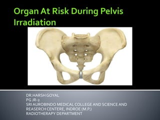
Organ at risk during pelvic irradiation
- 1. DR.HARSH GOYAL PG JR-2 SRI AUROBINDO MEDICAL COLLEGE AND SCIENCE AND REASERCH CENTERE, INDROE (M.P.) RADIOTHERAPY DEPARTMENT
- 2. These are normal tissues whose radiation may significantly influence the treatment planning or prescribed dose. It is divided into three classes. 1. Class I-Radiation lesions are fatal or result in severe morbidity.
- 3. 2. Class II-Radiation lesion result in mild to moderate morbidity. 3. Class III-Radiation lesion are mild, transient and reversible or result into no significant morbidity.
- 4. Organ at risk during pelvic radiotherapy are: 1. Bladder 2. Rectum 3. Urethra & Ureter 4. Small bowel 5. Vagina 6. Bone and bone marrow 7. Gonads
- 6. Radiation effects on the bladder have been documented during treatment of various pelvic malignancies including cervical cancers, prostate cancers, and bladder cancers. The urinary bladder and ureters are covered by urothelium—mucosa made of transitional epithelium of several cells.
- 7. For whole-bladder irradiation, doses in excess of 60 Gy, particularly with fraction sizes >2 Gy or accelerated radiation regimen, result in a significant risk of grade 3 or higher late toxicity. Risks are lower when the whole bladder receives 45 to 55 Gy followed by a boost to >60 Gy to a portion of the bladder.
- 8. Acute side effects of bladder irradiation are common and include urinary frequency, urgency, and dysuria. Late effects include hematuria, fistula, obstruction, ulceration, contracted bladder, vesicovaginal fistula, necrosis, spasm, reduced flow, and incontinence. Median onset of late complications after radiation is 13–20 months.
- 11. Prior pelvic surgery can result in increased risk of bladder toxicity as a direct result of bladder or urethral trauma or by denervation of the bladder, which can cause urinary hesitancy or retention, resulting in overflow incontinence. Patients receiving anticoagulants may be at greater risk of hematuria.
- 12. Cyclophosphamide, independently or with radiation, can cause chronic hemorrhagic cystitis, incontinence, contractions, and vesicoureteral reflux. Radiation-sensitizing chemotherapy may increase risk of acute and late bladder toxicity.
- 13. Recommended Dose–Volume Limits- for whole bladderV80 < 15%,V75<25%, V70<35%,V65<50%. For whole-bladder radiation, the reported risks of grade 3 or higher toxicity in doses of 50 to 60 Gy range from < 5% to 40%.
- 14. In early reactions-Symptomatic frequency and urgency are best treated with anticholinergic agents. These agents are thought to act primarily by inhibiting involuntary detrusor muscle contractions. Oxybutynin chloride is an antispasmodic that relaxes the bladder smooth muscle and may relieve the symptoms of frequency and urgency. Detrol has a greater inhibitory effect on bladder contraction and it has fewer side effects as compare to oxybutynin (eg, dryness of mouth), usual adult dose is 2 mg bd till symptomatic relief is obtained.
- 15. Phenazopyridine (Pyridium) can be used to provide symptomatic relief in dysuria. It is a azo dye, it acts directly on urinary tract mucosa to produce local analgesic effect. The usual adult dose of pyridium is 100 to 200 mg tds for 5 to 7 days. If dysuria associated with UTI, then proper antibiotic should be added.
- 16. Percutaneous posterior tibial nerve stimulation (PTNS):PTNS is a minimally invasive neuro modulation method designed to deliver retrograde electrical stimulation to the sacral nerve plexus through percutaneous electrical stimulation of the posterior tibial nerve. By using a battery-powered,hand-held stimulator and a 34-gauge needle electrode, the tibial nerve is accessed and stimulated. Patients receive a 30-minute weekly treatment for 12 weeks.
- 18. Botulinum toxin- Detrusor injections of botulinum toxin are approved by the FDA for the treatment of adults with over active bladder who do not adequately respond to anticholinergic medication. Most of the effects of botulinum toxin are thought to be the result of inhibition of the release of acetylcholine from the presynaptic nerve terminal, which prevents stimulation of the detrusor muscle. Recommended total dose is 100 Units injection across 20 sites into the detrusor muscles by cystoscope in every 12 weeks.
- 19. The primary treatment modality for hematuria is bladder irrigation.Intravesical treatments with silver nitrate, prostaglandins or formalin have also been used. Other treatment involves the injection of a sclerosing agent (1% ethoxysclerol) into the bleeding areas to control the severe hematuria in patients with hematuria that is not responding to simpler methods. More serious interventions can include embolization of the hypogastric arteries or urinary diversion and cystectomy.
- 20. Contracted bladder-Treated by reconstruction of bladder with a segment of small or large bowel (by sigmoid colon). Cystectomy- This procedure is used as a last resort for contracted bladder and involves removing the bladder and surgically constructing a replacement or an opening in the body (stoma) to attach a bag on the skin to collect urine.
- 21. Enterovesical fistula managed by surgically.The aim of operative management is to resect and reanastomose the offending bowel segment and to close the bladder. The treatment may involve single-stage or multistage procedures. The single stage procedure involves resection and primary anastomosis without a protective colostomy.
- 23. Urethral injury usually consists of stricture formation and Incontinence. For patients undergoing radiation to the prostate, most reports have documented increased risk of stricture and Incontinence in patients who have had previous transurethral resections of the prostate (TURP).
- 24. Therapeutic doses of radiation ranging from 60 to 70 Gy, patients with priorTURP demonstrated stricture rates of 10-20% compared to patients with out priorTURP who had stricture rates ranging from 0% to 5%.
- 25. Many factors can affect incontinence in addition to damage to the urethral sphincter including age,weight,comorbid medical conditions, bladder irritability, and pelvic floor weakening. Urethral damage causes stenosis, necrosis, and reflux from radiation. The most frequent cause of ureteral injury after treatment for cancer is a progressive disease.
- 26. Mild stenosis may be treated by placement of a ureteral stent. Other procedures to divert the urinary stream may be used including placement of ileal conduits. Nephrectomy may be required with recurrent urinary tract infections and a non-functional kidney.
- 28. Early injury to rectum at the cellular level is characterized by mucosal cell loss, acute inflammation, eosinophilic crypt abscesses, and endothelial swelling in the arterioles which can be seen following radiation to the rectum. A thickened and edematous lamina propria with patchy fibroblastic proliferation with decreased mitotic rate seen in the mucosa with radiotherapy.
- 29. According to quantec dose guidelines- recommended Dose–Volume Limits are- V50<50%,V60<35%,V65<25%,V70<20%. At 50 Gy doses (administered in 20 fractions over 1 month @ 2.5 Gy/#) in addition to mucosal cell injury and crypt shortening, infiltration of inflammatory cells occurs. The development of delayed tissue injury starts to be apparent as early as at 6 months post treatment and may gradually worsen.
- 30. The pathogenesis of the delayed injury is a result of the development of fibrosis in the stroma and in the blood vessels, causing ischemia. This fibrosis can lead to relative ischemia, and mucosal capillaries attempt to compensate for this develop telangiectasia with friable vessels that are prone to bleeding. More severe ischemia can lead to ulceration, perforation fistula, or abscess formation.
- 31. Acute symptoms of the rectum can be seen early in the course of radiation therapy for cancers in the pelvic region. Symptoms usually begin following 20Gy of standard fractionation. Acute proctitis usually with in three months of radiation. Early symptoms includes tenesmus, bleeding, and diarrhea.
- 32. Late reactions result in bleeding, ulcers, strictures, and fistula formation. Patients typically present with painless rectal bleeding. Other symptoms include evacuation difficulties, frequent elimination, fecal incontinence, and urgency which may also develop from alterations in anorectal function.
- 33. Grade 0- No symptoms and no intervention required. Grade 1 -Mild symptoms (mucus discharge or minimal bleeding & rectal discomfort) and self-limiting , not required analgesics, or other medications. Grade 2 –It is managed by conservatively, lifestyle not affected- Intermittent rectal bleeding not required regular use of pads, erythema seen in rectal lining on proctoscopy, diarrhea requiring medications.
- 34. Grade 3- Severe reactions occurs which alters patient lifestyle or rectal ulceration present- Rectal bleeding required regular use of pads and minor surgical intervention, rectal pain requiring narcotics. Grade 4- Life threatening reactions occurs which causes bowel obstruction, fistula formation may occur. Bleeding required hospitalization, surgical intervention required. Grade 5- Death directly related to radiation effects.
- 35. At sigmoidoscopy, a spectrum of mucosal changes can be seen including mucosal pallor or erythema, prominent telangiectasia,friability, or fistula. The onset of radiation proctitis is typically seen in 12– 18 months following treatment. Patient-related factors that may increase the risk of proctitis include hypertension, diabetes, and cerebrovascular disease that can all affect the vascular supply in the radiated field.
- 36. Acute symptoms of urgency and tenesmus can be treated with an antispasmodic such as Lomotil ( 5mg tds). Steroid enemas (predinisolone enema) also useful to decrease inflammation. Biopsy of mucosal changes should be discouraged if they occur in an area that likely received a high dose of radiation such as the anterior rectal wall. Biopsy may cause persistent inflammation, decrease healing, and precipitate fistula formation.
- 37. SUCRALFATE- It mechanically protects the gastrointestinal mucosa by forming a protective coating on inner surface of the bowel and stimulate healing by increasing angiogenesis. Administration of sucralfate enema given as 2 gm/day bd. METRONIDAZOLE- It is a antiprotozoal and immunomodulator drug which inhibits anaerobic Organism growth in rectum.The usual adult dose is 400 mg tds.
- 38. FORMALIN-Formalin is an aldehyde which induces coagulative tissue necrosis on contact. Endorectal formalin instillation has been used successfully in several studies. ENDOSCOPICTHERAPY-The goal of endoscopic therapy is to provide cessation of rectal bleeding, to decrease the need for transfusion or hospitalization, and thus improve the patient’s quality of life.These techniques should be considered after medical management fails, and the patient experiences persistent symptoms. These techniques includes endscopic application of clips, use of bending devices by endoscopy.
- 39. Balloon therapy- Patients with radiation proctitis can develop a lower gastrointestinal stricture with resultant obstructive symptoms. Mechanical dilation via an endoscopically passed balloon is a simple and effective treatment for patients with radiation-induced rectal strictures.
- 40. ARGON PLASMA COAGULATION-Argon plasma coagulation (APC) is a non-contact thermal method of coagulation and hemostasis. This modality utilizes a jet of sprayed argon gas, which is ionized by a high voltage spark into plasma. Once ionized, it produed coagulation and hemostasis in bledding mucosa.
- 41. PROCTECTOMY/PELVIC EXENTERATION-In patients with persistently refractive radiation proctitis, the most extreme intervention is complete rectal excision with possible removal of adjacent pelvic organs. This treatment can be considered the most definitive treatment for radiation proctitis, in that it removes the offending tissue and gives feacal diversion though a permanent ostomy and this treatment should be offered to patients who are refractory to all treatment.
- 43. Mucosa above the anal orifice is made of stratified squamous epithelial cells which show a rapid turnover and hence are very radiosensitive. This mucosa is involved in the pathology of the early radiation injury. Sustained radiation injury to muscle, stromal, and vascular cells are the cause of the delayed anal radiation injury.
- 44. Tolerance dose of the normal tissue is considered to be above 65Gy. Acute grade 3 toxicity with 2-Gy fractions is 40%, while it is 75% with 2.5-Gy fractions. Acute effects include epithelial discomfort which may be aggravated by radiation- induced diarrhea.
- 45. Epithelial effects follow a sequential progression from erythema to desquamation. Shallow erosions and ulcerations can develop which can lead to tenesmus. Late effects which includes strictures of the anus or ulceration, fibrosis required intervention.
- 46. Therapeutic interventions usually include antidiarrheal medications such as loperamide or codeine. Care of the skin includes cleansing ointments, Application of gentian violet,lidocaine which can help in manage symptoms. Biopsy of the anus following high dose radiation can result in non-healing ulceration. Avoidance of this procedure is preferred.
- 47. If a non-healing ulceration develops, conservative management is generally used initially.This involves stool softeners, sitz baths, and wound care. Some studies reports that use of hyperbaric oxygen is beneficial in the management of non healing ulcers. There are limited therapeutic interventions for incontinence with the primary treatment being colostomy.
- 49. The small bowel may incidentally irradiated during radiation therapy to the pelvis. It is particularly evident in the treatment of prostate cancer and in gynecological cancers. As a consequence of the radiation changes on the bowel, it may develop serious complications. prospective trials demonstrated that approximately 30% of postoperative patients with endometrial cancer who received radiation, experienced with acute diarrhea which persisted in approximately 10% of patients, up to 5 years after treatment.
- 50. FACTORS AFFECTING RISK- Use of concurrent chemotherapy increase acute toxicity like 5 FU, docetaxel, etoposide and bevacizumab. Previous surgery causes the development of adhesions that tend to fix the intestines, which may become involved in the radiation field. Patients with hypertension, diabetes mellitus, and generalized atherosclerosis are at an increased risk for vascular occlusive disease. patients with underlying inflammatory bowel disease may be at a higher risk for severe toxicity.
- 51. Recommended Dose–Volume Limits for small bowel isV45 <195 cc. TheTD5/5 andTD50/5 estimate for 1/3 small- bowel irradiation is 50 Gy and 60 Gy respectively.
- 54. Radiation induced small-bowel mucositis can be expressed as cramping and diarrhea and weight loss from interference with nutrient absorption, typically developing 1 to 2 weeks after the start of Radiation. Chronic small-bowel injury from RT can include persistent diarrhea, obstruction, ulceration, perforation and bleeding, majority of symptoms occur with in 3 years of post radiation.
- 55. Acute toxicity is managed by Diet- A low-residue diet and a diet low in fats and lactose are helpful. In more severe cases, an elemental diet may improve diarrhoea while maintaining nutrition. Drugs-Antispasmodics, anticholinergics and opiates can improve symptoms of pain and diarrhoea by reducing motility. Oral antibiotics may improve diarrhoea in patients with bacterial colonisation of the small bowel.
- 56. Total parenteral nutrition-Bowel rest and total parenteral nutrition (TPN) may be used for severe symptomatic disease and for enterocutaneous fistulas, although spontaneous closure is uncommon. Total parenteral nutrition is also valuable in malnourished patient prior of surgery.
- 57. Surgical management- Indications for surgery include bowel obstruction, perforation, abscess, intractable bleeding or diarrhoea, and occasionally malabsorption.
- 59. The testis is one of the most radiosensitive tissues in the body with a radiation dose as low as 15 cGy causing a significant depression in the sperm count. The testis may be directly in the radiation field or receive scattered dose from a nearby field. Recommended Dose–Volume Limits of testis is V3<50%.
- 60. Azoospermia after low doses (<2 Gy) takes 60-70 days due to damage to spermatogonia, after higher doses (>4 Gy), azoospermia develops much faster due to concurrent damage to spermatids. Low doses of radiation kill spermatogonia which are differentiating into spermatocytes. Therefore, low doses of radiation deplete the stem cells of these developing sperm which result in decreased sperm production during the first 50–60 days after radiation.
- 61. Complete recovery takes place with in 9–18 months after less than 1 Gy, 30 months for 2–3 Gy, and 5 or more years after 4–6 Gy. Direct testicular irradiation of 24 Gy results in ablation of the germinal epithelium which is responsible for sperm development and Leydig cell function which is responsible for testosterone,which is seriously affected in most men.
- 62. Symptoms of low testosterone includes: Loss of libido Erectile dysfunction Fatigue Decreased muscle mass Body and facial hair loss Difficulty in concentrating Depression Irritability Low sense of well-being
- 63. When testes present directly in the irradiation field, it cannot be protected. The dose from scattered irradiation from nearby beams, can be reduced by moving the gonad away from or by applying thick shielding cups directly over the scrotum.
- 64. (Front view of gonadal shield) The shield has aV cut for inserting the testis and to hold in position,wax coating is done on the inner surface of the shields. It makes the inner surface smooth and avoid back-scattered electron dose from the shield.
- 65. Androgen replacement therapy to enable normal pubertal development and future sexual function is required for patients with deficient testosterone production. Inj testesterone 200 mg (I.M.) in every 2 week should be given till regression of symptoms occurs. Pre treatment sperm banking may be preferable in patients who want continue their fertality.
- 67. The lining of the vagina is made of stratified squamous epithelium over a connective tissue lamina propria and longitudinal muscle fibers and elastic fibers. Radiosensitivity of the squamous epithelium is significant and early vaginal injury is marked by acute epithelial denudation with endothelial injury that may lead to thrombosis,edema,and smooth muscle necrosis.
- 68. Delayed injury involves severe fibrosis that may obliterate portions of the muscle and vasculature potentially resulting in vaginal stenosis and ulceration. Vaginal mucosa is reasonably tolerant to radiation. An irradiation tolerance level of the proximal vagina was suggested by Hintz in 1980.None of the patients treated to a maximum dose of 140 Gy developed severe complications or necrosis of the upper vagina.
- 69. The distal vagina (introitus) and posterior wall are more sensitive to radiation. Hintz suggested doses to the distal vagina not to be greater than 98 Gy. Serious complications include mucosal necrosis or fistula formation. Less serious complications included vaginal stenosis or shortening, formation of telangiectasia (which can lead to bleeding) or thinning of the vaginal mucosa, and dryness.
- 70. If soft tissue necrosis is observed, symptomatic management with antibiotics, debridement, estrogen cream, and gentle irrigation may heal the area. The strategy for prevention of vaginal stenosis or shortening has been to encourage sexual intercourse or use of vaginal dilators. Estrogen cream or systemic estrogen may also aid in the rejuvenation of cells and increase the elasticity of the vagina.
- 72. Radiation to the ovaries can damage oocytes and result in premature menopause because of ovarian failure of estrogen production. Oocytes undergo meiosis and are relatively radioresistant (single dose LD50 is 4 Gy) however, proliferating granulosal cells are very radiosensitive.
- 73. Therefore, a total dose of 24Gy (fractionated in 2-Gy single doses) leads to ablation of ovaries due to the loss of granulosa cells. Immediate ovarian failure will be produced by 16.5 Gy in females of 20 years, while 14 Gy is sufficient in 30-year-olds or in elderly group.
- 74. Early radiation injury then comprises of necrotic changes in proliferating granulosa cells, while early changes include microvascular thrombi and endothelial cell swelling. Late radiation effects include atrophy and fibrosis, with thick walled and hyaline arterioles and venules.
- 75. The effects on the ovary are dependent on the age of the patient and total dose to the ovary. Low doses of radiation (4–7 Gy in 1–4 fractions) can result in permanent menopause in women over 40 years of age. However, permanent sterility in young women may not result until a total dose of 20 Gy .
- 76. Symptoms of ovarian irradiation includes- Amenorrhea Hot flashes Night sweats Vaginal dryness Irritability Difficulty in concentrating Loss of libido Infertility
- 77. Harmonal therapy-Estrogen therapy can help prevent osteoporosis and relieve hot flashes and other symptoms of estrogen deficiency. Estrogens can be administered orally or transdermally. The usual adult dose for transdermal estradiol is 100-150 mcg and oral estradiol 2 to 4 mg.
- 78. Progestins should be administered cyclically, 10-14 days in each month, to prevent endometrial hyperplasia. The recommended regimens include medroxyprogesterone 10 mg daily for 10-12 days in each month. Preservation of fertility by egg storage (oocyte) is a useful technique prior to treatment.
- 80. Radiation can affect the microvasculature of the mature bone.This injury causes decreased blood supply to the periosteum which compromises osteoblastic function and can result in an insufficiency fracture (IF). Insufficiency fractures of bone occur as a result of physiological stress on bones with deficient elastic resistance. Irradiation can damage osteoblasts, osteocytes, and osteoclasts and leave an acellular matrix that appears radiographically normal.
- 81. Such radiation-induced atrophy reduces the number of functional and structural components of a tissue. These two processes can result in clinically and radiographically significant bone atrophy. In addition, previously irradiated atrophic bone is at risk for fracture, second malignancy, or infection, leading to true necrosis of bone.
- 82. Resulting injuries include atraumatic femoral neck fracture, and osteonecrosis of the femoral head or of the acetabulum. Abe et al. reported that asymptomatic Insufficiency Fracture was found in 34% of postmenopausal patients treated with adjuvant postoperative radiation for endometrial cancer.
- 83. The incidence of symptomatic pelvic fractures in several series of women treated for gynecologic malignancies ranges from 1.7% to 6% following doses of 46–50 Gy to the whole pelvis. Pain in the pelvic area is the initial complaint of patients.The average time to onset of symptoms is usually 11–12 months after radiation therapy.
- 84. CT scans can reveal radiological findings of IF, although MRI is currently the most sensitive modality for detecting these lesions. Most IF is multiple and the most common location for them is in the sacrum and pubic bones.
- 85. The tolerance doses for the femoral head have been estimated to be 52Gy for the TD 5/5 and 65 Gy for theTD 50/5. No fracture occurred below doses of 42Gy. Treatment for bone fractures following radiation has included calcium supplements, and pain management, and surgically.
- 87. The bone marrow is one of the most radiosensitive organs in the pelvis. Approximately 40% of the total body bone marrow reserve lies with in the pelvic bones. Hematologic toxicity can be seen acutely during radiation and exposure to radiation can result in long-term myelotoxicity.
- 88. The radiation dose, dose rate, and volume all affect the acute response of the bone marrow to radiation therapy. With exposure to large bone marrow volumes, neutropenia occurs in 2–3 weeks followed by thrombocytopenia and then anemia in 2–3 months.
- 89. The management of patients with bone marrow toxicity can be divided into supportive and preventive. Growth factor administration is now a common supportive measure in patients with white cell deficiencies. BloodTransfusion is typically reserved for patients with hemoglobin levels below 8 g/dl.
- 91. The aim of modern radiotherapy is to ensure a high level of accuracy in tumour targeting, to reduce normal tissue exposure, and to minimise side effects. Normal organ reactions depends on various factors, such as the type of radiotherapy, the size and site of the treatment field, and the dose delivered. A variety of different strategies have been proposed which includes improved radiotherapy delivery techniques, other aspects like timing of radiation delivery, patient positioning , pharmacological interventions, and non-pharmacological interventions who reduces radiation impact on normal tissue and to prevent side effects.
- 92. Radiotherapy delivery techniques- 3D conformal radiotherapy (3D-CRT) is intended to improve tumour targeting and reduce the amount of radiation to the surrounding tissues by aiming shaped radiotherapy beams from several different directions at the tumour. It uses pre-treatment imaging with computerised tomography (CT) to plan the radiotherapy treatment area in three dimensions (width,height and depth) matching the radiation beams to the 3D shape of the tumour.
- 93. Intensity-modulated radiotherapy (IMRT)- uses computerised methods to orientate multiple small beams of different intensities to the volume of tumour tissue that needs to be treated. IMRT potentially conform more precisely to the tumour than 3D-CRT as it allows the dose of radiation to be adjusted for different parts of the treatment area and can reduce exposure to organ at risk.
- 94. Image-guided radiotherapy (IGRT) IGRT includes imaging (e.g. CBCT) performed at pre- treatment and treatment delivery that improves or verifies the target accuracy of radiotherapy. It causes reduction in CTV to PTV margins thus reducing the volume of normal tissue receiving high radiation doses. MRI-based planning with its greater soft tissue definition can reduce the clinical target volume (CTV) by approximately 20% compared to CT-based planning and reduces radiation exposures to OARs.
- 95. Stereotactic body radiotherapy (SBRT) SBRT involves the use of a high and precise radiation dose in a small number of fractions. Radiotherapy beams are orientated from many different positions around the body to minimise the radiation dose to the surrounding tissues. SBRT is currently mainly used for small tumours of the brain, prostate, liver, lung and spinal cord.
- 96. Patient positioning- The position of a patient during radiotherapy delivery might influence the dose of radiation delivered to normal pelvic structures and subsequent GI injury. A systematic review of prospective and retrospective studies of patient positioning and the use of belly boards, it suggests that delivering radiotherapy to patients positioned in the prone position, and using positioning devices such as belly boards, displaces the small bowel away from the treatment field and can reduce the volume of small bowel radiation.
- 97. Timing of delivery (Physiological clocks)- According to Buchi day-night cycle is the core clock that influences response to anti-cancer treatments and the development of treatment side effects. It regulate the timing of physiological processes through gene expression exist in every organ and cell of the human body. Evidence from animal and human laboratory studies suggests that gastrointestinal cellular and mucosal proliferation being greatest in the morning and lowest in the evening.
- 98. As proliferating cells are most radiosensitive, it is plausible that radiotherapy delivered in the morning may be more likely to cause damage to gastrointestinal mucosal cells than radiotherapy delivered in the evening. Findings from a clinical study of women receiving radiotherapy for cervical cancer, which found that severe diarrhoea occurred less frequently with evening radiotherapy than morning radiotherapy, appear to support this theory (Shukla et al 2010).
- 99. Fractionation-Certain cancers such as prostate cancer have been shown to be more sensitive to fraction size. Therefore, increasing the fraction size (hypofractionation) per treatment, which allows the total dose to be delivered in fewer treatments, might improve the treatment outcome or therapeutic ratio but associated with late gastrointestinal and genitourinary toxicity.
- 100. Other interventions-Various surgical techniques, such as the surgical placement of absorbable mesh slings to exclude the small bowel from the field of radiation, have been proposed to reduce the gastrointestinal effects of pelvic radiotherapy. Using daily endorectal balloons filled with air or water or gel might be beneficial for men undergoing prostate radiotherapy.
- 101. Amifostine is thought to mediate a protective effect with in normal cells by free-radical scavenging, DNA protection and repair acceleration.The usual adult dose of amiofostine is 500 mg in 50 ml normal saline over 6 minute before 30 minute of radiotherapy. Antioxidants, such as vitamins C, D, and E, helps to reduce radiotherapy-induced injury by reducing antioxidant stress with in radiating tissue and facilitating tissue repair. Glutamine, a non-essential amino acid and other agents with antioxidant properties could also potentially to be protective.
- 102. Probiotics-Probiotic preparations contain live and defined micro-organisms , when administered in sufficiently large amounts, it causes increased secretion of protective mucins and stimulate the immune response. Nutrition- Malnutrition can occur as a consequence of radiotherapy-induced impaired gastrointestinal absorption and digestive functioning, and can cause radiation associated reactions. Dietary modification of fat, lactose, or fibre intake, or combinations of these dietary modifications probably reduces diarrhoea and plays a protective role in radiotherapy.