
Recommended
More Related Content
Similar to Liver Trauma Guide: Causes, Symptoms, Imaging & Treatment
Similar to Liver Trauma Guide: Causes, Symptoms, Imaging & Treatment (20)
Recently uploaded
Recently uploaded (20)
Liver Trauma Guide: Causes, Symptoms, Imaging & Treatment
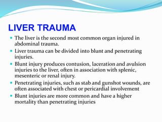
- 1. LIVER TRAUMA The liver is the second most common organ injured in abdominal trauma. Liver trauma can be divided into blunt and penetrating injuries. Blunt injury produces contusion, laceration and avulsion injuries to the liver, often in association with splenic, mesenteric or renal injury. Penetrating injuries, such as stab and gunshot wounds, are often associated with chest or pericardial involvement Blunt injuries are more common and have a higher mortality than penetrating injuries
- 2. Management of liver trauma Remember associated injuries At-risk groups Stabbing/gunshot in lower chest or upper abdomen Crush injury with multiple rib fractures Resuscitate Airway Breathing Circulation Assessment of injury CT chest and abdomen with contrast Laparotomy if haemodynamically unstable Treatment Correct coagulopathy Suture lacerations Resect if major vascular injury Packing if diffuse parenchymal injury
- 3. complications of liver trauma Intrahepatic haematoma Liver abscess Bile collection Biliary fistula Hepatic artery aneurysm Arteriovenous fistula Arteriobiliary fistula Liver failure
- 4. Liver infections and their treatment Treatment Causative agent Condition Supportive, anti-viral agents (lamivudine, interferon, ribavirin), Liver transplant for cirrhosis Hepatitis A, B, C Viral hepatitis Antibiotics (cephalosporin) Relieve obstruction Enteric bacteria Ascending cholangitis Antibiotics Aspiration Drainage Streptococcus milleri Escherichia coli Streptococcus faecalis Pyogenic liver abscess Metronidazole Entamoeba Amoebic liver abscess Mebendazole Resection/omentoplasty Echinococcus Hydatid liver disease
- 7. A primary cyst in the liver is composed of three layers: Adventitia (pericyst): consisting of compressed liver parenchyma and fibrous tissue induced by the expanding parasitic cyst..Laminated membrane (ectocyst): is elastic white covering, easily separable from the adventitia. Germinal epithelium (endocyst) – is a single layer of cells lining the inner aspects of the cyst and is the only living component, being responsible for the formation of the other layers as well as the hydatid fluid and brood capsules within the cyst. In some primary cysts, laminated membranes may eventually disintegrate and the brood capsules are freed and grow into daughter cysts. Sometimes the germinal Epithelium daughter cysts, which if left untreated may cause recurrence
- 9. Complications of hydatid cyst of the liver: Infection it is the most common complication and can be symptomatic. t is usually latent, subacute and is clinically translated by pains in the right hypochondrium, hepatomegaly, and fever Intrabiliary rupture of hydatid cyst, the rupture in the thorax ,The rupture in the peritoneum Rupture of the cyst in the peritoneal cavity is rare and generally followed by anaphylactic reactions. Intraperitoneal cysts may rupture spontaneously, due to increased intracystic pressure, or as a consequence of trauma, leading to the spread of hydatid fluid in the intraperitoneal cavity . Clinical feature After infection with Echinococcus granulosus, humans are usually asymptomatic for a long time.The growth of the cyst in the liver is variable, ranging from 1 mm to 5 mm in diameter per year. Most primary infections consist of a single cyst. The symptoms depend not only on the size and number of cysts, but also on the mass effect within the organ and upon surrounding structures
- 10. Non complicated cysts Hydatid cyst of the liver is silent and only diagnosed incidentally during abdominal investigation. The clinical signs appear gradually with the increase volume of the cyst. The most common symptom, is right upper quadrant or epigastric pain and findings on examination are an enlarged liver and a palpable mass. They may include non-specific pain, cough, low-grade fever, and the sensation of abdominal fullness. As the mass grows, the symptoms become more specific because the mass impinges on or obstructs specific organ . Patients may also present with complications of the cyst such as biliary communication, intraperitoneal rupture (spontaneous or post-traumatic) and intrathoracic or intrapericardial rupture Cyst rupture can be associated with anaphylaxis secondary to the highly antigenic content of the cyst fluid or may be silent and present with multiple intraperitoneal cysts With secondary infection, tender hepatomegaly, chills, and spiking temperatures occurs. Urticaria and erythema occur in cases of generalized anaphylactic reaction. With biliary rupture the classic triad of jaundice, biliary colic and urticaria occurs
- 11. investigation Serology and immunological tests Serological tests detect specific antibodies to the parasite and are the most commonly employed tools to diagnose past and recent infection with E. granulosus. Detection of IgG antibodies implies exposure to the parasite, while in active infection high titers of specific IgM and IgA antibodies are observed. Detection of circulating hydatid antigen in the serum is of use in monitoring after surgery and pharmacotherapy and in prognosis. ELISA is used most commonly, but alternate techniques are counter-immuno-electrophoresis and bacterial co-agglutination Elisa techniques have a high sensitivity above 90% and are useful in mass scale screening. The counter-immuno-electrophoresis has the highest specificity (100%) and high sensitivity (80 – 90%). CASONI TEST has been used most frequently in the past but is at present considered only of historical importance and has largely been abandoned because of low sensitivity
- 12. Imaging techniques Imaging modalities range from simple to complex and invasive. Ultrasonography (US) is the screening method of choice. It is currently the primary diagnostic technique and has diagnostic accuracy of 90%. CT scan is an important preoperative diagnostic tool to determine vascular,biliary or extra hepatic extension, to recognize complications, such as rupture and infections, and therefore to assess respectability The right lobe is the most frequently involved portion of the liver. Imaging findings in hepatic hydatid disease depend on the stage of cyst growth (whether the cyst is unilocular, contains daughter vesicles, contains daughter cysts, is partially calcified, or is completely calcified. Plain Radiographs: Plain radiographs of the abdomen and chest may reveal a thin rim of calcification delineating a cyst, or an elevated hemi diaphragm
- 13. Treatment Surgery remains the gold standard treatment for hydatid liver disease. The aim of surgical is to inactivate the parasite, to evacuate the cyst along with resection of the germinal layer, to prevent peritoneal spillage of scolices and to obliterate the residual cavity. However, surgery may be impractical in patients with multiple cysts localised in several organs and if surgical facilities are inadequate. The introduction of chemotherapy and of the PAIR technique (puncture-aspiration-injection-respiration) offers an alternative treatment, especially in inoperable patients and for cases with a high surgical risk. The principles of hydatid surgery are surgical procedures can be divided into two groups, a conservative group and a radical one. The conservative technique:are safe and simple, and are useful in uncomplicated hydatid cysts. Marsupialization was the most common used procedure because it is quick and safe their dis advantageis the high frequency of postoperative complications: being bile leak from a cyst-biliary communication, bilomas and bile peritonitis
- 14. :Radical surgical procedures 1. Cystectomy –. The procedure involves removal of hydatid cyst, comprising laminar layer, germinal layer and cyst contents (daughter cysts and brood capsules). The procedure is simple to perform and has low recurrence rates. 2. Pericystectomy –non-anatomical resection of cyst and surrounding compressed liver tissue. 3. Hepatic resections
- 15. PAIR (puncture, aspiration, injection, and reaspiration) is a percutaneous treatment technique for hydatid disease. In this minimally invasive method, a needle is introduced into the cyst under ultrasound guidance The World Health Organization guidelines for indications and contraindications of PAIR are as follows: Indications for PAIR lesion greater than or equal to 5 cm in diameter Cysts with daughter cysts and/or with membrane detachment Multiple cysts if accessible to puncture Infected cysts Patients who refuse surgery. Patients who relapse after surgery. Patients in whom surgery is contraindicated Patients who fail to respond to chemotherapy alone Children over 3 years. Pregnant women Contraindications for PAIR Non cooperative patients Inaccessible or risky location of the liver cyst Cyst in spine, brain, and/or heart Inactive or calcified lesion Cyst communicating with the biliary tree Patients should be followed clinically after PAIR treatment. Recurrence is increased in more complicated cysts, including those with multiple daughter cysts
- 16. medical treatment for hydatid disease of liver is based on benzoimidazole ,mebendazole and albendazole. It has been proposed that these agents contribute to clinical improvementof the disease by diminishing the size of the cyst. The factors for success seem to be the ability of the drug to penetrate the cyst wall and the persistence of adequate levels of the activemetabolites. Albendazole seems to be more effective owing to better penetration and absorption These agents have actually been used in several studies as a conservative treatment, leading to some decrease or stabilization of the cyst size, especially in cases with small cysts. However, their clinical efficacy still remains doubtful. They are used mainly for disseminated systemic disease, inoperable cases, and—combined with surgery—to prevent postoperative recurrence.. The different schedules for the treatment are: Inoperable cases - as primary treatment - 3 cycles Pre-operatively – to reduce the risk of recurrence 6 weeks continuous treatment Post-operatively to prevent recurrence in cases of intraoperative cyst spillage 3 cycles
- 17. LIVER TUMOURS Benign liver tumours Haemangiomas These are the most common liver lesions, and the reported incidence has increased with the widespread availability of diagnostic Ultrasound. Hepatic adenoma Hepatic adenomas are rare benign liver tumours. They occur mostly in women of child-bearing age. An association with sex hormones (including the oral contraceptive pill) is well recognised, and regression of symptomatic adenomas on withdrawal of hormone stimulation is well documented. Focal nodular hyperplasia This is an unusual benign condition of unknown aetiology in which there is a focal overgrowth of functioning liver tissue supported by fibrous stroma. Patients are usually middle-aged female. Contrast CT or/and MRI may show central scarring and evidence of a well- vascularised lesion.
- 18. Malignant liver tumours: Hepatocellular carcinoma (HCC) is one of the world’s most common cancers, and its incidence is expected to rise rapidly over the next decade due to the association with chronic liver disease, particularly HBV and HCV. Cholangiocarcinoma Bile duct cancers typically present with painless obstructive jaundice
- 19. liver metastases Staging and assessment with colorectal liver metastases General medical assessment CT, PET-CT or MRI of the abdomen/pelvis with contrast (?resectability) Chest CT Review histology of primary (? risk of local recurrence) Colonoscopy Liver function tests and tumour markers Prognostic factors in patients undergoing resection of colorectal liver metastases Stage of primary Time from primary resection Carcinoembryonic antigen (CEA) level Size of largest lesion Number of lesions