SlideShare a Scribd company logo
1 of 17
Download to read offline
90
Analytical and Quantitative Cytopathology and Histopathology®
0884-6812/21/4302-0090/$18.00/0 © Science Printers and Publishers, Inc.
Analytical and Quantitative Cytopathology and Histopathology®
OBJECTIVE: To establish a descriptive and comparative
anatomohistological analysis of mice and rats.
STUDY DESIGN: A total of 30 Mus musculus mice
and 15 Rattus norvegicus rats were used. All animals
were kept specific-pathogen-free and absent of surgical
interventions that could cause anatomical and physio-
logical changes.
RESULTS: The study observed species-specific anato-
mohistological characteristics, which will serve as a
basis to assist in choosing the most satisfactory research
model.
CONCLUSION: The evolution of knowledge in bio-
logical and medical areas are attributed to anatomical,
physiological, and immunological studies in animals,
contributing to the discovery of prophylactic measures
and disease treatments that affect humans and ani-
Descriptive Comparative Anatomohistological
Study of the Main Dissected Organs of
Mus musculus and Rattus norvegicus for
Experimental Model Research
Giorgio Silva-Santana, M.M., Licínio Esmeraldo Silva, M.D.,
Jemima Fuentes Ribeiro Silva, M.D., Alexia Gonçalves, M.M.,
Ana Luíza Mattos-Guaraldi, M.D., and Kátia Calvi Lenzi-Almeida, M.D.
From Health Sciences Center, Institute of Microbiology Paulo de Góes, Federal University of Rio de Janeiro, Rio de Janeiro, RJ, Brazil;
Department of Statistics, Institute of Mathematics and Statistics, and Department of Pathology, Medical College, Federal Fluminense
University, Niterói, RJ, Brazil; Department of Histology and Embryology, Biomedical Center, and Laboratory of Diphtheria and Coryne-
bacteria of Clinical Relevance, Faculty of Medical Sciences, University of the State of Rio de Janeiro, Rio de Janeiro, RJ, Brazil; the Collabo-
rating Centre for Reference and Research on Diphtheria/National Health Foundation/Ministry of Health, Brazil; and the Environmental
Science and Conservation Department, Medical College, Federal University of Rio de Janeiro, Macaé, Brazil.
Giorgio Silva-Santana is Doctoral Student, Health Sciences Center, Institute of Microbiology Paulo de Góes, Federal University of Rio de
Janeiro (ORCID ID: 0000-0002-3000-6513).
Licínio Esmeraldo Silva is Professor, Statistics Department, Institute of Mathematics and Statistics, Federal Fluminense University
(ORCID ID: 0000-0003-3861-2806).
Jemima Fuentes Ribeiro Silva is Professor, Histology and Embryology Department, Biomedical Center, University of the State of Rio de
Janeiro (ORCID ID: 0000-0001-7867-3131).
Alexia Gonçalves is Student, Histology and Embryology Department, Biomedical Center, University of the State of Rio de Janeiro.
Ana Luíza Mattos-Guaraldi is Professor, Health Sciences Center, Institute of Microbiology Paulo de Góes, Federal University of Rio de
Janeiro, and Laboratory of Diphtheria and Corynebacteria of Clinical Relevance, Faculty of Medical Sciences, University of the State of
Rio de Janeiro, and the Collaborating Center for Reference and Research on Diphtheria/National Health Foundation/Ministry of Health,
Brazil (ORCID ID: 0000-0003-2522-0416).
Kátia Calvi Lenzi-Almeida is Professor, Environmental Science and Conservation Department, Medical College, Federal University of
Rio de Janeiro, Macaé, and Department of Pathology, Medical College, Federal Fluminense University (ORCID ID: 0000-0003-1097-0927).
This study was financed in part by the National Council for Scientific and Technological Development (CNPq), Coordination for the
Improvement of Higher Level Personnel–Brazil (CAPES) (Finance Code 001), and the Research Support Foundation for the State of Rio
de Janeiro, Brazil (FAPERJ).
Address correspondence to:  Giorgio Silva-Santana, M.M., Universidade Federal do Rio de Janeiro, Instituto de Microbiologia Professor
Paulo de Góes, Centro de Ciências da Saúde (CCS), Cidade Universitária–Ilha do Fundão, Rio de Janeiro, Brazil (bio.sant@hotmail.com).
Financial Disclosure:  The authors have no connection to any companies or products mentioned in this article.
Volume 43, Number 2/April 2021 91
Descriptive Comparative Anatomohistological Study
mals. As mice and rats are the most used animals in
experimental studies, the anatomical and histological
differences between species should be carefully evalu-
ated in order to better apply the study model and avoid
unnecessary waste. (Anal Quant Cytopathol Hist-
pathol 2021;43:90–106)
Keywords:  anatomy; experimental model; histo­
logy; mice, laboratory; Mus musculus; rats, labora-
tory; Rattus norvegicus.
Using animals in laboratory research for biological
investigations arose from the study of compara-
tive diseases.1,2 These studies aimed to find sim-
ilarities in the origin and characteristics of the
pathological processes that affected the human
species in animals with the same conditions. Cur-
rently, implementing defined genetically appro-
priate and sanitary laboratory animals has aided
in new discoveries via experimental models, con-
tributing to the prevention of incurable diseases
such as cancers, AIDS, and multiple sclerosis and
also to the development of new surgical treat-
ment techniques. Other applications correspond
to vaccine development, monoclonal antibodies,
evaluation and control of biological products,
pharmacology, toxicology, bacteriology, virology,
and parasitology in addition to basic immunology
studies, immunopathology, organ transplants, and
immunosuppressive drug development.3,4 How-
ever, with technological advances, alternative in
vitro methods, such as cell culture, made it possi-
ble to obtain satisfactory results without relying
on laboratory animals. Nonetheless, animal models
still present advantages, such as providing infor-
mation about the body as a whole.5-8 In regard to
phylogenetic relationship or anatomical conformi-
ty, previous studies have shown that extrapolating
results to humans is not always reliable.7,9-11
Introduced as a laboratory animal in the 19th
century with the Asian species Mus musculus, mice
came to be, from that period on, the most used
animals in experimental studies, becoming an im­
portant experimental model for genetic research.4,12
The Swiss albino lineage is easily accepted and
features heavily in experimental studies due to
its small size, short gestation period, easy domes-
tication and maintenance, and the fact that they
are very prolific.4,13 Eventually, their 99% genet-
ic similarity with humans, allowing to establish
mechanisms involved in human genetic disorders,
and greater capacity of genetic modifications up to
97% of the total of its genes, ensured their labora-
tory use.4
Comprising 137 species from the Central Asia
regions, the genus Rattus have 2 species of great
laboratory importance: Rattus norvegicus (domestic
rat or brown rat) and Rattus rattus (black rat).14 Its
most commonly used species, the albino lineage
Wistar descendant from Rattus norvegicus, devel-
oped at the Wistar Institute in Philadelphia in 1906,
was the first to be implemented as a model organ-
ism at a time when researchers used primarily Mus
musculus mice.15 Heterogenic animals have been
used for various scientific purposes, such as stud-
ies on rheumatology, endocrinology, orthopedics,
and others.16
We consider it of great importance that each
laboratory knows the set of reference values of
its healthy animals according to species, lineage,
genre, and age in order to assist professionals in
their different studies. Thus, the present study
aims to address the general biology of Mus mus-
culus and Rattus norvegicus, establishing a descrip-
tive and comparative analysis of the animals’ main
organs.
Materials and Methods
Ethical Statement and Animals
All experimental procedures described in this
study were approved by the Animal Experimenta-
tion Ethics Committee (CEUA) of the Fluminense
Federal University (UFF) in accordance with Law
no. 11.794/2008 (Arouca law). All procedures used
followed the Brazilian Directive for the Care and
Use of Animals for Scientific and Didactic Pur-
poses of the National Council for Animal Experi-
mentation Control (CONCEA).
This study was conducted per the recommenda-
tions of the National Institutes of Health’s Guide
for the Care and Use of Laboratory Animals. This
study used data obtained from 30 male heterogenic
Mus musculus (Swiss) mice, aged between 60 and
90 days, selected from control groups in a project
approved by the CEUA of the Pro-Rectory of Re­
search, Graduate Programs and Innovation of the
UFF, under protocol no. 439/2013. This study also
used data collected from 15 male heterogenic Rat-
tus norvegicus (Wistar) rats, aged between 90 and
120 days, selected from control groups in a project
approved by the CEUA/UFF under protocol no.
035/2011.
All animals, procured from the Animal Core
Laboratory (NAL), were kept in strict health con-
92 Analytical and Quantitative Cytopathology and Histopathology®
Silva-Santana et al
trol and specific-pathogen-free (SPF). The animals
underwent no surgical intervention that could
cause anatomical changes, nor were they exposed
to any chemical or drug treatment that could alter
their natural physiological state.
Both animals were kept in the NAL/UFF vivar-
ium: the mice in collective cages in groups of 5
and the rats in collective cages in groups of 3.
Both species received commercial ration Nuvilab
CR-1, containing in their composition the nutri-
tional values required, and sterile water (auto-
claved) ad libitum, kept in 12-hour light-dark cy-
cles, with room temperature between 22°C (±2°C)
and 50% humidity, according to laboratory recom-
mendations.
Evaluation of Absolute and Relative Weight
The animals’ absolute weight was measured using
a precision scale (Marte AD2000, maximum load
210 g, sensitivity of 0.01 g).
The animals were euthanized with ketamine
(150 mg/kg) and xylazine (60 mg/kg). After con-
firmation of cardiac and respiratory arrest, absence
of corneal reflex, and drop in body temperature
<25°C,17 they underwent exsanguination by intra-
cardiac puncture for complete removal of blood in
the cardiac chambers, which could interfere with
the hearts’ final weight. Subsequently, a necropsy
was performed for the removal of organs: brain,
heart, lungs, liver, spleen, kidneys, stomach, small
intestine, and large intestine. The eyes were ex-
tracted applying light pressure around the orbital
cavity.
Organ weights were measured using a precision
scale (Sartorius BP 221S, maximum load 220 g,
sensitivity of 0.1 mg), with paired organs (eyes,
lungs, and kidneys) being weighed individually.
Evaluation of Organ Length and Width
The organs were evaluated for their anatomical
form, coloring, length, and width. Each organ was
anatomically positioned to measure length and
width using a titanium pachymeter (Mitutoyo, 6
inches, 150 mm), having as initial point the mid-
line. Subsequently, they were photographed (dig-
ital photographic camera, Sony DSC-W310) and
forwarded for anatomic-histological analysis.
Histology
The organs were sectioned symmetrically in half,
in vertical orientation, positioned in cassettes,
stored in formaldehyde at 10% with pH ~6–7 for
48 hours, and submitted to the dehydration pro-
cesses in growing concentrations of ethanol, diaph-
anization in xylol, and inclusion in paraffin. After-
wards, sections were made with 3 µm thickness
using a microtome (LAB-MR500), using the same
fixed in blade stained with hematoxylin and eosin
(H&E). The slides were observed using optical
microscope (model LX 500) and photographed
using an iVm 5000 camera by the ProgRes pro-
gram capture Pro 2.7.18-20
Statistical Analysis
The variables considered in the study were the
absolute and relative body weights of 2 animal
species and of its organs, besides the length and
width of each organ. The data were statistically
described by mean±standard deviation and es-
tablished the correlation between the weights of
paired organs by Pearson’s correlation coefficient.
Dispersion diagrams were used to allow visual-
izing the weights of the paired organs (lungs and
kidneys) and included the linear regression and
the coefficient of determination R2 of the linear
model. To compare body weights (absolute and
relative) as well as organ weights between spe-
cies, Student’s t test was used (in independent or
paired modality when the data presented normal-
ity), Mann-Whitney U test, or Wilcoxon signed-
rank test (in the absence of normality). Length and
width data comparison of each organ was made
by using the correspondent weights tests. Levene’s
test was carried out to investigate the difference
between numerical data variances of sets. The sta-
tistical analysis was set at 95% confidence interval
(CI) and p=0.05 (5%) level of significance, unless
stated otherwise. PASW version 18.0 acted as sup-
port for the statistical analyses.
Results
Analysis of the Weight, Length, and Width of the
Organs
The statistical description (mean±standard devia­
tion) of the rat and mouse organs’ weight, length,
and width can be observed in Tables I–II. As
expected, given the animal’s physical constitu-
tion, mice presented smaller measures when com-
pared with rats, which was not always verified
when evaluating the relative weights. The anal­
ysis of the relative weight of all organs revealed
statistically significant differences for mice and
rats in the heart, spleen, right and left kidney, right
lung, liver, and large intestine but no statistically
Volume 43, Number 2/April 2021 93
Descriptive Comparative Anatomohistological Study
significant evidence for left lung, brain, stomach,
and small intestine. Of the significant differences,
the relative weight of the mice’s organs exceeded
that of the rats’ organs, with greater expressive-
ness in the spleen, followed by the heart and other
organs (right and left kidney), with the liver and
large intestine showing smaller expressiveness
(Table I).
When analyzing the correlation of the weights
in paired organs, such as lungs and kidneys, both
species showed strong correlation (all larger than
0.800) (Figure 1). The relation between the Swiss
mice’s lung weight (right and left lung) presented
a coefficient correlation of r=0.960 (p<0.0001) and
r=0.906 (p<0.0001) between the kidney weight
(right and left kidney). For Wistar rats the observed
correlations were r=0.801 (p=0.0003) and r=0.857
(p<0.0001) for lungs and kidneys, respectively.
We found a statistically significant difference
between the Swiss mice’s right and left lung
weight (z=−3.411; value of p=0.001), with mean
difference of 0.057 g and 95% CI for mean differ-
ence of 0.054–0.060. We also observed a statistically
significant difference regarding the weight of the
Table I  Absolute and Relative Weight Values
		
Absolute weight
	 Comparison of species	
Relative weight
	 Comparison of species
Organ	Species	
(mean±SD)	 Statistical test	 p Value	 (mean±SD)	 Statistical test	 p Value
Body weight	 Mouse	 34.001±1.3031	 Student’s t test	 <0.0001*
(total)	 Rat	 189.39±1.7142	t=346.367;
	 		gl=46
Brain	 Mouse	 0.4415±0.05969	 Mann-Whitney	 <0.0001*	 0.0130±0.00143	 Mann-Whitney	 0.116
	 Rat	 2.7061±0.05916	 U=0			 0.0143±0.00021	 U=142
Eyes	 Mouse	 0.0224±0.00083	 Mann-Whitney	 <0.0001*	 0.0007±0.00002	 Mann-Whitney	 0.0003*
	 Rat	 0.1328±0.00811	 U=0			 0.0007±0.00004	 U=71
Left lung	 Mouse	 0.0801±0.01062	 Mann-Whitney	 <0.0001*	 0.0024±0.00026	 Mann-Whitney	 0.133
	 Rat	 0.4641±0.03702	 U=0			 0.0024±0.00018	 U=279.5
Right lung	 Mouse	 0.1372±0.01833	 Student’s t test	 <0.0001*	 0.0040±0.00042	 Student’s t test	 <0.0001*
	 Rat	 0.8127±0.03368	 t=–74.362; 			 0.0043±0.00014	 t=–5.566;
	 		gl=17.115				 gl=38.725
Heart	 Mouse	 0.1443±0.01734	 Mann-Whitney	 <0.0001*	 0.0042±0.00040	 Mann-Whitney	 0.0001*
	 Rat	 0.7203±0.01118	 U=0			 0.0038±0.00004	 U=81
Liver	 Mouse	 2.0446±0.02832	 Mann-Whitney	 <0.0001*	 0.0606±0.00176	 Mann-Whitney	 0.011**
	 Rat	 11.3733±0.02776	U=0			 0.0601±0.00042	U=106
Spleen	 Mouse	 0.1204±0.02476	 Student’s t test	 <0.0001*	 0.0035±0.00063	 Student’s t test	 <0.0001*
	 Rat	 0.4366±0.01833	 t=–51.978; 			 0.0023±0.00008	t=10.371;
			gl=40				 gl=28.257
Left kidney	 Mouse	 0.1982±0.00746	 Student’s t test	 <0.0001*	 0.0058±0.00014	 Mann-Whitney	 <0.0001*
	Rat	
1.0616±0.01314	 t=–244.004;			 0.0056±0.00005	 U=28
			
gl=16.679
Right Kidney	 Mouse	 0.2542±0.01965	 Mann-Whitney	 <0.0001*	 0.0075±0.00047	 Mann-Whitney	 <0.0001*
	 Rat	 1.3479±0.02656	 U=0			 0.0071±0.00010	 U=73.5
Stomach	 Mouse	 0.2823±0.01160	 Mann-Whitney	 <0.0001*	 0.0084±0.00022	 Mann-Whitney	 0.537
	 Rat	 1.5855±0.01120	 U=0			 0.0084±0.00005	 U=178.5
Small intestine	 Mouse	 1.5073±0.01160	 Mann-Whitney	 <0.0001*	 0.0402±0.01370	 Mann-Whitney	 0.077
	 Rat	 8.3373±0.01118	 U=0			 0.0440±0.00036	 U=151
Large intestine	 Mouse	 0.8676±0.01196	 Mann-Whitney	 <0.0001*	 0.0257±0.00074	 Mann-Whitney	 0.007*
	 Rat	 4.8105±0.01153	 U=0			 0.0254±0.00019	 U=101.5
*p<0.01.
**p<0.05.
gl = degree of freedom, SD = standard deviation.
94 Analytical and Quantitative Cytopathology and Histopathology®
Silva-Santana et al
mice’s right and left kidneys (z=−5.025; p<0.0001),
with mean difference of 0.056 g and 95% CI for
mean difference of 0.051–0.061. Regarding Wistar
rats, right and left lungs and kidneys were sta-
tistically different: lungs (p<0.0001), with mean
difference of 0.347 g and 95% CI for mean differ-
ence of 0.336–0.361, and kidneys (p<0.0001), with
mean difference of 0.286 g and 95% CI for mean
difference of 0.277–0.296.
The analysis revealed that, on average, the right
lung of mice is 71.32% heavier than the left lung,
and the right kidney is 28.26% heavier than the
left kidney. Similar data were verified in rats: on
average, the right lung is 75.12% heavier than the
left lung, and the right kidney is 26.97% heavier
than the left kidney.
The comparative analysis between the 2 species’
organ weights revealed significant statistical differ-
ences, with superior values for rats, as expected by
their proportion. The 95% confidence intervals in
Table III reinforce this observation.
Morphoanatomy and Histology
Mice have lissencephalic brains divided into 3 ana-
tomical parts: the hindbrain (rhombencephalon),
which connects the brain to the spinal cord; the
midbrain (mesencephalon), situated between the
other 2 parts; and the forebrain (prosencephalon),
comprising the cerebral cortex (telencephalon),
the diencephalon, and the olfactory bulb. The rat
brain, on the other hand, presents gyrification and
can be divided into the telencephalon (cortex), di-
encephalon, midbrain, bridge, cerebellum, and
bulb. The brain tissue consists of functional cells
(neurons) and support cells (macroglia and microg-
lia) (Figure 2).
In both species the eyes are paired with an almost
spherical shape. The inside out cornea consists of
the outer layer of stratified squamous epithelium
and stroma formed by collagen fibers, fibroblasts,
and some elastic fibers. The vitreous body fills
the space between the lens (almost spherical and
relatively large) and the retina. The lens consists
of laminated fibers formed by modified epithelial
cells, sealed per capsule. Formed by retinal pigment
epithelium, photoreceptor cell layer (containing
mainly rods), outer nuclear layer (containing the
receivers cell nuclei), and outer plexiform layer,
the retina contains intermediate bipolar, horizon­
tal, and amacrine neurons which cross through the
inner layer and the ganglion cell layer forming
the ganglion, where the cells form the axons of
the optic nerve, leading the visual impulses to the
brain. Lastly, the opaque sclera covers the poste-
rior of the eyes (Figure 3).
Their lungs are formed by the bronchi, divided
into bronchioles and alveoli wrapped by capil-
laries, and arteries and veins that follow the bron-
chial tree, with the walls of the pulmonary veins
containing striated heart muscle. While the left
lung consists of 1 lobe, the right lung is sub-
divided into cranial, middle, caudal, and accessory
(Figure 4).
In mammals the heart has 4 chambers: 2 atria
separated by an interatrial septum and 2 ventricles
separated by an interventricular septum. Both spe-
cies’ heart consists almost exclusively of myocytes
inside a fibrous leaf in transversal striated marks
(Figure 5).
The liver comprises 4 main lobes dorsally uni­
ted: a large middle lobe (subdivided by a fissure
in the left and right portions), a right lateral lobe
Table II  Length and Width Values
		 Length (mm)	 Width (mm)
Organ	 Species	(mean±SD)	(mean±SD)
Brain	 Mouse	 13.52±0.54	 12.49±0.44
	 Rat	 23.63±0.63	 16.73±0.45
Eyes	 Mouse	 3.5±0 (constant radius)
	 Rat	 7.0±0 (constant radius)
Left lung	 Mouse	 11.26±0.56	 5.22±0.35
	 Rat	 23.04±1.64	 10.67±1.79
Right lung	 Mouse	 13.45±1.19	 6.16±0.28
	 Rat	 26.09±1.82	 12.04±0.43
Heart	 Mouse	 8.08±0.2	 6±0.11
	 Rat
	 12.54±0.4
	 10.01±0.1
Liver	 Mouse	 28.66±0.98	 24.46±5.17
	 Rat	 37.37±1.27	 42.03±1.03
Spleen	 Mouse	 15.2±0.7	 4.64±0.41
	 Rat	 31.89±1.32	 6.77±0.39
Left kidney	 Mouse	 10.79±0.83	 6.28±0.42
	 Rat	 15.65±2.07	 8.57±0.33
Right kidney	 Mouse	 10.74±0.66	 6.1±0.43
	 Rat	 16.92±0.97	 9.1±0.57
Stomach	 Mouse	 13.02±1.12	 5.72±1.01
	 Rat	 25.69±1.32	 13.23±1.02
Small intestine	 Mouse	 371.99±1.07	 1.87±0.61
	 Rat	 506.06±0.85	 5.09±1.03
Large intestine	 Mouse	 116.02±1.12	 2.88±0.91
	 Rat	 222.46±1.32	 8.09±1.03
SD = standard deviation.
Volume 43, Number 2/April 2021 95
Descriptive Comparative Anatomohistological Study
(divided horizontally into anterior and posterior
portions), and a left lateral lobe and caudal lobe.
The latter consists of 2 portions: 1 dorsally, resem-
bling a leaf, and 1 ventrally to the esophagus in
the smallest stomach curve, the surface of which
forms the papillary process. Blood feeds the liver
through interlobular branches of hepatic arteries
and hepatic veins, which are open for the sinu-
soids. The gallbladder sits at the base of the deep
bifurcation of the middle lobe next to the point
of origin of the falciform ligament. Hepatocytes
(parenchymal cells) are placed on plates which
radiate from the central vein to the lobular pe-
riphery (Figure 6).
The spleen is elongated and triangular in cross-
section, with a posterolateral face (diaphragmatic),
a posteromedial face (renal), an anteromedial face
(gastric), and an antero-lower face (colic). Its tis-
sue is formed by parenchyma (splenic pulp), con­
stituted by a differentiated cellular arrangement
into a white pulp (a marginal zone of lower cell
density) and a red pulp (found between the nod-
ules and which constitutes the major part of the
parenchyma) (Figure 7).
Their kidneys are shaped like bean grains with
a dark red coloration. The nephron consists of the
glomerulus surrounded by a Bowman’s capsule,
parietal cells of which appear flattened in the vas­
cular pole and cuboid on the glomerular urinary
pole. Found mainly in the cortex, the proximal
tubules are formed by cuboidal cells with promi-
nent brush-like microvilli. The distal tubules reen-
ter in the cortex and present a cuboid epithelium
similar to that of proximal tubules but deprived of
microvilli (Figure 8).
In rodents the stomach divides into aglandular
and glandular, separated by a pleated margin or
limiting crest (margo plicatus). The glandless por-
tion is of an opaque whitish coloration, coated by
keratinized stratified pavement epithelium. The
glandular portion is translucent, colored reddish-
pink, and coated with simple cubic epithelium.
The mucosa of the glandular stomach encompasses
(1) the cardiac region, located around the gastric
Figure 1  Correlation between weights of the organ pairs. (A) Lungs (right and left) of mice, (B) kidneys (right and left) of mice, (C) lungs
(right and left) of rats, and (D) kidneys (right and left) of rats.
96 Analytical and Quantitative Cytopathology and Histopathology®
Silva-Santana et al
opening of the esophagus extending to the edge
of the pleated margin, containing mucous cells
called cardiac glands, (2) the fundic region (ven-
tral side of the stomach), composed mainly of
glandular mucosa containing fundic glands and
granular eosinophilic parietal cells, which pro-
duce hydrochloric acid, and (3) the pyloric region
extending to the pylorus (sphincter between the
stomach and small intestine), formed by pyloric
mucous glands (Figure 9).
Both species’ intestine is slender and thick, hav-
ing 3 layers in all its length (mucosa with sub-
mucosa, muscular, and serosa). The mucosal epi-
thelium of the small intestine shows scattered
Paneth’s cells, goblet cells, and absorbent cells
forming microvilli, which become smaller and in
greater quantity from the duodenum to the ileum
(Figure 9). The large intestine comprises the ce-
cum, colon, and rectum. The cecum consists of a
corpuscle and an apex, which is more elongated
and less saccular in mice; its mucosa forms trans-
versal folds. The colon has an ascending, a trans­
versal, and a descending part; its mucous mem-
brane contains goblet cells in greater proportion
than in the small intestine, which form crypts with
no villi (Figure 10).
Discussion
Surgical research using laboratory animals has
expanded in recent decades as a result of better
anesthetic support, improved surgery monitoring
infrastructure, and the incessant search for models
capable of reproducing morbid conditions to solve
human diseases.21 Researchers must consider the
Table III  Comparison Between Weights of the Organs of Mice and Rats
	 AHV (Levene’s test)	 CBOWB-M/R	
DBW-A/O
Organ	 Species	 MW (g)	 F statistic	 p Value	 AWD (g)	 SWD (g)	 t Statistic	 gl	 p Value	 (95% CI)
Brain	 Mouse	 0.442	
0.313	 0.579	 2.265	 0.0192	 118.169	 40  	 <0.0001**	 2.226–2.303
	 Rat	 2.706
Eyes	 Mouse	 0.022	
116.055	 <0.0001*	 0.110	 0.0021	 52.601	 14.165	 <0.0001**	 0.106–0.115
	 Rat	 0.133
Left lung	 Mouse	 0.077	
13.519	 0.001*	 0.387	 0.0097	 40.005	 14.751	 <0.0001**	 0.367–0.408
	 Rat	 0.464
Right lung	 Mouse	 0.132	
13.546	 0.001*	 0.681	 0.0092	 74.362	 17.115	 <0.0001**	 0.662–0.700
	 Rat	 0.813
Heart	 Mouse	 0.139	
0.086	 0.770	 0.582	 0.0039	 149.742	 40  	 <0.0001**	 0.574–0.590
	 Rat	 0.720
Liver	 Mouse	 2.045	
0.210	 0.649	 9.329	 0.0091	 1029.905	 40  	 <0.0001**	 9.310–9.347
	 Rat	11.373
Spleen	 Mouse	 0.113	
0.251	 0.619	 0.324	 0.0062	 51.978	 40  	 <0.0001**	 0.311–0.336
	 Rat	 0.437
Left kidney	 Mouse	 0.196	
6.663	 0.014*	 0.866	 0.0035	 244.004	 16.679	 <0.0001**	 0.858–0.873
	 Rat	 1.062
Right kidney	 Mouse	 0.246	
14.944	 0.0004*	 1.102	 0.0070	 157.667	 15.080	 <0.0001**	 1.087–1.117
	 Rat	 1.348
Stomach	 Mouse	 0.282	
0.126	 0.725	 1.303	 0.0037	 352.956	 40	 <0.0001**	 1.296–1.311
	 Rat	 1.586
Small intestine	 Mouse	 1.507	
0.161	 0.691	 6.830	 0.0037	 1850.916	 40	 <0.0001**	 6.823–6.837
	 Rat	 8.337
Large intestine	 Mouse	 0.868	
0.155	 0.695	 3.943	 0.0038	 1036.818	 40	 <0.0001**	 3.935–3.951
	 Rat	 4.811
*Indicates that variances are unequal (p<0.05).
**Indicates statistical differences highly significant (p<0.01).
AHV = analysis of the homogeneity of variances, AWD = average weight difference, CBOWB-M/R = comparison between organ weights between mice and
rats, DBW-A/O (95% CI) = difference between the weights of animal organs (95% confidence interval), gl = degree of freedom, MW = middle-weight,
SWD = standard weight difference error.
Volume 43, Number 2/April 2021 97
Descriptive Comparative Anatomohistological Study
general biology, anatomy, histology, and physiol-
ogy of laboratory animals used in research before
choosing the most appropriate experimental mod­
el. In the course of this article we observed some
Figure 2  Anatomy and histology of the mouse and rat brain. (A–B) Brain of mouse and (E–F) brain of rat (bar=5 mm). Dorsal surfaces
(A and E) and ventral surfaces (B and F) of the cerebral hemispheres. (1) Olfactory bulb, (2) forebrain (prosencephalon), (3) midbrain
(mesencephalon), (4) flocculus, (5) vermis, (6) pyriform cortex, (7) rhombencephalon, (8) longitudinal fissure, (9) cerebral cortex,
(10) caudal colliculus, (11) transverse fissure, (12) optic nerve, (13) olfactory bulb, (14) lateral olfactory tract, (15) optic chiasma,
(16) mamillary bodies, (17) cerebral crus, (18) pons, (19) ventral medial fissure, (20) trapezoid body, (21) paraflocculus, (22) lateral ventral
sulcus, (23) medullary pyramid, (24) medulla oblongata, and (25) spinal medulla. (C–D) Mouse brain tissue, 4× and 40×, respectively
(bar=500 μm). (G–H) Rat brain tissue, 20× and 40×, respectively (bar=500 μm). (1) Cerebral cortex, (2) molecular layer, (3) Purkinje
cells, (4) granular layer, (5) white matter, and (6) sulcus.
Figure 3  Anatomy and histology of the mouse and rat eyes. (A) Eyes (right and left) of mouse and (E) eyes (right and left) of rat (bar=5
mm). (1) Eye globe, (2) optic nerve. (B–D) Tissues that make up eyes of mice, 4×, 10×, and 20×, respectively (bar=500 μm). (F–H) Tissues
that make up eyes of rats, 4×, 10×, and 40×, respectively (bar=500 μm). (1) Cornea, (2) anterior chamber, (3) iris, (4) ciliary body,
(5) lens, (6) vitreous body, (7) retina, (8) sclera, (9) choroid, (10) lamina vitrea, (11) pigmented epithelium, (12) roads and cones, (13) outer
nuclear layer, (14) outer plexiform layer, (15) inner nuclear layer, (16) inner plexiform layer, and (17) ganglion cell.
98 Analytical and Quantitative Cytopathology and Histopathology®
Silva-Santana et al
Mice have lissencephalic brains (devoid of folds),
where the caudate nucleus and the putamen form
important anatomical and histological features
specific to Mus musculus and Rattus norvegicus.
Figure 4  Anatomy and histology of the mouse and rat lungs (right and left). (A–B) Lungs (right and left) of mouse: (A) ventral surface and
(B) dorsal surface. (F–G) Lungs (right and left) of rat: (F) ventral surface and (G) dorsal surface (bar = 5 mm). (1) Left lung, (2) right cranial
lobe, (3) right middle lobe, (4) right caudate lobe, and (5) accessory lobe. (C–E) Pulmonary tissue of mice, 4×, 10×, and 20×, respectively
(bar=500 μm). (H–J) Pulmonary tissue of rat, 4×, 10×, and 20×, respectively (bar=500 μm). (1) Pleura, (2) alveolus, (3) alveolar septum,
(4) bronchioles, (5) pulmonary vein, and (6) bronchial coating epithelial cells.
Figure 5  Anatomy and histology of the mouse and rat heart. (A) Mouse heart and (D) rat heart (bar = 5 mm). (1) Aorta, (2) left auricle,
(3) left ventricle, (4) right auricle, (5) right ventricle, and (6) conoventricular vein. (B–C) Myocardium of the mouse, 10× and 40×,
respectively (bar=500 μm). (E–F) Myocardium of the rat, 10× and 40×, respectively (bar=500 μm). (1) Myocardial fibers exhibit cross
striations formed by alternating segments, (2) myocyte nuclei, and (3) vein.
Volume 43, Number 2/April 2021 99
Descriptive Comparative Anatomohistological Study
Figure 6  Anatomy and histology of the mouse and rat liver. (A) Visceral surface of the mouse liver and (D) visceral surface of the rat liver
(bar=5 mm). (1) Left lateral lobe, (2) caudate lobe, (3) right lateral lobe, (4) right middle lobe, (5) gallbladder, (6) papillary process, and
(7) left middle lobe. (B–C) Hepatic tissue of mice, 10× and 20×, respectively (bar=500 μm). (E–F) Hepatic tissue of rats, 10× and 40×,
respectively (bar=500 μm). (1) Central vein, (2) sinusoid, (3) hepatocyte, (4) portal triad: portal vein, (5) hepatic artery, and (6) bile duct.
Figure 7  Anatomy and histology of the mouse and rat spleen. (A–B) Spleen of a mouse and (E–F) spleen of a rat (bar=5 mm).
(1) Colic surface, (2) gastric surface, (3) diaphragmatic surface, and (4) renal surface. (C–D) Splenic tissue of a mouse, 4× and 20×,
respectively (bar=500 μm), and (G–H) splenic tissue of a rat, 20× and 40×, respectively (bar=500 μm). (1) White pulp, (2) red pulp, and
(3) central artery.
100 Analytical and Quantitative Cytopathology and Histopathology®
Silva-Santana et al
Figure 8  Anatomy and histology of the mouse and rat kidney. (A) Kidneys (right and left) of a mouse and (D) Kidneys (right and left) of a
rat (bar=5 mm). (1) Papilla, (2) medulla, and (3) cortex. (B–C) Renal cortex of a mouse, 10× and 20×, respectively (bar=500 μm).
(E–F) Renal cortex of a rat, 10× and 40×, respectively (bar=500 μm). (1) Glomerulus, (2) Bowman’s space, (3) renal tubules, (4) capillary,
and (5) mesentery.
Figure 9  Anatomy and histology of the mice and rat stomach and small intestine. (A) Stomach and small intestine of a mouse and
(E) stomach and small intestine of a rat (bar=5 mm). (1) Stomach, (2) duodenum, (3) jejunum, and (4) ileus. (B–D) Stomach of a mouse,
4×, 10×, and 40×, respectively (bar=500 μm). (F–H) Jejunum of a rat, 4×, 10×, and 40×, respectively (bar=500 μm). (1) Mucosa of
the aglandular stomach, (2) main cells, (2a) parietal cells, (3) limiting crest, (4) mucosa of the glandular stomach, (5) villi, (6) absorbent
columnar cells, (7) lamina propria, (8) crypts, (9) submucosa, (9a) muscularis mucosae, (10) muscular layer, (10a) internal circular
muscular layer, (10b) outer longitudinal muscular layer, (11) adventitia, and (12) cardiac gland.
Volume 43, Number 2/April 2021 101
Descriptive Comparative Anatomohistological Study
In mice the rhombencephalon is responsible
for muscle balance and control and autonomous
functions such as heart and respiratory rate.23,24
The midbrain contains structures responsible for
receiving and interpreting visual and auditory
signals.22,24 The hypothalamus controls endocrine
functions and the survival instincts (fight or flight,
reproduction); the thalamus relays sensory infor-
mation to the primary areas of the cortex.23,24 The
fissures and folds that form the cortex, although
abundant in rats and other higher mammals, are
few in mice.23-25 The genes responsible for neural
development in humans and mice are 90% identi-
cal, making the use of these animals in studies of
mental illness of great importance.24
The eyes of both species comprise structures
that nourish, protect, and lubricate the eyeball,
including conjunctiva, eyelids, nictitating mem-
brane (third eyelid), Harderian gland, and Mei-
bomian glands.22-24 Being nocturnal animals, their
retina present peculiarities: a central round area or
“horizontal ray” increases its visual acuity, it also
lacks macula. Under weak light the mouse pupil
a continuous structure (caudate putamen). Com-
monly, the brain of both species can be divided
into 3 anatomical parts: rhombencephalon, mid-
brain, and forebrain. The rhombencephalon, com-
posed of marrow, pons, and cerebellum, connects
the brain to the spinal cord (oblong). The midbrain,
sitting between the hindbrain and the prosenceph-
alon, consists of the tectum, integument, and the
cerebral peduncle. The forebrain encompasses the
cerebral cortex (telencephalon), the basal ganglia,
septum, epithalamus, thalamus, and hypothalamus
(diencephalon) and the olfactory bulb. In the cere-
bral cortex the right and left sides are joined by the
corpus callosum, a thick band of nerve fibers.22,23
The brain tissue consists of functional cells (neu-
rons) and the support cells, macroglia, and microg-
lia. Macroglia are oligodendrocytes (producers of
myelin and astrocytes) present in both gray and
white matter. Ependymal ciliated cells line the
walls of the brain ventricles and can react posi-
tively with astrocytes. The epithelium of the cho-
roid plexus forms microvilli and reacts positively
with epithelial markers.23,24
Figure 10  Anatomy and histology of the mouse and rat large intestine. (A) Large intestine of a mouse and (D) large intestine of a rat
(bar=5 mm). (1) Cecum, (2) colon, and (3) rectum. (B–C) Cecum of a mouse, 10× and 40×, respectively (bar=500 μm). (E–F) Colon of a
rat, 10× and 40×, respectively (bar=500 μm). (1) Serosa (visceral peritoneum), (2) muscular layer, (3) fine submucosa, (4) submucosa,
(5) crypt elongation, (6) Lieberkühn’s glands, (7) goblet cells, and (8) lymphatic follicle.
102 Analytical and Quantitative Cytopathology and Histopathology®
Silva-Santana et al
and left lung weight, directly correlated to the
number of lobes. In mice the right lung can be
71.32% heavier than the left; in rats this differ-
ence can reach 75.12%. Another important ana-
tomical difference is the presence of the accessory
lobe in rats and its absence in mice.
In mammals the heart is divided into 4 cham-
bers: 2 atria separated by an interatrial septum
and 2 ventricles separated by an interventricular
septum.22-24 Between the interatrial septum and
interventricular septum exists a small septal seg-
ment known as the atrioventricular septum (rela­
tively thick and mainly muscular, a result of the
displacement of the atrioventricular valves), placed
between the subaortic segment leaving the left
ventricle.23,24 At the junction between the atria and
ventricles (AVJ) we find 2 valves: the left AVJ has
a mitral valve with 2 separate leaflets (bicuspid
valve), while the right AVJ comprises a valve with
3 separate leaflets (tricuspid valve).22-24 The inter-
nal cover of the ventricles is characterized by
the presence of numerous myocardial protrusions
(trabeculae).23,24 Inside of the apical cavity of the
ventricles, beyond trabeculation and chordae ten­
dineae, we can distinguish fine structures similar
to tendon cords linked to the papillary muscle
(trabeculae tendineae).22-24
The heart consists almost exclusively of myo-
cytes specialized in the conduction system (Pur­
kinje cells) inside a fibrous leaf. The pulmonary
veins join in confluence, entering through a single
foramen on the dorsal wall of the left atrium.23,24
The superior relative weight of the mouse heart
may be correlated with the arrangement of myo-
cardial fibers, with greater quantity of myocardio-
cytes observed in histology. Another important
factor that may explain these results is that the
mouse’s heart rate at rest (~500–780 bpm) is higher
than the rat’s (~250–480 bpm).4,36,37
The liver occupies one-third of the anterior re-
gion of the abdominal cavity, presenting a surface
covered by a capsule, formed by conjunctive tis-
sue.23-25 The blood flows from the perilobular re-
gion to the central vein, following to the cava vein
via large hepatic veins. The hepatocytes are dis­
tributed on plates radiating from the central vein
to the lobular periphery; the sinusoidal coating
cells (endothelial cells, Kupffer cells, fixed macro-
phages linked to sinusoid wall cells, and granular
lymphocytes with natural killer activity) show ac-
cumulation of cytoplasmic lipid and vitamin A, he-
matopoietic cells, bile duct cells, connective tissue
widens to 1.2 mm in diameter, while in strong
light it contracts about 0.2 mm diameter for half
a second.24,26,27 It can have 2 types of photorecep-
tors: rods, which detect light and darkness, and
cones for green and blue colors. As blue cones
are sensitive to short wavelengths, both animals
can see ultraviolet.4,28 Each neural cell on the
rodent’s retina has a greater number of photore­
ceptors than that in humans, i.e., the ganglionic
receptor fields of the rodent’s cells are greater
than those of the human fovea,29 which increases
sensitivity and reduces acuity. Present only in
albino mice, the Bowman’s membrane has both
pigmentation and retinal pigment epithelium.23,24
The mouse sclera is opaque, covering from the
back of the eyes to the choroids, with no tapetum
lucidum.23-25 The ciliary body, iris, and choroid
form the external fibrous vascular tunic (uvea)
containing pigment, except in albino species.23-25
The lens consists of rolled fibers formed by modi-
fied epithelial cells, enclosed per capsule, allowing
for almost all visible light and 50% of ultravio-
let.23,30 Its poorly developed ciliary muscle renders
it impossible to change the shape of the lens26,31
unless administered atropine drops, which relax
the lens keeping its focus, but these are inconclu-
sive data.31,32 Lastly, the nictitating membrane
forms a translucent conjunctiva that, together with
the Harderian gland, surrounds and protects the
optic nerve.23,24
Located in the thoracic cavity, covered by the
parietal pleura, the left lung consists of a single
lobe, while the right lung consists of 4 lobes (cra-
nial, middle, caudal, and accessory), with stud-
ies describing at least 9 standards for pulmonary
lobulation.23,24,33,34 Two bronchi form an intrapul-
monary route with bifurcations ending in bron­
chioles. The smallest bronchial airways in mice
are the terminal bronchioles, which evolve into al-
veolar ducts (alveolar sacs and alveoli). The alve-
olar epithelium consists of lung cells type I and cu-
boidal cell type II. Lungs of healthy mice present
few lymphoid tissues (bronchus-associated lym-
phoid tissue [BALT] associated with bronchial tis-
sue), becoming well developed only by pathogen-
ic infections.23,24,35 Blood enters the lungs from the
systemic circulation through the bronchial arter-
ies, and venous blood exits through the pulmo-
nary arteries of the heart. Arteries and veins end
on the bronchial tree, and the walls of the pulmo-
nary veins contain cardiac muscles (striated).23,24
We found a significant difference between right
Volume 43, Number 2/April 2021 103
Descriptive Comparative Anatomohistological Study
and macrophages that, when stimulated by an
antigen, can store plasma cells. In continuations
of the periarteriolar lymphoid sheath, found in bi-
furcations of the central arterioles, the follicles
comprise mainly B lymphocytes, follicular den-
dritic cells, and T CD4+ cells in smaller number;
commonly, T CD8+ cells are absent.39,43 When
stimulated by antigens, follicles can form germinal
centers with a greater presence of follicular macro-
phages.39,40 Lastly, situated in the interface of red
pulp with the periarteriolar lymphoid sheath and
the follicles, the marginal zone consists of reticular
fibrils, macrophages, dendritic cells, and medium
B cells.41,44 The macrophages of the marginal zone
assist in removing microorganisms and virus, rec-
ognizing receptors (TLRs) and recognizing bac-
teria.39,44 The marginal zone B cells are a unique
subset of noncirculating B cells that have an IgM+/
IgD− phenotype as opposed to follicular B cells,
which are IgM+/IgD+.39,43
Kidneys lie parallel in the dorsal part of the
abdominal cavity; the right kidney is slightly high-
er (cranially) than the left kidney.22-24 The right
kidney is usually larger than the left, and the kid-
neys of males are relatively larger than those of
females.24 This article corroborates these findings,
as the right kidney of mice showed, on average,
28.26% more weight than the left, with similar
results observed in the right kidney of rats, on
average 26.97% heavier than the left.
In rats, the kidney is unilobed with a single
papilla, formed by the cortex and medulla. The
cortex contains cortical tubular labyrinths (mainly
proximal convoluted tubules) and medullary rays
that extend from the external medulla.23 The me-
dulla is subdivided into an outer zone, with one
external and internal band and an internal zone
forming the papilla. Its functional unit is the neph-
ron, which consists of the glomerulus (covered by
Bowman’s capsule) wound around the proximal
and distal tubules, the descending and ascending
portions of the Henle loop and the straight por-
tions.24 The nephrons connect into the collecting
ducts, which emerge from the papillary ducts,22,23
which in turn open at the tip of the renal papilla
in the renal pelvis.24 The renal pelvis is lined with
transitional cell epithelium, and its continuation
forms the ureter.22 The renal papilla in rats can
be long and protrudes into the initial portion of
the ureter.23 Most rat species may exhibit sexual
dimorphism under the influence of testosterone
(the female parietal epithelial cells are typically
cells, and blood vessel wall cells (adventitial cells
and smooth muscle).23,24 A characteristic usually
found in hepatic cells of rats and mice is anisocy-
tosis and anisokaryosis (variations in the size of
cells and nuclei).24
Observed only in mice, the gallbladder sits at
the base of the deep bifurcation of the middle lobe
next to the point of origin of the falciform liga-
ment.23,24,38 Both the hepatic duct of the liver and
the cystic duct of the gallbladder join to form the
common bile duct—the standard hepatic pattern,
although studies have described at least 13 dif-
ferent ones.33 In rats the periportal biliary system
consists of a network of canaliculi that end in a
common bile duct.21
Of friable consistency and purple coloration,
the spleen rests on the left dorsocranial region of
the abdominal cavity, posterior to the stomach
and above the upper pole of the left kidney.22,23,39
Both the greater intensity of the color purple found
in the spleens of rats and the superior relative
weight of the mouse spleen can be attributed to
the venous sinuses (hematopoietic tissue)—they
are bigger and more abundant in rats, generating
numerous anastomoses (sinus) that provide larger
areas of red pulp, unlike the venous sinuses in
mice.39 Mice showed a greater proportion of
white pulp than did rats.39,40 Finally, extramedul-
lary hematopoiesis is common in the red pulp of
rodents, with greater prevalence in the spleens of
mice than that in rats.39
This organ acts as an immunological system
and blood reservoir, producing and maturing B
and T lymphocytes, integrating, in some cases,
the reticuloendothelial system and participating
in the hematopoiesis and eryptosis process (reno-
vation of red blood cells).39 The red pulp consists
of reticular tissue formed of splenic cords (or
Cords of Billroth) and venous sinuses (splenic
sinuses), which go to the blood tissue (capillaries
and sinusoids); the leukocytes perform its selec-
tions and destruction, if they present anomalies,
are old or injured. Besides serving as a leukocyte
and platelet deposit, the macrophages contained
therein are responsible for the phagocytosis of
microorganisms.24,39,41 The white flesh comprises
a periarteriolar lymphoid sheath, follicles, and
marginal zone.41,42 The internal periarteriolar lym-
phoid sheath consists predominantly of T CD4+
cells, T CD8+ cells in smaller number, dendritic
cells, and migrating B cells; its outer part consists
of small/medium lymphocytes (B and T cells)
104 Analytical and Quantitative Cytopathology and Histopathology®
Silva-Santana et al
thelium lining and a fibrovascular stroma called
“own blade,” separated from the submucosa by
mucosal muscular lamina (a thin layer of smooth
muscle).23,24 The submucosa consists of connec-
tive tissue involving blood vessels, lymphatic ves-
sels, and nerves. The muscular layer consists of
the muscle inner circular layer, and the serosa com-
prise the thin layer of the peritoneum.23,24,38
Its lymphoid tissue (gut-associated lymphoid tis-
sue) forms the scattered nodules, the submucosa,
and the lamina propria. Located over the mesen­
teric accessory of the small intestine are the aggre-
gated lymphoid nodules (Peyer’s patches); on the
surface of the epithelium of the antimesenteric
portion we find antigen-producing M cells.45 In the
large intestine the Peyer’s patches can be antimes-
enteric or not. The mucosal epithelium usually
has absorption cells, with luminal membrane cells
forming microvilli, and a greater number of goblet
cells than in the small intestine, forming crypts.
Rats show a greater number of Paneth cells, which
contain eosinophils (granulocytes) with lysozyme
and antimicrobial peptides. Polypeptide entero-
endocrine cells appear scattered along the gastro-
intestinal tract, and caveolae cells are responsible
for producing intestinal chemoreceptors.23,24 The
entrance of the ileum forms the sacculus rotundus
and the exit of the colon forms the ampulla coli.24
Different from higher mammals, the mucosal
surface of the small intestine of rats has no folds
(plicae). Instead, the mucosa forms villi from epi-
thelium and lamina propria, forming the inner
intestinal lumen23,24,38; the villi, each containing 1
lymphatic vessel, decreases in number from the
duodenum to the ileum.23,24 Between the villi, pro-
trusions emerge in the opposite direction under
the surface of the mucosa, forming crypts or intes-
tinal glands.23,24,38
Both species have a functional cecum, large in
size, with sacs forming a fermentation tank where
specialized microorganisms degrade cell walls
formed by cellulose. In rats the cecum has a cor-
puscle and an apex; its mucosa forms transversal
folds.45 The colon has an ascending, a transversal,
and a descending part and a mucosa consisting of
an ascending and transversal part, which forms
transversal folds. The descending colon and rectum
consist of longitudinal protuberances prominent in
the lumen, formed by the mucosa and submucosa.
The muscular mucosa is more prominent in the
rectum than in the colon. From rectum to anus,
the superficial epithelium becomes squamous and
flattened and the male parietal epithelium can be
both cuboidal and flattened).22,24
Regardless of the genus, the parietal cells of
Bowman’s capsule are flattened in the vascular
pole and cuboidal in the urinary pole of the glom-
eruli.23,24 The proximal tubules, found mainly in the
cortex, consist of cuboidal cells with a prominent
brush-like border (microvilli).22,23 The descend-
ing and ascending portions of the Henle loop are
found in the marrow, lined with flattened epithe­
lium that resembles the endothelium of blood
vessels.24 The distal tubules reenter the cortex and
consist of a cuboidal epithelium similar to that
of proximal tubules but devoid of microvilli.22
The straight portion of the distal tubules leads to
dense macula in the vascular pole of the glomer-
ulus, where specialized cells produce renin.24 In
rats, the renal vasculature resembles other spe-
cies: the branches of the renal artery fashion the
arcuate arteries on the corticomedullary border.23
The interlobular branches of the arched arteries
supply the afferent arterioles of the glomeruli,22,24
which in turn provide the cortex with blood and
form the downward straight vessel, responsible for
supplying blood to the marrow.23,24 The ascending
vessel collects venous blood, while spontaneous
vacuolation occurs in the arched interlobular veins,
probably of lysosomal origin, in the renal tubular
epithelium of the external medulla.22,23
In rodents, the stomach divides into glandular
and nonglandular.24,38,45 The nonglandular stom-
ach usually has thin, transparent walls lined with
keratinized, stratified squamous epithelium, serv-
ing for storage and food digestion. The glandular
stomach has thick walls lined with epithelium;
the blade itself consists of tubular gastric glands
containing cells that secrete mucus and pepsino-
gen, main cells, and hydrochloric acid–producing
cells (parietal cells).21,23,24,45 The initial portion of
the duodenum has special tubular alveolar glands
known as Brunner glands.23,24 One or more pan-
creatic ducts and the common bile duct end in
the duodenal papilla.24 Models used to replicate
stomach ulcers should consider inducing ulcers in
the nonsecretory mucosa; such studies must un­
dergo rigorous evaluation, as producing ulcers
in the rat’s gastric mucosa is a difficult task, and
most proposed methods frequently end in animal
suffering.21
The intestine is slender and thick, having 3 lay-
ers in all its length (mucosa with submucosa, mus-
cular, and serosa).23,24,38 The mucosa has an epi­
Volume 43, Number 2/April 2021 105
Descriptive Comparative Anatomohistological Study
Criação e experimentação. Rio de Janeiro, Fiocruz, 2006, p
388. Available at http:/
/books.scielo.org/id/sfwtj. Accessed
July 25, 2019
  4.  Chorilli M, Michelin DC, Salgado HRN: Animais de labora-
tório: o camundongo. Rev Ciênc Farm Básica Apl 2007;28(1):
11-23
  5.  Heywood R: The use of animals in testing. ATLA 1987;
14(1987)
  6.  Ribeiro SML, Campos P, Tirapegui J: Rat as an experimental
animal: History, biological data and critical analysis of its
usage. Rev Farm Bioquim Univ Säo Paulo 1995;31(1):21-28
  7.  Salén JCW: Animal models: Principles and problems. In The
Experimental Animal in Biomedical Research: Care, Hus-
bandry and Well-Being: An Overview by Species. Edited by
BE Rollin, ML Kessel. Boston, CRC Press, 1995, p 560
  8.  Snitkoff GG: Testes biológicos. In A Ciência e a Prática da
Farmácia. Edited by GAR Remington. Rio de Janeiro, Gua-
nabara Koogan, 2004, pp 556-568
  9.  Calabrese EJ: Principles of Animal Extrapolation. Michigan,
Lewis Publishers, 1991
10. Fagundes DJ, Taha MO: Animal disease model: Choice’s
criteria and current animals specimens. Acta Cir Bras 2004;
19(1):59-65
11.  Lynette AH: Responsible conduct with animals in research.
Oxford, Oxford University Press, 1998
12.  Santos BF: Camundongos mutantes mais utilizados. In Ani-
mais de Laboratório: Criação e Experimentação. Edited by
A Andrade, SC Pinto, RS Oliveira. Rio de Janeiro, Fiocruz,
2002, pp 139-142
13. Santos BF: Criação e manejo de camundongos. In Animais
de Laboratório: Criação e Experimentação. Edited by A
Andrade, SC Pinto, RS Oliveira. Rio de Janeiro, Fiocruz,
2002, pp 115-118
14.  Cesarino JL, Gontijo JAR, Zapparoli A: Environment in an ex-
perimental animal facility and the species Rattus norvegicus:
Review. Rev Elet Farm 2011;8(2):25-32
15.  Clause BT: The Wistar Institute Archives: Rats (not mice) and
history. Mendel Newsl 1998;7:2-7
16.  Andersen ML, D’Almeida V, Ko GM, Kawakami R, Martins
PJF, Magalhães LE, Tufik S: Eutanásia. In Princípios Éticos
e Práticos do Uso de Animais de Experimentação. Edited
by ML Andersen, V D’Almeida, GM Ko, R Kawakami, PJF
Martins. São Paulo, UNIFESP–Universidade Federal de São
Paulo, 2004, pp 71-79
17.  Lima JBA, Skare TL, Malafaia O, Ribas-Filho JM, Michaelis
T, Ribas FM, Macedo RAC: Sepsis inducing syndrome of
multiple organ dysfunction: An experimental study in rats.
Arq Bras Cir Dig 2011;24(2):95-102
18.  Silva-Santana G, Lenzi-Almeida KC, Fernandes-Santos C,
Couto DS, Paes-De-Almeida EC, Aguiar-Alves F: Mice
infection by methicillin-resistant Staphylococcus aureus from
different colonization sites in humans resulting in diffusion
to multiple organs. J Clin Exp Pathol 2016;6:1000283
19.  Silva-Santana G, Lenzi-Almeida KC, Lopes VGS, Aguiar-
Alves F: Atypical manifestation in infection by methicillin-
resistant Staphylococcus aureus carrier SCCmec IV and Panton-
Valentine Leukocidin-producer in experimental sepsis
model. Afr J Microbiol Res 2017;11(18):724-728
stratified, presenting sebaceous modified glands
(circum-anal gland) around the anus.23,24,38
Conclusion
Except for some veterinary and zoology books,
few are the studies focusing on the specific char-
acteristics between laboratory animals. Today we
can dispose of more refined experimental models,
maintaining and intensifying specific phenotypic
and genotypic characteristics, since the animal’s
genome results from directed mating. The greater
the similarity between the animal’s physiologi-
cal, anatomical, and organic characteristics with
humans, the greater its applicability in studies
and the reliability of results, even if establishing
reliable general rules to validate the extrapolation
from one species to another is infeasible.
Mice and rats are the species most used in
research. They are better known scientifically and
present advantages over other species, such as
smaller physical size and greater weight, easy
mobility, and lower maintenance cost. Currently,
multiple lineages of mice and rats destined to dif-
ferent research purposes exist. However, studies
using animals require careful research planning,
knowledge of the country’s laws and guidelines,
ethical principles, and, above all, being up-to-date
on previous studies in the same area so as to avoid
repetitive tests and inconclusive results, which
would lead to waste, thus allowing to choose the
most suitable species.
With technological advances, alternative re-
search methods, such as in vitro, are being devel-
oped, but experimental models using laboratory
animals still pose the advantage of obtaining in-
formation about the organism as a whole.
Acknowledgements
We would like to thank the Pathology Program at
Fluminense Federal University (UFF), the Institute
of Microbiology Paulo de Góes at Federal Univer-
sity of Rio de Janeiro, and the Laboratory of Diph-
theria and Corynebacteria of Clinical Relevance at
the University of the State of Rio de Janeiro (UERJ).
References
  1.  Feijó AGS, Braga LMGM, Pitrez PMC: Animais na pesquisa
e no ensino: Aspectos éticos e técnicos. Porto Alegre, Edi­
PUCRS, 2010, p 421
  2.  Neves SMP: Manual de cuidados e procedimentos com ani-
mais de laboratório do Biotério de Produção e Experimen-
tação da FCF-IQ/USP. São Paulo, FCF-IQ/USP, 2013, p 216
 3. Andrade A, Pinto SC, Oliveira RS: Animais de laboratório:
106 Analytical and Quantitative Cytopathology and Histopathology®
Silva-Santana et al
34.  Kittel B, Ruehl-Fehlert C, Morawietz G, Klapwijk J, Elwell
MR, Lenz B, O’Sullivan MG, Roth DR, Wadsworth PF:
Revised guides for organ sampling and trimming in rats and
mice: Part 2. A joint publication of the RITA and NACAD
groups. Exp Toxic Pathol 2004;55(6):413-431
35.  Souza JB, Oliveira MT, Nascimento ER, Verícimo MA,
Barreto ML: Mycoplasma pulmonis, murine respiratory my-
coplasmosis agent: review. Arch Vet Sci 2016;21(4):8-25
36.  Ko GM, DeLuca RR: Camundongo. In Cuidados e Manejos
de Animais de Laboratório. Edited by V Lapchik, V Mattara-
ia, G Ko. São Paulo, Atheneu, 2009, pp 137-167
37.  Oliveira FS, Rangel JA, Batista WS, Gameiro LS, Oliveira
GM: Influence of paternal aggression in the physical/emo-
tional development of Swiss Webster mice in laboratory
environment. RESBCAL 2014;2(4):244-253
38.  Ruehl-Fehlert C, Kittel B, Morawietz G, Deslex P, Keenan
C, Mahrt CR, Nolte T, Robinson M, Stuart BP, Deschl U: Re-
vised guides for organ sampling and trimming in rats and
mice: Part 1: A joint publication of the RITA and NACAD
groups. Exp Toxic Pathol 2003;55(2–3):91-106
39.  Cesta MF: Normal structure, function, and histology of the
spleen. Toxicol Pathol 2006;34(5):455-465
40.  Ward JM, Mann PC, Morishima H, Frith CH: Thymus,
spleen, and lymph nodes. In Pathology of the Mouse. Edited
by RR Maronpot. Vienna, Illinois, Cache River Press, 1999,
pp 333-360
41.  Veerman AJP, van Ewijk W: White pulp compartments in
the spleen of rats and mice. A light and electron microscopic
study of lymphoid and non-lymphoid cell types in T- and
B-areas. Cell Tissue Res 1975;156(4):417-441
42.  Saito H, Yokoi Y, Watanabe S, Tajima J, Kuroda H, Namihisa
T: Reticular meshwork of the spleen in rats studied by elec-
tron microscopy. Am J Anat 1988;181(3):235-252
43.  van Rees EP, Sminia T, Dijkstra CD: Structure and develop-
ment of the lymphoid organs. In Pathobiology of the aging
mouse. Edited by U Mohr, DL Dungworth, CC Capen, WW
Carlton, JP Sundberg, JM Ward. Washington, ILSI Press,
1996, pp 173-187
44.  Mebius RE, Kraal G: Structure and function of the spleen.
Nat Rev Immunol 2005;5(8):606-616
45.  Kararli TT: Comparison of the gastrointestinal anatomy,
physiology, and biochemistry of humans and commonly
used laboratory animals. Biopharm Drug Dispos 1995;16(5):
351-380
20.  Silva-Santana G, Lenzi-Almeida KC, Lopes VGS, Aguiar-
Alves F: Biofilm formation in catheter-related infections
by Panton-Valentine leukocidin-producing Staphylococcus
aureus. Int Microbiol 2016;19(4):199-207
21.  Schanaider A, Silva PC: The use of animals in experimental
surgery. Acta Cir Bras 2004;19(4):441-447
22.  Morawietz G, Ruehl-Fehlert C, Kittel B, Bube A, Keane
K, Halm S, Heuser A, Hellmann J; RITA Group, NACAD
Group: Revised guides for organ sampling and trimming in
rats and mice: Part 3. Exp Toxic Pathol 2004;55(6):433-449
23.  Treuting PM, Dintzis SM, Montine KS: Comparative anato-
my and histology. In A Mouse and Human Atlas. Edited by
PM Treuting, SM Dintzis, KS Montine. Elsevier, Academic
Press, 2012, pp 457
24.  Krinke GJ: Normative histology of organs. In The Laboratory
Mouse. Edited by HJ Hedrich, G Bullock. Elsevier, Academic
Press, 2004, pp 133-166
25.  Cook MJ: The Anatomy of the Laboratory Mouse. Elsevi-
er/Academic Press, 1965. Adapted for the Web by Mouse
Genome Informatics, Bar Harbor, Maine, The Jackson Lab-
oratory, 2008
26.  Lashley KS: Studies of cerebral function in learning. VIII.
A reanalysis of data on mass action in the visual cortex.
J Comp Neurol 1932;54:77-84
27.  Remtulia S, Hallett P: A schematic eye for the mouse, and
comparisons with the rat. Vision Res 1985;25(1):21-31
28.  Jacob L, Beecken V, Bartunik LJ, Rose M, Bartunik HD: Puri-
fication and crystallization of yeast hexokinase isoenzymes.
Characterization of different forms by chromatofocusing.
J Chromatogr 1991;587(1):85-92
29.  Brown JE, Rojas JA: Rat retinal ganglion cells: receptive
field organization and maintained activity. J Neurophysiol
1965;28(6):1073-1090
30.  Gorgels TG, van Norren D: Spectral transmittance of the rat
lens. Vision Res 1992;32(8):1509-1512
31.  Woolf D: A comparative cytological study of the ciliary mus-
cle. Anat Rec 1956;124(2):145-163
32.  Artal P, Herreros de Tejada P, Muñoz Tedó C, Green DG:
Retinal image quality in the rodent eye. Vis Neurosci 1998;
15(4):597-605
33.  Hummel KP, Richardson EL, Fekete E: Anatomy. In Biology
of the Laboratory Mouse. Edited by EL Green, EU Fahey.
New York, Dover Publications, 1975, pp 247-307

More Related Content

What's hot

Alternate animal experiments ( Basic Introduction )
Alternate animal experiments ( Basic Introduction )Alternate animal experiments ( Basic Introduction )
Alternate animal experiments ( Basic Introduction )Deepak Joshi
 
Practical experiments in lab. animals
Practical experiments in lab. animalsPractical experiments in lab. animals
Practical experiments in lab. animalsmazin Alrubaiee
 
2009Microbial Contaminations of Laboratory Mice
2009Microbial Contaminations of Laboratory Mice2009Microbial Contaminations of Laboratory Mice
2009Microbial Contaminations of Laboratory MiceDavid Chung -Tiang Liang
 
Medical research in_animal1_(1)
Medical research in_animal1_(1)Medical research in_animal1_(1)
Medical research in_animal1_(1)hedleymfb
 
Animals Used For Medical Research
Animals Used For Medical ResearchAnimals Used For Medical Research
Animals Used For Medical ResearchMerci Cueva
 
Jnnd endogenous archeological sciences luisetto m. n. almukthar et al 2018 I...
Jnnd endogenous archeological sciences luisetto m. n. almukthar et al 2018  I...Jnnd endogenous archeological sciences luisetto m. n. almukthar et al 2018  I...
Jnnd endogenous archeological sciences luisetto m. n. almukthar et al 2018 I...M. Luisetto Pharm.D.Spec. Pharmacology
 
Rodents used in Drug Research
Rodents used in Drug ResearchRodents used in Drug Research
Rodents used in Drug ResearchMadhuraNewrekar
 
Experimental Models of Infectious Diseaase
Experimental Models of Infectious DiseaaseExperimental Models of Infectious Diseaase
Experimental Models of Infectious DiseaaseKaren Slattery
 
Research Methods: Ethics II (Animal Research)
Research Methods: Ethics II (Animal Research)Research Methods: Ethics II (Animal Research)
Research Methods: Ethics II (Animal Research)Brian Piper
 
Animal Experimentation- Contemporary Issue
Animal Experimentation- Contemporary IssueAnimal Experimentation- Contemporary Issue
Animal Experimentation- Contemporary IssueChandan Saha
 
Alternative to Animal Experiment Models
Alternative to Animal Experiment ModelsAlternative to Animal Experiment Models
Alternative to Animal Experiment ModelsDr Jayant Rai
 
Experiment modelling of Auto-immune diseases
Experiment modelling of Auto-immune diseasesExperiment modelling of Auto-immune diseases
Experiment modelling of Auto-immune diseasesPratik Parikh
 
Research associate resume
Research associate resumeResearch associate resume
Research associate resumeShravida Shetty
 

What's hot (20)

Animal models in toxicological studies
Animal models in toxicological studiesAnimal models in toxicological studies
Animal models in toxicological studies
 
Alternate animal experiments ( Basic Introduction )
Alternate animal experiments ( Basic Introduction )Alternate animal experiments ( Basic Introduction )
Alternate animal experiments ( Basic Introduction )
 
S1 animal model
S1 animal modelS1 animal model
S1 animal model
 
Practical experiments in lab. animals
Practical experiments in lab. animalsPractical experiments in lab. animals
Practical experiments in lab. animals
 
2004 MHV
2004 MHV2004 MHV
2004 MHV
 
2009Microbial Contaminations of Laboratory Mice
2009Microbial Contaminations of Laboratory Mice2009Microbial Contaminations of Laboratory Mice
2009Microbial Contaminations of Laboratory Mice
 
Medical research in_animal1_(1)
Medical research in_animal1_(1)Medical research in_animal1_(1)
Medical research in_animal1_(1)
 
Chicken patagoniensis
Chicken patagoniensisChicken patagoniensis
Chicken patagoniensis
 
Animals Used For Medical Research
Animals Used For Medical ResearchAnimals Used For Medical Research
Animals Used For Medical Research
 
Jnnd endogenous archeological sciences luisetto m. n. almukthar et al 2018 I...
Jnnd endogenous archeological sciences luisetto m. n. almukthar et al 2018  I...Jnnd endogenous archeological sciences luisetto m. n. almukthar et al 2018  I...
Jnnd endogenous archeological sciences luisetto m. n. almukthar et al 2018 I...
 
Rodents used in Drug Research
Rodents used in Drug ResearchRodents used in Drug Research
Rodents used in Drug Research
 
Experimental Models of Infectious Diseaase
Experimental Models of Infectious DiseaaseExperimental Models of Infectious Diseaase
Experimental Models of Infectious Diseaase
 
Animal care and ethics
Animal care and ethicsAnimal care and ethics
Animal care and ethics
 
Research Methods: Ethics II (Animal Research)
Research Methods: Ethics II (Animal Research)Research Methods: Ethics II (Animal Research)
Research Methods: Ethics II (Animal Research)
 
Animal models
Animal modelsAnimal models
Animal models
 
Animal Experimentation- Contemporary Issue
Animal Experimentation- Contemporary IssueAnimal Experimentation- Contemporary Issue
Animal Experimentation- Contemporary Issue
 
Alternative to Animal Experiment Models
Alternative to Animal Experiment ModelsAlternative to Animal Experiment Models
Alternative to Animal Experiment Models
 
Experiment modelling of Auto-immune diseases
Experiment modelling of Auto-immune diseasesExperiment modelling of Auto-immune diseases
Experiment modelling of Auto-immune diseases
 
Research associate resume
Research associate resumeResearch associate resume
Research associate resume
 
Animal experiments
Animal experimentsAnimal experiments
Animal experiments
 

Similar to Descriptive Comparative Anatomohistological Study of the Main Dissected Organs of Mus musculus and Rattus norvegicus for Experimental Model Research

Experimental animal models ( lab animals )
Experimental animal models ( lab animals )Experimental animal models ( lab animals )
Experimental animal models ( lab animals )ebrahem elalfy
 
Academic aspect of Animal Research and its Application
Academic aspect of Animal Research and its ApplicationAcademic aspect of Animal Research and its Application
Academic aspect of Animal Research and its ApplicationSharadindu Shil
 
Michael Festing - MedicReS World Congress 2011
Michael Festing - MedicReS World Congress 2011Michael Festing - MedicReS World Congress 2011
Michael Festing - MedicReS World Congress 2011MedicReS
 
Ethics of Using animals in Research
Ethics of Using animals in ResearchEthics of Using animals in Research
Ethics of Using animals in ResearchRabie Fayed
 
A preliminary survey of gastrointestinal parasites of animals in
A preliminary survey of gastrointestinal parasites of animals inA preliminary survey of gastrointestinal parasites of animals in
A preliminary survey of gastrointestinal parasites of animals inAlexander Decker
 
A preliminary survey of gastrointestinal parasites of animals in
A preliminary survey of gastrointestinal parasites of animals inA preliminary survey of gastrointestinal parasites of animals in
A preliminary survey of gastrointestinal parasites of animals inAlexander Decker
 
Study of virulence genes in vancomycin resistant Enterococci (vre) from anima...
Study of virulence genes in vancomycin resistant Enterococci (vre) from anima...Study of virulence genes in vancomycin resistant Enterococci (vre) from anima...
Study of virulence genes in vancomycin resistant Enterococci (vre) from anima...Innspub Net
 
Bioethics presentation
Bioethics presentationBioethics presentation
Bioethics presentationShahab Karam
 
Ethical issues related to animal biotechnology
Ethical issues related to animal biotechnologyEthical issues related to animal biotechnology
Ethical issues related to animal biotechnologyKAUSHAL SAHU
 
Animal Diversity ........ Introduction
        Animal Diversity ........ Introduction         Animal Diversity ........ Introduction
Animal Diversity ........ Introduction AnzaDar3
 
Alternatives to animal testing
Alternatives to animal testingAlternatives to animal testing
Alternatives to animal testingGandla Sowmya
 
Animal Testing: Rationale for conducting studies, CPCSEA Guidelines
Animal Testing: Rationale for conducting studies, CPCSEA Guidelines Animal Testing: Rationale for conducting studies, CPCSEA Guidelines
Animal Testing: Rationale for conducting studies, CPCSEA Guidelines Parul Institute of Pharmacy
 
Suggested guidelines for_immunohistochemical_techn
Suggested guidelines for_immunohistochemical_technSuggested guidelines for_immunohistochemical_techn
Suggested guidelines for_immunohistochemical_technEduardo J Kwiecien
 
Animal Experimentation for Cosmetics
Animal Experimentation for Cosmetics Animal Experimentation for Cosmetics
Animal Experimentation for Cosmetics v2zq
 
Pharmacology Laboratory Animals.pptx
Pharmacology Laboratory Animals.pptxPharmacology Laboratory Animals.pptx
Pharmacology Laboratory Animals.pptxabhiirode
 
ANIMAL HANDLING AND ETHICS.ppt
ANIMAL HANDLING AND ETHICS.pptANIMAL HANDLING AND ETHICS.ppt
ANIMAL HANDLING AND ETHICS.pptlaxmiyadav165230
 
Effects of acupuncture on point pericardium 6 on hydromorphone induced nausea...
Effects of acupuncture on point pericardium 6 on hydromorphone induced nausea...Effects of acupuncture on point pericardium 6 on hydromorphone induced nausea...
Effects of acupuncture on point pericardium 6 on hydromorphone induced nausea...Amanda Maijer
 

Similar to Descriptive Comparative Anatomohistological Study of the Main Dissected Organs of Mus musculus and Rattus norvegicus for Experimental Model Research (20)

Experimental animal models ( lab animals )
Experimental animal models ( lab animals )Experimental animal models ( lab animals )
Experimental animal models ( lab animals )
 
Academic aspect of Animal Research and its Application
Academic aspect of Animal Research and its ApplicationAcademic aspect of Animal Research and its Application
Academic aspect of Animal Research and its Application
 
Michael Festing - MedicReS World Congress 2011
Michael Festing - MedicReS World Congress 2011Michael Festing - MedicReS World Congress 2011
Michael Festing - MedicReS World Congress 2011
 
2016 CV
2016 CV2016 CV
2016 CV
 
Ethics of Using animals in Research
Ethics of Using animals in ResearchEthics of Using animals in Research
Ethics of Using animals in Research
 
A preliminary survey of gastrointestinal parasites of animals in
A preliminary survey of gastrointestinal parasites of animals inA preliminary survey of gastrointestinal parasites of animals in
A preliminary survey of gastrointestinal parasites of animals in
 
A preliminary survey of gastrointestinal parasites of animals in
A preliminary survey of gastrointestinal parasites of animals inA preliminary survey of gastrointestinal parasites of animals in
A preliminary survey of gastrointestinal parasites of animals in
 
Study of virulence genes in vancomycin resistant Enterococci (vre) from anima...
Study of virulence genes in vancomycin resistant Enterococci (vre) from anima...Study of virulence genes in vancomycin resistant Enterococci (vre) from anima...
Study of virulence genes in vancomycin resistant Enterococci (vre) from anima...
 
Animal research
Animal researchAnimal research
Animal research
 
Bioethics presentation
Bioethics presentationBioethics presentation
Bioethics presentation
 
Ethical issues related to animal biotechnology
Ethical issues related to animal biotechnologyEthical issues related to animal biotechnology
Ethical issues related to animal biotechnology
 
Animalsc
AnimalscAnimalsc
Animalsc
 
Animal Diversity ........ Introduction
        Animal Diversity ........ Introduction         Animal Diversity ........ Introduction
Animal Diversity ........ Introduction
 
Alternatives to animal testing
Alternatives to animal testingAlternatives to animal testing
Alternatives to animal testing
 
Animal Testing: Rationale for conducting studies, CPCSEA Guidelines
Animal Testing: Rationale for conducting studies, CPCSEA Guidelines Animal Testing: Rationale for conducting studies, CPCSEA Guidelines
Animal Testing: Rationale for conducting studies, CPCSEA Guidelines
 
Suggested guidelines for_immunohistochemical_techn
Suggested guidelines for_immunohistochemical_technSuggested guidelines for_immunohistochemical_techn
Suggested guidelines for_immunohistochemical_techn
 
Animal Experimentation for Cosmetics
Animal Experimentation for Cosmetics Animal Experimentation for Cosmetics
Animal Experimentation for Cosmetics
 
Pharmacology Laboratory Animals.pptx
Pharmacology Laboratory Animals.pptxPharmacology Laboratory Animals.pptx
Pharmacology Laboratory Animals.pptx
 
ANIMAL HANDLING AND ETHICS.ppt
ANIMAL HANDLING AND ETHICS.pptANIMAL HANDLING AND ETHICS.ppt
ANIMAL HANDLING AND ETHICS.ppt
 
Effects of acupuncture on point pericardium 6 on hydromorphone induced nausea...
Effects of acupuncture on point pericardium 6 on hydromorphone induced nausea...Effects of acupuncture on point pericardium 6 on hydromorphone induced nausea...
Effects of acupuncture on point pericardium 6 on hydromorphone induced nausea...
 

More from ANALYTICAL AND QUANTITATIVE CYTOPATHOLOGY AND HISTOPATHOLOGY

More from ANALYTICAL AND QUANTITATIVE CYTOPATHOLOGY AND HISTOPATHOLOGY (20)

Subcutaneous Epstein-Barr Virus–Positive Diffuse Large B Cell Lymphoma Follow...
Subcutaneous Epstein-Barr Virus–Positive Diffuse Large B Cell Lymphoma Follow...Subcutaneous Epstein-Barr Virus–Positive Diffuse Large B Cell Lymphoma Follow...
Subcutaneous Epstein-Barr Virus–Positive Diffuse Large B Cell Lymphoma Follow...
 
Prophylactic Effects of Losartan in Intestinal Ischemia-Reperfusion Injury Model
Prophylactic Effects of Losartan in Intestinal Ischemia-Reperfusion Injury ModelProphylactic Effects of Losartan in Intestinal Ischemia-Reperfusion Injury Model
Prophylactic Effects of Losartan in Intestinal Ischemia-Reperfusion Injury Model
 
Association Between Telomerase Reverse Transcriptase Promoter Mutations and M...
Association Between Telomerase Reverse Transcriptase Promoter Mutations and M...Association Between Telomerase Reverse Transcriptase Promoter Mutations and M...
Association Between Telomerase Reverse Transcriptase Promoter Mutations and M...
 
Preparation and Properties of Chitosan-Based Thermosensitive Hydrogel and Its...
Preparation and Properties of Chitosan-Based Thermosensitive Hydrogel and Its...Preparation and Properties of Chitosan-Based Thermosensitive Hydrogel and Its...
Preparation and Properties of Chitosan-Based Thermosensitive Hydrogel and Its...
 
Evaluation of the Bond Strength of Resin-Modified Glass Ionomer Enhanced with...
Evaluation of the Bond Strength of Resin-Modified Glass Ionomer Enhanced with...Evaluation of the Bond Strength of Resin-Modified Glass Ionomer Enhanced with...
Evaluation of the Bond Strength of Resin-Modified Glass Ionomer Enhanced with...
 
Ultrasound Findings of Different Subtypes of Ovarian Borderline Tumors
Ultrasound Findings of Different Subtypes of Ovarian Borderline TumorsUltrasound Findings of Different Subtypes of Ovarian Borderline Tumors
Ultrasound Findings of Different Subtypes of Ovarian Borderline Tumors
 
Effect of Graft Application and Nebivolol Treatment on Tibial Bone Defect in ...
Effect of Graft Application and Nebivolol Treatment on Tibial Bone Defect in ...Effect of Graft Application and Nebivolol Treatment on Tibial Bone Defect in ...
Effect of Graft Application and Nebivolol Treatment on Tibial Bone Defect in ...
 
Cytogenetic Risk and Hemocyte Account for the Age-Related Poor Prognosis in A...
Cytogenetic Risk and Hemocyte Account for the Age-Related Poor Prognosis in A...Cytogenetic Risk and Hemocyte Account for the Age-Related Poor Prognosis in A...
Cytogenetic Risk and Hemocyte Account for the Age-Related Poor Prognosis in A...
 
Effect of Intracoronary Application of Nicorandil and Tirofiban on No-Reflow ...
Effect of Intracoronary Application of Nicorandil and Tirofiban on No-Reflow ...Effect of Intracoronary Application of Nicorandil and Tirofiban on No-Reflow ...
Effect of Intracoronary Application of Nicorandil and Tirofiban on No-Reflow ...
 
Kit-Positive Cells in the Murine Common Bile Duct
Kit-Positive Cells in the Murine Common Bile DuctKit-Positive Cells in the Murine Common Bile Duct
Kit-Positive Cells in the Murine Common Bile Duct
 
Effect of Resveratrol on the Changes in the Cerebellum in Traumatic Brain Injury
Effect of Resveratrol on the Changes in the Cerebellum in Traumatic Brain InjuryEffect of Resveratrol on the Changes in the Cerebellum in Traumatic Brain Injury
Effect of Resveratrol on the Changes in the Cerebellum in Traumatic Brain Injury
 
Comparison of Antibacterial Activities of Cavity Disinfectants
Comparison of Antibacterial Activities of Cavity DisinfectantsComparison of Antibacterial Activities of Cavity Disinfectants
Comparison of Antibacterial Activities of Cavity Disinfectants
 
Effect of miR-21 on Oral Squamous Cell Carcinoma Cell Proliferation and Apopt...
Effect of miR-21 on Oral Squamous Cell Carcinoma Cell Proliferation and Apopt...Effect of miR-21 on Oral Squamous Cell Carcinoma Cell Proliferation and Apopt...
Effect of miR-21 on Oral Squamous Cell Carcinoma Cell Proliferation and Apopt...
 
Prolonged Simvastatin Treatment Provided a Decrease in Apoptotic, Inflammator...
Prolonged Simvastatin Treatment Provided a Decrease in Apoptotic, Inflammator...Prolonged Simvastatin Treatment Provided a Decrease in Apoptotic, Inflammator...
Prolonged Simvastatin Treatment Provided a Decrease in Apoptotic, Inflammator...
 
Effect of Deltamethrin Toxicity on Rat Retina and Examination of FAS and NOS ...
Effect of Deltamethrin Toxicity on Rat Retina and Examination of FAS and NOS ...Effect of Deltamethrin Toxicity on Rat Retina and Examination of FAS and NOS ...
Effect of Deltamethrin Toxicity on Rat Retina and Examination of FAS and NOS ...
 
Silenced microRNA-135b-5p Inhibits Tongue Squamous Cell Carcinoma Proliferati...
Silenced microRNA-135b-5p Inhibits Tongue Squamous Cell Carcinoma Proliferati...Silenced microRNA-135b-5p Inhibits Tongue Squamous Cell Carcinoma Proliferati...
Silenced microRNA-135b-5p Inhibits Tongue Squamous Cell Carcinoma Proliferati...
 
Immunohistochemical Analysis of Hypoxia-Inducible Factor 1-Alpha and Ki-67 Ex...
Immunohistochemical Analysis of Hypoxia-Inducible Factor 1-Alpha and Ki-67 Ex...Immunohistochemical Analysis of Hypoxia-Inducible Factor 1-Alpha and Ki-67 Ex...
Immunohistochemical Analysis of Hypoxia-Inducible Factor 1-Alpha and Ki-67 Ex...
 
Changes in the Bladder After Spinal Cord Injury and Expression of VEGF and AP...
Changes in the Bladder After Spinal Cord Injury and Expression of VEGF and AP...Changes in the Bladder After Spinal Cord Injury and Expression of VEGF and AP...
Changes in the Bladder After Spinal Cord Injury and Expression of VEGF and AP...
 
Protective Effect of Sildenafil on the Heart in Hepatic Ischemia/Reperfusion ...
Protective Effect of Sildenafil on the Heart in Hepatic Ischemia/Reperfusion ...Protective Effect of Sildenafil on the Heart in Hepatic Ischemia/Reperfusion ...
Protective Effect of Sildenafil on the Heart in Hepatic Ischemia/Reperfusion ...
 
Three-Dimensional Investigation of the Effects of Ectodermal Dysplasia on the...
Three-Dimensional Investigation of the Effects of Ectodermal Dysplasia on the...Three-Dimensional Investigation of the Effects of Ectodermal Dysplasia on the...
Three-Dimensional Investigation of the Effects of Ectodermal Dysplasia on the...
 

Recently uploaded

Call Girls Service Surat Samaira ❤️🍑 8250192130 👄 Independent Escort Service ...
Call Girls Service Surat Samaira ❤️🍑 8250192130 👄 Independent Escort Service ...Call Girls Service Surat Samaira ❤️🍑 8250192130 👄 Independent Escort Service ...
Call Girls Service Surat Samaira ❤️🍑 8250192130 👄 Independent Escort Service ...CALL GIRLS
 
Russian Call Girls in Pune Riya 9907093804 Short 1500 Night 6000 Best call gi...
Russian Call Girls in Pune Riya 9907093804 Short 1500 Night 6000 Best call gi...Russian Call Girls in Pune Riya 9907093804 Short 1500 Night 6000 Best call gi...
Russian Call Girls in Pune Riya 9907093804 Short 1500 Night 6000 Best call gi...Miss joya
 
Bangalore Call Girls Nelamangala Number 7001035870 Meetin With Bangalore Esc...
Bangalore Call Girls Nelamangala Number 7001035870  Meetin With Bangalore Esc...Bangalore Call Girls Nelamangala Number 7001035870  Meetin With Bangalore Esc...
Bangalore Call Girls Nelamangala Number 7001035870 Meetin With Bangalore Esc...narwatsonia7
 
Call Girls Yelahanka Bangalore 📲 9907093804 💞 Full Night Enjoy
Call Girls Yelahanka Bangalore 📲 9907093804 💞 Full Night EnjoyCall Girls Yelahanka Bangalore 📲 9907093804 💞 Full Night Enjoy
Call Girls Yelahanka Bangalore 📲 9907093804 💞 Full Night Enjoynarwatsonia7
 
Call Girl Number in Panvel Mumbai📲 9833363713 💞 Full Night Enjoy
Call Girl Number in Panvel Mumbai📲 9833363713 💞 Full Night EnjoyCall Girl Number in Panvel Mumbai📲 9833363713 💞 Full Night Enjoy
Call Girl Number in Panvel Mumbai📲 9833363713 💞 Full Night Enjoybabeytanya
 
(Rocky) Jaipur Call Girl - 9521753030 Escorts Service 50% Off with Cash ON De...
(Rocky) Jaipur Call Girl - 9521753030 Escorts Service 50% Off with Cash ON De...(Rocky) Jaipur Call Girl - 9521753030 Escorts Service 50% Off with Cash ON De...
(Rocky) Jaipur Call Girl - 9521753030 Escorts Service 50% Off with Cash ON De...indiancallgirl4rent
 
Call Girls Horamavu WhatsApp Number 7001035870 Meeting With Bangalore Escorts
Call Girls Horamavu WhatsApp Number 7001035870 Meeting With Bangalore EscortsCall Girls Horamavu WhatsApp Number 7001035870 Meeting With Bangalore Escorts
Call Girls Horamavu WhatsApp Number 7001035870 Meeting With Bangalore Escortsvidya singh
 
Bangalore Call Girls Majestic 📞 9907093804 High Profile Service 100% Safe
Bangalore Call Girls Majestic 📞 9907093804 High Profile Service 100% SafeBangalore Call Girls Majestic 📞 9907093804 High Profile Service 100% Safe
Bangalore Call Girls Majestic 📞 9907093804 High Profile Service 100% Safenarwatsonia7
 
Bangalore Call Girls Hebbal Kempapura Number 7001035870 Meetin With Bangalor...
Bangalore Call Girls Hebbal Kempapura Number 7001035870  Meetin With Bangalor...Bangalore Call Girls Hebbal Kempapura Number 7001035870  Meetin With Bangalor...
Bangalore Call Girls Hebbal Kempapura Number 7001035870 Meetin With Bangalor...narwatsonia7
 
Call Girls Colaba Mumbai ❤️ 9920874524 👈 Cash on Delivery
Call Girls Colaba Mumbai ❤️ 9920874524 👈 Cash on DeliveryCall Girls Colaba Mumbai ❤️ 9920874524 👈 Cash on Delivery
Call Girls Colaba Mumbai ❤️ 9920874524 👈 Cash on Deliverynehamumbai
 
Russian Call Girls in Bangalore Manisha 7001305949 Independent Escort Service...
Russian Call Girls in Bangalore Manisha 7001305949 Independent Escort Service...Russian Call Girls in Bangalore Manisha 7001305949 Independent Escort Service...
Russian Call Girls in Bangalore Manisha 7001305949 Independent Escort Service...narwatsonia7
 
♛VVIP Hyderabad Call Girls Chintalkunta🖕7001035870🖕Riya Kappor Top Call Girl ...
♛VVIP Hyderabad Call Girls Chintalkunta🖕7001035870🖕Riya Kappor Top Call Girl ...♛VVIP Hyderabad Call Girls Chintalkunta🖕7001035870🖕Riya Kappor Top Call Girl ...
♛VVIP Hyderabad Call Girls Chintalkunta🖕7001035870🖕Riya Kappor Top Call Girl ...astropune
 
VIP Call Girls Pune Vrinda 9907093804 Short 1500 Night 6000 Best call girls S...
VIP Call Girls Pune Vrinda 9907093804 Short 1500 Night 6000 Best call girls S...VIP Call Girls Pune Vrinda 9907093804 Short 1500 Night 6000 Best call girls S...
VIP Call Girls Pune Vrinda 9907093804 Short 1500 Night 6000 Best call girls S...Miss joya
 
Artifacts in Nuclear Medicine with Identifying and resolving artifacts.
Artifacts in Nuclear Medicine with Identifying and resolving artifacts.Artifacts in Nuclear Medicine with Identifying and resolving artifacts.
Artifacts in Nuclear Medicine with Identifying and resolving artifacts.MiadAlsulami
 
Premium Call Girls Cottonpet Whatsapp 7001035870 Independent Escort Service
Premium Call Girls Cottonpet Whatsapp 7001035870 Independent Escort ServicePremium Call Girls Cottonpet Whatsapp 7001035870 Independent Escort Service
Premium Call Girls Cottonpet Whatsapp 7001035870 Independent Escort Servicevidya singh
 
Call Girls Service Jaipur Grishma WhatsApp ❤8445551418 VIP Call Girls Jaipur
Call Girls Service Jaipur Grishma WhatsApp ❤8445551418 VIP Call Girls JaipurCall Girls Service Jaipur Grishma WhatsApp ❤8445551418 VIP Call Girls Jaipur
Call Girls Service Jaipur Grishma WhatsApp ❤8445551418 VIP Call Girls Jaipurparulsinha
 
Aspirin presentation slides by Dr. Rewas Ali
Aspirin presentation slides by Dr. Rewas AliAspirin presentation slides by Dr. Rewas Ali
Aspirin presentation slides by Dr. Rewas AliRewAs ALI
 
💎VVIP Kolkata Call Girls Parganas🩱7001035870🩱Independent Girl ( Ac Rooms Avai...
💎VVIP Kolkata Call Girls Parganas🩱7001035870🩱Independent Girl ( Ac Rooms Avai...💎VVIP Kolkata Call Girls Parganas🩱7001035870🩱Independent Girl ( Ac Rooms Avai...
💎VVIP Kolkata Call Girls Parganas🩱7001035870🩱Independent Girl ( Ac Rooms Avai...Taniya Sharma
 

Recently uploaded (20)

sauth delhi call girls in Bhajanpura 🔝 9953056974 🔝 escort Service
sauth delhi call girls in Bhajanpura 🔝 9953056974 🔝 escort Servicesauth delhi call girls in Bhajanpura 🔝 9953056974 🔝 escort Service
sauth delhi call girls in Bhajanpura 🔝 9953056974 🔝 escort Service
 
Call Girls Service Surat Samaira ❤️🍑 8250192130 👄 Independent Escort Service ...
Call Girls Service Surat Samaira ❤️🍑 8250192130 👄 Independent Escort Service ...Call Girls Service Surat Samaira ❤️🍑 8250192130 👄 Independent Escort Service ...
Call Girls Service Surat Samaira ❤️🍑 8250192130 👄 Independent Escort Service ...
 
Russian Call Girls in Pune Riya 9907093804 Short 1500 Night 6000 Best call gi...
Russian Call Girls in Pune Riya 9907093804 Short 1500 Night 6000 Best call gi...Russian Call Girls in Pune Riya 9907093804 Short 1500 Night 6000 Best call gi...
Russian Call Girls in Pune Riya 9907093804 Short 1500 Night 6000 Best call gi...
 
Bangalore Call Girls Nelamangala Number 7001035870 Meetin With Bangalore Esc...
Bangalore Call Girls Nelamangala Number 7001035870  Meetin With Bangalore Esc...Bangalore Call Girls Nelamangala Number 7001035870  Meetin With Bangalore Esc...
Bangalore Call Girls Nelamangala Number 7001035870 Meetin With Bangalore Esc...
 
Call Girls Yelahanka Bangalore 📲 9907093804 💞 Full Night Enjoy
Call Girls Yelahanka Bangalore 📲 9907093804 💞 Full Night EnjoyCall Girls Yelahanka Bangalore 📲 9907093804 💞 Full Night Enjoy
Call Girls Yelahanka Bangalore 📲 9907093804 💞 Full Night Enjoy
 
Call Girl Number in Panvel Mumbai📲 9833363713 💞 Full Night Enjoy
Call Girl Number in Panvel Mumbai📲 9833363713 💞 Full Night EnjoyCall Girl Number in Panvel Mumbai📲 9833363713 💞 Full Night Enjoy
Call Girl Number in Panvel Mumbai📲 9833363713 💞 Full Night Enjoy
 
(Rocky) Jaipur Call Girl - 9521753030 Escorts Service 50% Off with Cash ON De...
(Rocky) Jaipur Call Girl - 9521753030 Escorts Service 50% Off with Cash ON De...(Rocky) Jaipur Call Girl - 9521753030 Escorts Service 50% Off with Cash ON De...
(Rocky) Jaipur Call Girl - 9521753030 Escorts Service 50% Off with Cash ON De...
 
Call Girls Horamavu WhatsApp Number 7001035870 Meeting With Bangalore Escorts
Call Girls Horamavu WhatsApp Number 7001035870 Meeting With Bangalore EscortsCall Girls Horamavu WhatsApp Number 7001035870 Meeting With Bangalore Escorts
Call Girls Horamavu WhatsApp Number 7001035870 Meeting With Bangalore Escorts
 
Bangalore Call Girls Majestic 📞 9907093804 High Profile Service 100% Safe
Bangalore Call Girls Majestic 📞 9907093804 High Profile Service 100% SafeBangalore Call Girls Majestic 📞 9907093804 High Profile Service 100% Safe
Bangalore Call Girls Majestic 📞 9907093804 High Profile Service 100% Safe
 
Bangalore Call Girls Hebbal Kempapura Number 7001035870 Meetin With Bangalor...
Bangalore Call Girls Hebbal Kempapura Number 7001035870  Meetin With Bangalor...Bangalore Call Girls Hebbal Kempapura Number 7001035870  Meetin With Bangalor...
Bangalore Call Girls Hebbal Kempapura Number 7001035870 Meetin With Bangalor...
 
Russian Call Girls in Delhi Tanvi ➡️ 9711199012 💋📞 Independent Escort Service...
Russian Call Girls in Delhi Tanvi ➡️ 9711199012 💋📞 Independent Escort Service...Russian Call Girls in Delhi Tanvi ➡️ 9711199012 💋📞 Independent Escort Service...
Russian Call Girls in Delhi Tanvi ➡️ 9711199012 💋📞 Independent Escort Service...
 
Call Girls Colaba Mumbai ❤️ 9920874524 👈 Cash on Delivery
Call Girls Colaba Mumbai ❤️ 9920874524 👈 Cash on DeliveryCall Girls Colaba Mumbai ❤️ 9920874524 👈 Cash on Delivery
Call Girls Colaba Mumbai ❤️ 9920874524 👈 Cash on Delivery
 
Russian Call Girls in Bangalore Manisha 7001305949 Independent Escort Service...
Russian Call Girls in Bangalore Manisha 7001305949 Independent Escort Service...Russian Call Girls in Bangalore Manisha 7001305949 Independent Escort Service...
Russian Call Girls in Bangalore Manisha 7001305949 Independent Escort Service...
 
♛VVIP Hyderabad Call Girls Chintalkunta🖕7001035870🖕Riya Kappor Top Call Girl ...
♛VVIP Hyderabad Call Girls Chintalkunta🖕7001035870🖕Riya Kappor Top Call Girl ...♛VVIP Hyderabad Call Girls Chintalkunta🖕7001035870🖕Riya Kappor Top Call Girl ...
♛VVIP Hyderabad Call Girls Chintalkunta🖕7001035870🖕Riya Kappor Top Call Girl ...
 
VIP Call Girls Pune Vrinda 9907093804 Short 1500 Night 6000 Best call girls S...
VIP Call Girls Pune Vrinda 9907093804 Short 1500 Night 6000 Best call girls S...VIP Call Girls Pune Vrinda 9907093804 Short 1500 Night 6000 Best call girls S...
VIP Call Girls Pune Vrinda 9907093804 Short 1500 Night 6000 Best call girls S...
 
Artifacts in Nuclear Medicine with Identifying and resolving artifacts.
Artifacts in Nuclear Medicine with Identifying and resolving artifacts.Artifacts in Nuclear Medicine with Identifying and resolving artifacts.
Artifacts in Nuclear Medicine with Identifying and resolving artifacts.
 
Premium Call Girls Cottonpet Whatsapp 7001035870 Independent Escort Service
Premium Call Girls Cottonpet Whatsapp 7001035870 Independent Escort ServicePremium Call Girls Cottonpet Whatsapp 7001035870 Independent Escort Service
Premium Call Girls Cottonpet Whatsapp 7001035870 Independent Escort Service
 
Call Girls Service Jaipur Grishma WhatsApp ❤8445551418 VIP Call Girls Jaipur
Call Girls Service Jaipur Grishma WhatsApp ❤8445551418 VIP Call Girls JaipurCall Girls Service Jaipur Grishma WhatsApp ❤8445551418 VIP Call Girls Jaipur
Call Girls Service Jaipur Grishma WhatsApp ❤8445551418 VIP Call Girls Jaipur
 
Aspirin presentation slides by Dr. Rewas Ali
Aspirin presentation slides by Dr. Rewas AliAspirin presentation slides by Dr. Rewas Ali
Aspirin presentation slides by Dr. Rewas Ali
 
💎VVIP Kolkata Call Girls Parganas🩱7001035870🩱Independent Girl ( Ac Rooms Avai...
💎VVIP Kolkata Call Girls Parganas🩱7001035870🩱Independent Girl ( Ac Rooms Avai...💎VVIP Kolkata Call Girls Parganas🩱7001035870🩱Independent Girl ( Ac Rooms Avai...
💎VVIP Kolkata Call Girls Parganas🩱7001035870🩱Independent Girl ( Ac Rooms Avai...
 

Descriptive Comparative Anatomohistological Study of the Main Dissected Organs of Mus musculus and Rattus norvegicus for Experimental Model Research

  • 1. 90 Analytical and Quantitative Cytopathology and Histopathology® 0884-6812/21/4302-0090/$18.00/0 © Science Printers and Publishers, Inc. Analytical and Quantitative Cytopathology and Histopathology® OBJECTIVE: To establish a descriptive and comparative anatomohistological analysis of mice and rats. STUDY DESIGN: A total of 30 Mus musculus mice and 15 Rattus norvegicus rats were used. All animals were kept specific-pathogen-free and absent of surgical interventions that could cause anatomical and physio- logical changes. RESULTS: The study observed species-specific anato- mohistological characteristics, which will serve as a basis to assist in choosing the most satisfactory research model. CONCLUSION: The evolution of knowledge in bio- logical and medical areas are attributed to anatomical, physiological, and immunological studies in animals, contributing to the discovery of prophylactic measures and disease treatments that affect humans and ani- Descriptive Comparative Anatomohistological Study of the Main Dissected Organs of Mus musculus and Rattus norvegicus for Experimental Model Research Giorgio Silva-Santana, M.M., Licínio Esmeraldo Silva, M.D., Jemima Fuentes Ribeiro Silva, M.D., Alexia Gonçalves, M.M., Ana Luíza Mattos-Guaraldi, M.D., and Kátia Calvi Lenzi-Almeida, M.D. From Health Sciences Center, Institute of Microbiology Paulo de Góes, Federal University of Rio de Janeiro, Rio de Janeiro, RJ, Brazil; Department of Statistics, Institute of Mathematics and Statistics, and Department of Pathology, Medical College, Federal Fluminense University, Niterói, RJ, Brazil; Department of Histology and Embryology, Biomedical Center, and Laboratory of Diphtheria and Coryne- bacteria of Clinical Relevance, Faculty of Medical Sciences, University of the State of Rio de Janeiro, Rio de Janeiro, RJ, Brazil; the Collabo- rating Centre for Reference and Research on Diphtheria/National Health Foundation/Ministry of Health, Brazil; and the Environmental Science and Conservation Department, Medical College, Federal University of Rio de Janeiro, Macaé, Brazil. Giorgio Silva-Santana is Doctoral Student, Health Sciences Center, Institute of Microbiology Paulo de Góes, Federal University of Rio de Janeiro (ORCID ID: 0000-0002-3000-6513). Licínio Esmeraldo Silva is Professor, Statistics Department, Institute of Mathematics and Statistics, Federal Fluminense University (ORCID ID: 0000-0003-3861-2806). Jemima Fuentes Ribeiro Silva is Professor, Histology and Embryology Department, Biomedical Center, University of the State of Rio de Janeiro (ORCID ID: 0000-0001-7867-3131). Alexia Gonçalves is Student, Histology and Embryology Department, Biomedical Center, University of the State of Rio de Janeiro. Ana Luíza Mattos-Guaraldi is Professor, Health Sciences Center, Institute of Microbiology Paulo de Góes, Federal University of Rio de Janeiro, and Laboratory of Diphtheria and Corynebacteria of Clinical Relevance, Faculty of Medical Sciences, University of the State of Rio de Janeiro, and the Collaborating Center for Reference and Research on Diphtheria/National Health Foundation/Ministry of Health, Brazil (ORCID ID: 0000-0003-2522-0416). Kátia Calvi Lenzi-Almeida is Professor, Environmental Science and Conservation Department, Medical College, Federal University of Rio de Janeiro, Macaé, and Department of Pathology, Medical College, Federal Fluminense University (ORCID ID: 0000-0003-1097-0927). This study was financed in part by the National Council for Scientific and Technological Development (CNPq), Coordination for the Improvement of Higher Level Personnel–Brazil (CAPES) (Finance Code 001), and the Research Support Foundation for the State of Rio de Janeiro, Brazil (FAPERJ). Address correspondence to:  Giorgio Silva-Santana, M.M., Universidade Federal do Rio de Janeiro, Instituto de Microbiologia Professor Paulo de Góes, Centro de Ciências da Saúde (CCS), Cidade Universitária–Ilha do Fundão, Rio de Janeiro, Brazil (bio.sant@hotmail.com). Financial Disclosure:  The authors have no connection to any companies or products mentioned in this article.
  • 2. Volume 43, Number 2/April 2021 91 Descriptive Comparative Anatomohistological Study mals. As mice and rats are the most used animals in experimental studies, the anatomical and histological differences between species should be carefully evalu- ated in order to better apply the study model and avoid unnecessary waste. (Anal Quant Cytopathol Hist- pathol 2021;43:90–106) Keywords:  anatomy; experimental model; histo­ logy; mice, laboratory; Mus musculus; rats, labora- tory; Rattus norvegicus. Using animals in laboratory research for biological investigations arose from the study of compara- tive diseases.1,2 These studies aimed to find sim- ilarities in the origin and characteristics of the pathological processes that affected the human species in animals with the same conditions. Cur- rently, implementing defined genetically appro- priate and sanitary laboratory animals has aided in new discoveries via experimental models, con- tributing to the prevention of incurable diseases such as cancers, AIDS, and multiple sclerosis and also to the development of new surgical treat- ment techniques. Other applications correspond to vaccine development, monoclonal antibodies, evaluation and control of biological products, pharmacology, toxicology, bacteriology, virology, and parasitology in addition to basic immunology studies, immunopathology, organ transplants, and immunosuppressive drug development.3,4 How- ever, with technological advances, alternative in vitro methods, such as cell culture, made it possi- ble to obtain satisfactory results without relying on laboratory animals. Nonetheless, animal models still present advantages, such as providing infor- mation about the body as a whole.5-8 In regard to phylogenetic relationship or anatomical conformi- ty, previous studies have shown that extrapolating results to humans is not always reliable.7,9-11 Introduced as a laboratory animal in the 19th century with the Asian species Mus musculus, mice came to be, from that period on, the most used animals in experimental studies, becoming an im­ portant experimental model for genetic research.4,12 The Swiss albino lineage is easily accepted and features heavily in experimental studies due to its small size, short gestation period, easy domes- tication and maintenance, and the fact that they are very prolific.4,13 Eventually, their 99% genet- ic similarity with humans, allowing to establish mechanisms involved in human genetic disorders, and greater capacity of genetic modifications up to 97% of the total of its genes, ensured their labora- tory use.4 Comprising 137 species from the Central Asia regions, the genus Rattus have 2 species of great laboratory importance: Rattus norvegicus (domestic rat or brown rat) and Rattus rattus (black rat).14 Its most commonly used species, the albino lineage Wistar descendant from Rattus norvegicus, devel- oped at the Wistar Institute in Philadelphia in 1906, was the first to be implemented as a model organ- ism at a time when researchers used primarily Mus musculus mice.15 Heterogenic animals have been used for various scientific purposes, such as stud- ies on rheumatology, endocrinology, orthopedics, and others.16 We consider it of great importance that each laboratory knows the set of reference values of its healthy animals according to species, lineage, genre, and age in order to assist professionals in their different studies. Thus, the present study aims to address the general biology of Mus mus- culus and Rattus norvegicus, establishing a descrip- tive and comparative analysis of the animals’ main organs. Materials and Methods Ethical Statement and Animals All experimental procedures described in this study were approved by the Animal Experimenta- tion Ethics Committee (CEUA) of the Fluminense Federal University (UFF) in accordance with Law no. 11.794/2008 (Arouca law). All procedures used followed the Brazilian Directive for the Care and Use of Animals for Scientific and Didactic Pur- poses of the National Council for Animal Experi- mentation Control (CONCEA). This study was conducted per the recommenda- tions of the National Institutes of Health’s Guide for the Care and Use of Laboratory Animals. This study used data obtained from 30 male heterogenic Mus musculus (Swiss) mice, aged between 60 and 90 days, selected from control groups in a project approved by the CEUA of the Pro-Rectory of Re­ search, Graduate Programs and Innovation of the UFF, under protocol no. 439/2013. This study also used data collected from 15 male heterogenic Rat- tus norvegicus (Wistar) rats, aged between 90 and 120 days, selected from control groups in a project approved by the CEUA/UFF under protocol no. 035/2011. All animals, procured from the Animal Core Laboratory (NAL), were kept in strict health con-
  • 3. 92 Analytical and Quantitative Cytopathology and Histopathology® Silva-Santana et al trol and specific-pathogen-free (SPF). The animals underwent no surgical intervention that could cause anatomical changes, nor were they exposed to any chemical or drug treatment that could alter their natural physiological state. Both animals were kept in the NAL/UFF vivar- ium: the mice in collective cages in groups of 5 and the rats in collective cages in groups of 3. Both species received commercial ration Nuvilab CR-1, containing in their composition the nutri- tional values required, and sterile water (auto- claved) ad libitum, kept in 12-hour light-dark cy- cles, with room temperature between 22°C (±2°C) and 50% humidity, according to laboratory recom- mendations. Evaluation of Absolute and Relative Weight The animals’ absolute weight was measured using a precision scale (Marte AD2000, maximum load 210 g, sensitivity of 0.01 g). The animals were euthanized with ketamine (150 mg/kg) and xylazine (60 mg/kg). After con- firmation of cardiac and respiratory arrest, absence of corneal reflex, and drop in body temperature <25°C,17 they underwent exsanguination by intra- cardiac puncture for complete removal of blood in the cardiac chambers, which could interfere with the hearts’ final weight. Subsequently, a necropsy was performed for the removal of organs: brain, heart, lungs, liver, spleen, kidneys, stomach, small intestine, and large intestine. The eyes were ex- tracted applying light pressure around the orbital cavity. Organ weights were measured using a precision scale (Sartorius BP 221S, maximum load 220 g, sensitivity of 0.1 mg), with paired organs (eyes, lungs, and kidneys) being weighed individually. Evaluation of Organ Length and Width The organs were evaluated for their anatomical form, coloring, length, and width. Each organ was anatomically positioned to measure length and width using a titanium pachymeter (Mitutoyo, 6 inches, 150 mm), having as initial point the mid- line. Subsequently, they were photographed (dig- ital photographic camera, Sony DSC-W310) and forwarded for anatomic-histological analysis. Histology The organs were sectioned symmetrically in half, in vertical orientation, positioned in cassettes, stored in formaldehyde at 10% with pH ~6–7 for 48 hours, and submitted to the dehydration pro- cesses in growing concentrations of ethanol, diaph- anization in xylol, and inclusion in paraffin. After- wards, sections were made with 3 µm thickness using a microtome (LAB-MR500), using the same fixed in blade stained with hematoxylin and eosin (H&E). The slides were observed using optical microscope (model LX 500) and photographed using an iVm 5000 camera by the ProgRes pro- gram capture Pro 2.7.18-20 Statistical Analysis The variables considered in the study were the absolute and relative body weights of 2 animal species and of its organs, besides the length and width of each organ. The data were statistically described by mean±standard deviation and es- tablished the correlation between the weights of paired organs by Pearson’s correlation coefficient. Dispersion diagrams were used to allow visual- izing the weights of the paired organs (lungs and kidneys) and included the linear regression and the coefficient of determination R2 of the linear model. To compare body weights (absolute and relative) as well as organ weights between spe- cies, Student’s t test was used (in independent or paired modality when the data presented normal- ity), Mann-Whitney U test, or Wilcoxon signed- rank test (in the absence of normality). Length and width data comparison of each organ was made by using the correspondent weights tests. Levene’s test was carried out to investigate the difference between numerical data variances of sets. The sta- tistical analysis was set at 95% confidence interval (CI) and p=0.05 (5%) level of significance, unless stated otherwise. PASW version 18.0 acted as sup- port for the statistical analyses. Results Analysis of the Weight, Length, and Width of the Organs The statistical description (mean±standard devia­ tion) of the rat and mouse organs’ weight, length, and width can be observed in Tables I–II. As expected, given the animal’s physical constitu- tion, mice presented smaller measures when com- pared with rats, which was not always verified when evaluating the relative weights. The anal­ ysis of the relative weight of all organs revealed statistically significant differences for mice and rats in the heart, spleen, right and left kidney, right lung, liver, and large intestine but no statistically
  • 4. Volume 43, Number 2/April 2021 93 Descriptive Comparative Anatomohistological Study significant evidence for left lung, brain, stomach, and small intestine. Of the significant differences, the relative weight of the mice’s organs exceeded that of the rats’ organs, with greater expressive- ness in the spleen, followed by the heart and other organs (right and left kidney), with the liver and large intestine showing smaller expressiveness (Table I). When analyzing the correlation of the weights in paired organs, such as lungs and kidneys, both species showed strong correlation (all larger than 0.800) (Figure 1). The relation between the Swiss mice’s lung weight (right and left lung) presented a coefficient correlation of r=0.960 (p<0.0001) and r=0.906 (p<0.0001) between the kidney weight (right and left kidney). For Wistar rats the observed correlations were r=0.801 (p=0.0003) and r=0.857 (p<0.0001) for lungs and kidneys, respectively. We found a statistically significant difference between the Swiss mice’s right and left lung weight (z=−3.411; value of p=0.001), with mean difference of 0.057 g and 95% CI for mean differ- ence of 0.054–0.060. We also observed a statistically significant difference regarding the weight of the Table I  Absolute and Relative Weight Values Absolute weight Comparison of species Relative weight Comparison of species Organ Species (mean±SD) Statistical test p Value (mean±SD) Statistical test p Value Body weight Mouse 34.001±1.3031 Student’s t test <0.0001* (total) Rat 189.39±1.7142 t=346.367; gl=46 Brain Mouse 0.4415±0.05969 Mann-Whitney <0.0001* 0.0130±0.00143 Mann-Whitney 0.116 Rat 2.7061±0.05916 U=0 0.0143±0.00021 U=142 Eyes Mouse 0.0224±0.00083 Mann-Whitney <0.0001* 0.0007±0.00002 Mann-Whitney 0.0003* Rat 0.1328±0.00811 U=0 0.0007±0.00004 U=71 Left lung Mouse 0.0801±0.01062 Mann-Whitney <0.0001* 0.0024±0.00026 Mann-Whitney 0.133 Rat 0.4641±0.03702 U=0 0.0024±0.00018 U=279.5 Right lung Mouse 0.1372±0.01833 Student’s t test <0.0001* 0.0040±0.00042 Student’s t test <0.0001* Rat 0.8127±0.03368 t=–74.362; 0.0043±0.00014 t=–5.566; gl=17.115 gl=38.725 Heart Mouse 0.1443±0.01734 Mann-Whitney <0.0001* 0.0042±0.00040 Mann-Whitney 0.0001* Rat 0.7203±0.01118 U=0 0.0038±0.00004 U=81 Liver Mouse 2.0446±0.02832 Mann-Whitney <0.0001* 0.0606±0.00176 Mann-Whitney 0.011** Rat 11.3733±0.02776 U=0 0.0601±0.00042 U=106 Spleen Mouse 0.1204±0.02476 Student’s t test <0.0001* 0.0035±0.00063 Student’s t test <0.0001* Rat 0.4366±0.01833 t=–51.978; 0.0023±0.00008 t=10.371; gl=40 gl=28.257 Left kidney Mouse 0.1982±0.00746 Student’s t test <0.0001* 0.0058±0.00014 Mann-Whitney <0.0001* Rat 1.0616±0.01314 t=–244.004; 0.0056±0.00005 U=28 gl=16.679 Right Kidney Mouse 0.2542±0.01965 Mann-Whitney <0.0001* 0.0075±0.00047 Mann-Whitney <0.0001* Rat 1.3479±0.02656 U=0 0.0071±0.00010 U=73.5 Stomach Mouse 0.2823±0.01160 Mann-Whitney <0.0001* 0.0084±0.00022 Mann-Whitney 0.537 Rat 1.5855±0.01120 U=0 0.0084±0.00005 U=178.5 Small intestine Mouse 1.5073±0.01160 Mann-Whitney <0.0001* 0.0402±0.01370 Mann-Whitney 0.077 Rat 8.3373±0.01118 U=0 0.0440±0.00036 U=151 Large intestine Mouse 0.8676±0.01196 Mann-Whitney <0.0001* 0.0257±0.00074 Mann-Whitney 0.007* Rat 4.8105±0.01153 U=0 0.0254±0.00019 U=101.5 *p<0.01. **p<0.05. gl = degree of freedom, SD = standard deviation.
  • 5. 94 Analytical and Quantitative Cytopathology and Histopathology® Silva-Santana et al mice’s right and left kidneys (z=−5.025; p<0.0001), with mean difference of 0.056 g and 95% CI for mean difference of 0.051–0.061. Regarding Wistar rats, right and left lungs and kidneys were sta- tistically different: lungs (p<0.0001), with mean difference of 0.347 g and 95% CI for mean differ- ence of 0.336–0.361, and kidneys (p<0.0001), with mean difference of 0.286 g and 95% CI for mean difference of 0.277–0.296. The analysis revealed that, on average, the right lung of mice is 71.32% heavier than the left lung, and the right kidney is 28.26% heavier than the left kidney. Similar data were verified in rats: on average, the right lung is 75.12% heavier than the left lung, and the right kidney is 26.97% heavier than the left kidney. The comparative analysis between the 2 species’ organ weights revealed significant statistical differ- ences, with superior values for rats, as expected by their proportion. The 95% confidence intervals in Table III reinforce this observation. Morphoanatomy and Histology Mice have lissencephalic brains divided into 3 ana- tomical parts: the hindbrain (rhombencephalon), which connects the brain to the spinal cord; the midbrain (mesencephalon), situated between the other 2 parts; and the forebrain (prosencephalon), comprising the cerebral cortex (telencephalon), the diencephalon, and the olfactory bulb. The rat brain, on the other hand, presents gyrification and can be divided into the telencephalon (cortex), di- encephalon, midbrain, bridge, cerebellum, and bulb. The brain tissue consists of functional cells (neurons) and support cells (macroglia and microg- lia) (Figure 2). In both species the eyes are paired with an almost spherical shape. The inside out cornea consists of the outer layer of stratified squamous epithelium and stroma formed by collagen fibers, fibroblasts, and some elastic fibers. The vitreous body fills the space between the lens (almost spherical and relatively large) and the retina. The lens consists of laminated fibers formed by modified epithelial cells, sealed per capsule. Formed by retinal pigment epithelium, photoreceptor cell layer (containing mainly rods), outer nuclear layer (containing the receivers cell nuclei), and outer plexiform layer, the retina contains intermediate bipolar, horizon­ tal, and amacrine neurons which cross through the inner layer and the ganglion cell layer forming the ganglion, where the cells form the axons of the optic nerve, leading the visual impulses to the brain. Lastly, the opaque sclera covers the poste- rior of the eyes (Figure 3). Their lungs are formed by the bronchi, divided into bronchioles and alveoli wrapped by capil- laries, and arteries and veins that follow the bron- chial tree, with the walls of the pulmonary veins containing striated heart muscle. While the left lung consists of 1 lobe, the right lung is sub- divided into cranial, middle, caudal, and accessory (Figure 4). In mammals the heart has 4 chambers: 2 atria separated by an interatrial septum and 2 ventricles separated by an interventricular septum. Both spe- cies’ heart consists almost exclusively of myocytes inside a fibrous leaf in transversal striated marks (Figure 5). The liver comprises 4 main lobes dorsally uni­ ted: a large middle lobe (subdivided by a fissure in the left and right portions), a right lateral lobe Table II  Length and Width Values Length (mm) Width (mm) Organ Species (mean±SD) (mean±SD) Brain Mouse 13.52±0.54 12.49±0.44 Rat 23.63±0.63 16.73±0.45 Eyes Mouse 3.5±0 (constant radius) Rat 7.0±0 (constant radius) Left lung Mouse 11.26±0.56 5.22±0.35 Rat 23.04±1.64 10.67±1.79 Right lung Mouse 13.45±1.19 6.16±0.28 Rat 26.09±1.82 12.04±0.43 Heart Mouse 8.08±0.2 6±0.11 Rat 12.54±0.4 10.01±0.1 Liver Mouse 28.66±0.98 24.46±5.17 Rat 37.37±1.27 42.03±1.03 Spleen Mouse 15.2±0.7 4.64±0.41 Rat 31.89±1.32 6.77±0.39 Left kidney Mouse 10.79±0.83 6.28±0.42 Rat 15.65±2.07 8.57±0.33 Right kidney Mouse 10.74±0.66 6.1±0.43 Rat 16.92±0.97 9.1±0.57 Stomach Mouse 13.02±1.12 5.72±1.01 Rat 25.69±1.32 13.23±1.02 Small intestine Mouse 371.99±1.07 1.87±0.61 Rat 506.06±0.85 5.09±1.03 Large intestine Mouse 116.02±1.12 2.88±0.91 Rat 222.46±1.32 8.09±1.03 SD = standard deviation.
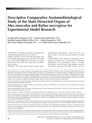
  • 6. Volume 43, Number 2/April 2021 95 Descriptive Comparative Anatomohistological Study (divided horizontally into anterior and posterior portions), and a left lateral lobe and caudal lobe. The latter consists of 2 portions: 1 dorsally, resem- bling a leaf, and 1 ventrally to the esophagus in the smallest stomach curve, the surface of which forms the papillary process. Blood feeds the liver through interlobular branches of hepatic arteries and hepatic veins, which are open for the sinu- soids. The gallbladder sits at the base of the deep bifurcation of the middle lobe next to the point of origin of the falciform ligament. Hepatocytes (parenchymal cells) are placed on plates which radiate from the central vein to the lobular pe- riphery (Figure 6). The spleen is elongated and triangular in cross- section, with a posterolateral face (diaphragmatic), a posteromedial face (renal), an anteromedial face (gastric), and an antero-lower face (colic). Its tis- sue is formed by parenchyma (splenic pulp), con­ stituted by a differentiated cellular arrangement into a white pulp (a marginal zone of lower cell density) and a red pulp (found between the nod- ules and which constitutes the major part of the parenchyma) (Figure 7). Their kidneys are shaped like bean grains with a dark red coloration. The nephron consists of the glomerulus surrounded by a Bowman’s capsule, parietal cells of which appear flattened in the vas­ cular pole and cuboid on the glomerular urinary pole. Found mainly in the cortex, the proximal tubules are formed by cuboidal cells with promi- nent brush-like microvilli. The distal tubules reen- ter in the cortex and present a cuboid epithelium similar to that of proximal tubules but deprived of microvilli (Figure 8). In rodents the stomach divides into aglandular and glandular, separated by a pleated margin or limiting crest (margo plicatus). The glandless por- tion is of an opaque whitish coloration, coated by keratinized stratified pavement epithelium. The glandular portion is translucent, colored reddish- pink, and coated with simple cubic epithelium. The mucosa of the glandular stomach encompasses (1) the cardiac region, located around the gastric Figure 1  Correlation between weights of the organ pairs. (A) Lungs (right and left) of mice, (B) kidneys (right and left) of mice, (C) lungs (right and left) of rats, and (D) kidneys (right and left) of rats.
  • 7. 96 Analytical and Quantitative Cytopathology and Histopathology® Silva-Santana et al opening of the esophagus extending to the edge of the pleated margin, containing mucous cells called cardiac glands, (2) the fundic region (ven- tral side of the stomach), composed mainly of glandular mucosa containing fundic glands and granular eosinophilic parietal cells, which pro- duce hydrochloric acid, and (3) the pyloric region extending to the pylorus (sphincter between the stomach and small intestine), formed by pyloric mucous glands (Figure 9). Both species’ intestine is slender and thick, hav- ing 3 layers in all its length (mucosa with sub- mucosa, muscular, and serosa). The mucosal epi- thelium of the small intestine shows scattered Paneth’s cells, goblet cells, and absorbent cells forming microvilli, which become smaller and in greater quantity from the duodenum to the ileum (Figure 9). The large intestine comprises the ce- cum, colon, and rectum. The cecum consists of a corpuscle and an apex, which is more elongated and less saccular in mice; its mucosa forms trans- versal folds. The colon has an ascending, a trans­ versal, and a descending part; its mucous mem- brane contains goblet cells in greater proportion than in the small intestine, which form crypts with no villi (Figure 10). Discussion Surgical research using laboratory animals has expanded in recent decades as a result of better anesthetic support, improved surgery monitoring infrastructure, and the incessant search for models capable of reproducing morbid conditions to solve human diseases.21 Researchers must consider the Table III  Comparison Between Weights of the Organs of Mice and Rats AHV (Levene’s test) CBOWB-M/R DBW-A/O Organ Species MW (g) F statistic p Value AWD (g) SWD (g) t Statistic gl p Value (95% CI) Brain Mouse 0.442 0.313 0.579 2.265 0.0192 118.169 40   <0.0001** 2.226–2.303 Rat 2.706 Eyes Mouse 0.022 116.055 <0.0001* 0.110 0.0021 52.601 14.165 <0.0001** 0.106–0.115 Rat 0.133 Left lung Mouse 0.077 13.519 0.001* 0.387 0.0097 40.005 14.751 <0.0001** 0.367–0.408 Rat 0.464 Right lung Mouse 0.132 13.546 0.001* 0.681 0.0092 74.362 17.115 <0.0001** 0.662–0.700 Rat 0.813 Heart Mouse 0.139 0.086 0.770 0.582 0.0039 149.742 40   <0.0001** 0.574–0.590 Rat 0.720 Liver Mouse 2.045 0.210 0.649 9.329 0.0091 1029.905 40   <0.0001** 9.310–9.347 Rat 11.373 Spleen Mouse 0.113 0.251 0.619 0.324 0.0062 51.978 40   <0.0001** 0.311–0.336 Rat 0.437 Left kidney Mouse 0.196 6.663 0.014* 0.866 0.0035 244.004 16.679 <0.0001** 0.858–0.873 Rat 1.062 Right kidney Mouse 0.246 14.944 0.0004* 1.102 0.0070 157.667 15.080 <0.0001** 1.087–1.117 Rat 1.348 Stomach Mouse 0.282 0.126 0.725 1.303 0.0037 352.956 40 <0.0001** 1.296–1.311 Rat 1.586 Small intestine Mouse 1.507 0.161 0.691 6.830 0.0037 1850.916 40 <0.0001** 6.823–6.837 Rat 8.337 Large intestine Mouse 0.868 0.155 0.695 3.943 0.0038 1036.818 40 <0.0001** 3.935–3.951 Rat 4.811 *Indicates that variances are unequal (p<0.05). **Indicates statistical differences highly significant (p<0.01). AHV = analysis of the homogeneity of variances, AWD = average weight difference, CBOWB-M/R = comparison between organ weights between mice and rats, DBW-A/O (95% CI) = difference between the weights of animal organs (95% confidence interval), gl = degree of freedom, MW = middle-weight, SWD = standard weight difference error.
  • 8. Volume 43, Number 2/April 2021 97 Descriptive Comparative Anatomohistological Study general biology, anatomy, histology, and physiol- ogy of laboratory animals used in research before choosing the most appropriate experimental mod­ el. In the course of this article we observed some Figure 2  Anatomy and histology of the mouse and rat brain. (A–B) Brain of mouse and (E–F) brain of rat (bar=5 mm). Dorsal surfaces (A and E) and ventral surfaces (B and F) of the cerebral hemispheres. (1) Olfactory bulb, (2) forebrain (prosencephalon), (3) midbrain (mesencephalon), (4) flocculus, (5) vermis, (6) pyriform cortex, (7) rhombencephalon, (8) longitudinal fissure, (9) cerebral cortex, (10) caudal colliculus, (11) transverse fissure, (12) optic nerve, (13) olfactory bulb, (14) lateral olfactory tract, (15) optic chiasma, (16) mamillary bodies, (17) cerebral crus, (18) pons, (19) ventral medial fissure, (20) trapezoid body, (21) paraflocculus, (22) lateral ventral sulcus, (23) medullary pyramid, (24) medulla oblongata, and (25) spinal medulla. (C–D) Mouse brain tissue, 4× and 40×, respectively (bar=500 μm). (G–H) Rat brain tissue, 20× and 40×, respectively (bar=500 μm). (1) Cerebral cortex, (2) molecular layer, (3) Purkinje cells, (4) granular layer, (5) white matter, and (6) sulcus. Figure 3  Anatomy and histology of the mouse and rat eyes. (A) Eyes (right and left) of mouse and (E) eyes (right and left) of rat (bar=5 mm). (1) Eye globe, (2) optic nerve. (B–D) Tissues that make up eyes of mice, 4×, 10×, and 20×, respectively (bar=500 μm). (F–H) Tissues that make up eyes of rats, 4×, 10×, and 40×, respectively (bar=500 μm). (1) Cornea, (2) anterior chamber, (3) iris, (4) ciliary body, (5) lens, (6) vitreous body, (7) retina, (8) sclera, (9) choroid, (10) lamina vitrea, (11) pigmented epithelium, (12) roads and cones, (13) outer nuclear layer, (14) outer plexiform layer, (15) inner nuclear layer, (16) inner plexiform layer, and (17) ganglion cell.
  • 9. 98 Analytical and Quantitative Cytopathology and Histopathology® Silva-Santana et al Mice have lissencephalic brains (devoid of folds), where the caudate nucleus and the putamen form important anatomical and histological features specific to Mus musculus and Rattus norvegicus. Figure 4  Anatomy and histology of the mouse and rat lungs (right and left). (A–B) Lungs (right and left) of mouse: (A) ventral surface and (B) dorsal surface. (F–G) Lungs (right and left) of rat: (F) ventral surface and (G) dorsal surface (bar = 5 mm). (1) Left lung, (2) right cranial lobe, (3) right middle lobe, (4) right caudate lobe, and (5) accessory lobe. (C–E) Pulmonary tissue of mice, 4×, 10×, and 20×, respectively (bar=500 μm). (H–J) Pulmonary tissue of rat, 4×, 10×, and 20×, respectively (bar=500 μm). (1) Pleura, (2) alveolus, (3) alveolar septum, (4) bronchioles, (5) pulmonary vein, and (6) bronchial coating epithelial cells. Figure 5  Anatomy and histology of the mouse and rat heart. (A) Mouse heart and (D) rat heart (bar = 5 mm). (1) Aorta, (2) left auricle, (3) left ventricle, (4) right auricle, (5) right ventricle, and (6) conoventricular vein. (B–C) Myocardium of the mouse, 10× and 40×, respectively (bar=500 μm). (E–F) Myocardium of the rat, 10× and 40×, respectively (bar=500 μm). (1) Myocardial fibers exhibit cross striations formed by alternating segments, (2) myocyte nuclei, and (3) vein.
  • 10. Volume 43, Number 2/April 2021 99 Descriptive Comparative Anatomohistological Study Figure 6  Anatomy and histology of the mouse and rat liver. (A) Visceral surface of the mouse liver and (D) visceral surface of the rat liver (bar=5 mm). (1) Left lateral lobe, (2) caudate lobe, (3) right lateral lobe, (4) right middle lobe, (5) gallbladder, (6) papillary process, and (7) left middle lobe. (B–C) Hepatic tissue of mice, 10× and 20×, respectively (bar=500 μm). (E–F) Hepatic tissue of rats, 10× and 40×, respectively (bar=500 μm). (1) Central vein, (2) sinusoid, (3) hepatocyte, (4) portal triad: portal vein, (5) hepatic artery, and (6) bile duct. Figure 7  Anatomy and histology of the mouse and rat spleen. (A–B) Spleen of a mouse and (E–F) spleen of a rat (bar=5 mm). (1) Colic surface, (2) gastric surface, (3) diaphragmatic surface, and (4) renal surface. (C–D) Splenic tissue of a mouse, 4× and 20×, respectively (bar=500 μm), and (G–H) splenic tissue of a rat, 20× and 40×, respectively (bar=500 μm). (1) White pulp, (2) red pulp, and (3) central artery.
  • 11. 100 Analytical and Quantitative Cytopathology and Histopathology® Silva-Santana et al Figure 8  Anatomy and histology of the mouse and rat kidney. (A) Kidneys (right and left) of a mouse and (D) Kidneys (right and left) of a rat (bar=5 mm). (1) Papilla, (2) medulla, and (3) cortex. (B–C) Renal cortex of a mouse, 10× and 20×, respectively (bar=500 μm). (E–F) Renal cortex of a rat, 10× and 40×, respectively (bar=500 μm). (1) Glomerulus, (2) Bowman’s space, (3) renal tubules, (4) capillary, and (5) mesentery. Figure 9  Anatomy and histology of the mice and rat stomach and small intestine. (A) Stomach and small intestine of a mouse and (E) stomach and small intestine of a rat (bar=5 mm). (1) Stomach, (2) duodenum, (3) jejunum, and (4) ileus. (B–D) Stomach of a mouse, 4×, 10×, and 40×, respectively (bar=500 μm). (F–H) Jejunum of a rat, 4×, 10×, and 40×, respectively (bar=500 μm). (1) Mucosa of the aglandular stomach, (2) main cells, (2a) parietal cells, (3) limiting crest, (4) mucosa of the glandular stomach, (5) villi, (6) absorbent columnar cells, (7) lamina propria, (8) crypts, (9) submucosa, (9a) muscularis mucosae, (10) muscular layer, (10a) internal circular muscular layer, (10b) outer longitudinal muscular layer, (11) adventitia, and (12) cardiac gland.
  • 12. Volume 43, Number 2/April 2021 101 Descriptive Comparative Anatomohistological Study In mice the rhombencephalon is responsible for muscle balance and control and autonomous functions such as heart and respiratory rate.23,24 The midbrain contains structures responsible for receiving and interpreting visual and auditory signals.22,24 The hypothalamus controls endocrine functions and the survival instincts (fight or flight, reproduction); the thalamus relays sensory infor- mation to the primary areas of the cortex.23,24 The fissures and folds that form the cortex, although abundant in rats and other higher mammals, are few in mice.23-25 The genes responsible for neural development in humans and mice are 90% identi- cal, making the use of these animals in studies of mental illness of great importance.24 The eyes of both species comprise structures that nourish, protect, and lubricate the eyeball, including conjunctiva, eyelids, nictitating mem- brane (third eyelid), Harderian gland, and Mei- bomian glands.22-24 Being nocturnal animals, their retina present peculiarities: a central round area or “horizontal ray” increases its visual acuity, it also lacks macula. Under weak light the mouse pupil a continuous structure (caudate putamen). Com- monly, the brain of both species can be divided into 3 anatomical parts: rhombencephalon, mid- brain, and forebrain. The rhombencephalon, com- posed of marrow, pons, and cerebellum, connects the brain to the spinal cord (oblong). The midbrain, sitting between the hindbrain and the prosenceph- alon, consists of the tectum, integument, and the cerebral peduncle. The forebrain encompasses the cerebral cortex (telencephalon), the basal ganglia, septum, epithalamus, thalamus, and hypothalamus (diencephalon) and the olfactory bulb. In the cere- bral cortex the right and left sides are joined by the corpus callosum, a thick band of nerve fibers.22,23 The brain tissue consists of functional cells (neu- rons) and the support cells, macroglia, and microg- lia. Macroglia are oligodendrocytes (producers of myelin and astrocytes) present in both gray and white matter. Ependymal ciliated cells line the walls of the brain ventricles and can react posi- tively with astrocytes. The epithelium of the cho- roid plexus forms microvilli and reacts positively with epithelial markers.23,24 Figure 10  Anatomy and histology of the mouse and rat large intestine. (A) Large intestine of a mouse and (D) large intestine of a rat (bar=5 mm). (1) Cecum, (2) colon, and (3) rectum. (B–C) Cecum of a mouse, 10× and 40×, respectively (bar=500 μm). (E–F) Colon of a rat, 10× and 40×, respectively (bar=500 μm). (1) Serosa (visceral peritoneum), (2) muscular layer, (3) fine submucosa, (4) submucosa, (5) crypt elongation, (6) Lieberkühn’s glands, (7) goblet cells, and (8) lymphatic follicle.
  • 13. 102 Analytical and Quantitative Cytopathology and Histopathology® Silva-Santana et al and left lung weight, directly correlated to the number of lobes. In mice the right lung can be 71.32% heavier than the left; in rats this differ- ence can reach 75.12%. Another important ana- tomical difference is the presence of the accessory lobe in rats and its absence in mice. In mammals the heart is divided into 4 cham- bers: 2 atria separated by an interatrial septum and 2 ventricles separated by an interventricular septum.22-24 Between the interatrial septum and interventricular septum exists a small septal seg- ment known as the atrioventricular septum (rela­ tively thick and mainly muscular, a result of the displacement of the atrioventricular valves), placed between the subaortic segment leaving the left ventricle.23,24 At the junction between the atria and ventricles (AVJ) we find 2 valves: the left AVJ has a mitral valve with 2 separate leaflets (bicuspid valve), while the right AVJ comprises a valve with 3 separate leaflets (tricuspid valve).22-24 The inter- nal cover of the ventricles is characterized by the presence of numerous myocardial protrusions (trabeculae).23,24 Inside of the apical cavity of the ventricles, beyond trabeculation and chordae ten­ dineae, we can distinguish fine structures similar to tendon cords linked to the papillary muscle (trabeculae tendineae).22-24 The heart consists almost exclusively of myo- cytes specialized in the conduction system (Pur­ kinje cells) inside a fibrous leaf. The pulmonary veins join in confluence, entering through a single foramen on the dorsal wall of the left atrium.23,24 The superior relative weight of the mouse heart may be correlated with the arrangement of myo- cardial fibers, with greater quantity of myocardio- cytes observed in histology. Another important factor that may explain these results is that the mouse’s heart rate at rest (~500–780 bpm) is higher than the rat’s (~250–480 bpm).4,36,37 The liver occupies one-third of the anterior re- gion of the abdominal cavity, presenting a surface covered by a capsule, formed by conjunctive tis- sue.23-25 The blood flows from the perilobular re- gion to the central vein, following to the cava vein via large hepatic veins. The hepatocytes are dis­ tributed on plates radiating from the central vein to the lobular periphery; the sinusoidal coating cells (endothelial cells, Kupffer cells, fixed macro- phages linked to sinusoid wall cells, and granular lymphocytes with natural killer activity) show ac- cumulation of cytoplasmic lipid and vitamin A, he- matopoietic cells, bile duct cells, connective tissue widens to 1.2 mm in diameter, while in strong light it contracts about 0.2 mm diameter for half a second.24,26,27 It can have 2 types of photorecep- tors: rods, which detect light and darkness, and cones for green and blue colors. As blue cones are sensitive to short wavelengths, both animals can see ultraviolet.4,28 Each neural cell on the rodent’s retina has a greater number of photore­ ceptors than that in humans, i.e., the ganglionic receptor fields of the rodent’s cells are greater than those of the human fovea,29 which increases sensitivity and reduces acuity. Present only in albino mice, the Bowman’s membrane has both pigmentation and retinal pigment epithelium.23,24 The mouse sclera is opaque, covering from the back of the eyes to the choroids, with no tapetum lucidum.23-25 The ciliary body, iris, and choroid form the external fibrous vascular tunic (uvea) containing pigment, except in albino species.23-25 The lens consists of rolled fibers formed by modi- fied epithelial cells, enclosed per capsule, allowing for almost all visible light and 50% of ultravio- let.23,30 Its poorly developed ciliary muscle renders it impossible to change the shape of the lens26,31 unless administered atropine drops, which relax the lens keeping its focus, but these are inconclu- sive data.31,32 Lastly, the nictitating membrane forms a translucent conjunctiva that, together with the Harderian gland, surrounds and protects the optic nerve.23,24 Located in the thoracic cavity, covered by the parietal pleura, the left lung consists of a single lobe, while the right lung consists of 4 lobes (cra- nial, middle, caudal, and accessory), with stud- ies describing at least 9 standards for pulmonary lobulation.23,24,33,34 Two bronchi form an intrapul- monary route with bifurcations ending in bron­ chioles. The smallest bronchial airways in mice are the terminal bronchioles, which evolve into al- veolar ducts (alveolar sacs and alveoli). The alve- olar epithelium consists of lung cells type I and cu- boidal cell type II. Lungs of healthy mice present few lymphoid tissues (bronchus-associated lym- phoid tissue [BALT] associated with bronchial tis- sue), becoming well developed only by pathogen- ic infections.23,24,35 Blood enters the lungs from the systemic circulation through the bronchial arter- ies, and venous blood exits through the pulmo- nary arteries of the heart. Arteries and veins end on the bronchial tree, and the walls of the pulmo- nary veins contain cardiac muscles (striated).23,24 We found a significant difference between right
  • 14. Volume 43, Number 2/April 2021 103 Descriptive Comparative Anatomohistological Study and macrophages that, when stimulated by an antigen, can store plasma cells. In continuations of the periarteriolar lymphoid sheath, found in bi- furcations of the central arterioles, the follicles comprise mainly B lymphocytes, follicular den- dritic cells, and T CD4+ cells in smaller number; commonly, T CD8+ cells are absent.39,43 When stimulated by antigens, follicles can form germinal centers with a greater presence of follicular macro- phages.39,40 Lastly, situated in the interface of red pulp with the periarteriolar lymphoid sheath and the follicles, the marginal zone consists of reticular fibrils, macrophages, dendritic cells, and medium B cells.41,44 The macrophages of the marginal zone assist in removing microorganisms and virus, rec- ognizing receptors (TLRs) and recognizing bac- teria.39,44 The marginal zone B cells are a unique subset of noncirculating B cells that have an IgM+/ IgD− phenotype as opposed to follicular B cells, which are IgM+/IgD+.39,43 Kidneys lie parallel in the dorsal part of the abdominal cavity; the right kidney is slightly high- er (cranially) than the left kidney.22-24 The right kidney is usually larger than the left, and the kid- neys of males are relatively larger than those of females.24 This article corroborates these findings, as the right kidney of mice showed, on average, 28.26% more weight than the left, with similar results observed in the right kidney of rats, on average 26.97% heavier than the left. In rats, the kidney is unilobed with a single papilla, formed by the cortex and medulla. The cortex contains cortical tubular labyrinths (mainly proximal convoluted tubules) and medullary rays that extend from the external medulla.23 The me- dulla is subdivided into an outer zone, with one external and internal band and an internal zone forming the papilla. Its functional unit is the neph- ron, which consists of the glomerulus (covered by Bowman’s capsule) wound around the proximal and distal tubules, the descending and ascending portions of the Henle loop and the straight por- tions.24 The nephrons connect into the collecting ducts, which emerge from the papillary ducts,22,23 which in turn open at the tip of the renal papilla in the renal pelvis.24 The renal pelvis is lined with transitional cell epithelium, and its continuation forms the ureter.22 The renal papilla in rats can be long and protrudes into the initial portion of the ureter.23 Most rat species may exhibit sexual dimorphism under the influence of testosterone (the female parietal epithelial cells are typically cells, and blood vessel wall cells (adventitial cells and smooth muscle).23,24 A characteristic usually found in hepatic cells of rats and mice is anisocy- tosis and anisokaryosis (variations in the size of cells and nuclei).24 Observed only in mice, the gallbladder sits at the base of the deep bifurcation of the middle lobe next to the point of origin of the falciform liga- ment.23,24,38 Both the hepatic duct of the liver and the cystic duct of the gallbladder join to form the common bile duct—the standard hepatic pattern, although studies have described at least 13 dif- ferent ones.33 In rats the periportal biliary system consists of a network of canaliculi that end in a common bile duct.21 Of friable consistency and purple coloration, the spleen rests on the left dorsocranial region of the abdominal cavity, posterior to the stomach and above the upper pole of the left kidney.22,23,39 Both the greater intensity of the color purple found in the spleens of rats and the superior relative weight of the mouse spleen can be attributed to the venous sinuses (hematopoietic tissue)—they are bigger and more abundant in rats, generating numerous anastomoses (sinus) that provide larger areas of red pulp, unlike the venous sinuses in mice.39 Mice showed a greater proportion of white pulp than did rats.39,40 Finally, extramedul- lary hematopoiesis is common in the red pulp of rodents, with greater prevalence in the spleens of mice than that in rats.39 This organ acts as an immunological system and blood reservoir, producing and maturing B and T lymphocytes, integrating, in some cases, the reticuloendothelial system and participating in the hematopoiesis and eryptosis process (reno- vation of red blood cells).39 The red pulp consists of reticular tissue formed of splenic cords (or Cords of Billroth) and venous sinuses (splenic sinuses), which go to the blood tissue (capillaries and sinusoids); the leukocytes perform its selec- tions and destruction, if they present anomalies, are old or injured. Besides serving as a leukocyte and platelet deposit, the macrophages contained therein are responsible for the phagocytosis of microorganisms.24,39,41 The white flesh comprises a periarteriolar lymphoid sheath, follicles, and marginal zone.41,42 The internal periarteriolar lym- phoid sheath consists predominantly of T CD4+ cells, T CD8+ cells in smaller number, dendritic cells, and migrating B cells; its outer part consists of small/medium lymphocytes (B and T cells)
  • 15. 104 Analytical and Quantitative Cytopathology and Histopathology® Silva-Santana et al thelium lining and a fibrovascular stroma called “own blade,” separated from the submucosa by mucosal muscular lamina (a thin layer of smooth muscle).23,24 The submucosa consists of connec- tive tissue involving blood vessels, lymphatic ves- sels, and nerves. The muscular layer consists of the muscle inner circular layer, and the serosa com- prise the thin layer of the peritoneum.23,24,38 Its lymphoid tissue (gut-associated lymphoid tis- sue) forms the scattered nodules, the submucosa, and the lamina propria. Located over the mesen­ teric accessory of the small intestine are the aggre- gated lymphoid nodules (Peyer’s patches); on the surface of the epithelium of the antimesenteric portion we find antigen-producing M cells.45 In the large intestine the Peyer’s patches can be antimes- enteric or not. The mucosal epithelium usually has absorption cells, with luminal membrane cells forming microvilli, and a greater number of goblet cells than in the small intestine, forming crypts. Rats show a greater number of Paneth cells, which contain eosinophils (granulocytes) with lysozyme and antimicrobial peptides. Polypeptide entero- endocrine cells appear scattered along the gastro- intestinal tract, and caveolae cells are responsible for producing intestinal chemoreceptors.23,24 The entrance of the ileum forms the sacculus rotundus and the exit of the colon forms the ampulla coli.24 Different from higher mammals, the mucosal surface of the small intestine of rats has no folds (plicae). Instead, the mucosa forms villi from epi- thelium and lamina propria, forming the inner intestinal lumen23,24,38; the villi, each containing 1 lymphatic vessel, decreases in number from the duodenum to the ileum.23,24 Between the villi, pro- trusions emerge in the opposite direction under the surface of the mucosa, forming crypts or intes- tinal glands.23,24,38 Both species have a functional cecum, large in size, with sacs forming a fermentation tank where specialized microorganisms degrade cell walls formed by cellulose. In rats the cecum has a cor- puscle and an apex; its mucosa forms transversal folds.45 The colon has an ascending, a transversal, and a descending part and a mucosa consisting of an ascending and transversal part, which forms transversal folds. The descending colon and rectum consist of longitudinal protuberances prominent in the lumen, formed by the mucosa and submucosa. The muscular mucosa is more prominent in the rectum than in the colon. From rectum to anus, the superficial epithelium becomes squamous and flattened and the male parietal epithelium can be both cuboidal and flattened).22,24 Regardless of the genus, the parietal cells of Bowman’s capsule are flattened in the vascular pole and cuboidal in the urinary pole of the glom- eruli.23,24 The proximal tubules, found mainly in the cortex, consist of cuboidal cells with a prominent brush-like border (microvilli).22,23 The descend- ing and ascending portions of the Henle loop are found in the marrow, lined with flattened epithe­ lium that resembles the endothelium of blood vessels.24 The distal tubules reenter the cortex and consist of a cuboidal epithelium similar to that of proximal tubules but devoid of microvilli.22 The straight portion of the distal tubules leads to dense macula in the vascular pole of the glomer- ulus, where specialized cells produce renin.24 In rats, the renal vasculature resembles other spe- cies: the branches of the renal artery fashion the arcuate arteries on the corticomedullary border.23 The interlobular branches of the arched arteries supply the afferent arterioles of the glomeruli,22,24 which in turn provide the cortex with blood and form the downward straight vessel, responsible for supplying blood to the marrow.23,24 The ascending vessel collects venous blood, while spontaneous vacuolation occurs in the arched interlobular veins, probably of lysosomal origin, in the renal tubular epithelium of the external medulla.22,23 In rodents, the stomach divides into glandular and nonglandular.24,38,45 The nonglandular stom- ach usually has thin, transparent walls lined with keratinized, stratified squamous epithelium, serv- ing for storage and food digestion. The glandular stomach has thick walls lined with epithelium; the blade itself consists of tubular gastric glands containing cells that secrete mucus and pepsino- gen, main cells, and hydrochloric acid–producing cells (parietal cells).21,23,24,45 The initial portion of the duodenum has special tubular alveolar glands known as Brunner glands.23,24 One or more pan- creatic ducts and the common bile duct end in the duodenal papilla.24 Models used to replicate stomach ulcers should consider inducing ulcers in the nonsecretory mucosa; such studies must un­ dergo rigorous evaluation, as producing ulcers in the rat’s gastric mucosa is a difficult task, and most proposed methods frequently end in animal suffering.21 The intestine is slender and thick, having 3 lay- ers in all its length (mucosa with submucosa, mus- cular, and serosa).23,24,38 The mucosa has an epi­
  • 16. Volume 43, Number 2/April 2021 105 Descriptive Comparative Anatomohistological Study Criação e experimentação. Rio de Janeiro, Fiocruz, 2006, p 388. Available at http:/ /books.scielo.org/id/sfwtj. Accessed July 25, 2019   4.  Chorilli M, Michelin DC, Salgado HRN: Animais de labora- tório: o camundongo. Rev Ciênc Farm Básica Apl 2007;28(1): 11-23   5.  Heywood R: The use of animals in testing. ATLA 1987; 14(1987)   6.  Ribeiro SML, Campos P, Tirapegui J: Rat as an experimental animal: History, biological data and critical analysis of its usage. Rev Farm Bioquim Univ Säo Paulo 1995;31(1):21-28   7.  Salén JCW: Animal models: Principles and problems. In The Experimental Animal in Biomedical Research: Care, Hus- bandry and Well-Being: An Overview by Species. Edited by BE Rollin, ML Kessel. Boston, CRC Press, 1995, p 560   8.  Snitkoff GG: Testes biológicos. In A Ciência e a Prática da Farmácia. Edited by GAR Remington. Rio de Janeiro, Gua- nabara Koogan, 2004, pp 556-568   9.  Calabrese EJ: Principles of Animal Extrapolation. Michigan, Lewis Publishers, 1991 10. Fagundes DJ, Taha MO: Animal disease model: Choice’s criteria and current animals specimens. Acta Cir Bras 2004; 19(1):59-65 11.  Lynette AH: Responsible conduct with animals in research. Oxford, Oxford University Press, 1998 12.  Santos BF: Camundongos mutantes mais utilizados. In Ani- mais de Laboratório: Criação e Experimentação. Edited by A Andrade, SC Pinto, RS Oliveira. Rio de Janeiro, Fiocruz, 2002, pp 139-142 13. Santos BF: Criação e manejo de camundongos. In Animais de Laboratório: Criação e Experimentação. Edited by A Andrade, SC Pinto, RS Oliveira. Rio de Janeiro, Fiocruz, 2002, pp 115-118 14.  Cesarino JL, Gontijo JAR, Zapparoli A: Environment in an ex- perimental animal facility and the species Rattus norvegicus: Review. Rev Elet Farm 2011;8(2):25-32 15.  Clause BT: The Wistar Institute Archives: Rats (not mice) and history. Mendel Newsl 1998;7:2-7 16.  Andersen ML, D’Almeida V, Ko GM, Kawakami R, Martins PJF, Magalhães LE, Tufik S: Eutanásia. In Princípios Éticos e Práticos do Uso de Animais de Experimentação. Edited by ML Andersen, V D’Almeida, GM Ko, R Kawakami, PJF Martins. São Paulo, UNIFESP–Universidade Federal de São Paulo, 2004, pp 71-79 17.  Lima JBA, Skare TL, Malafaia O, Ribas-Filho JM, Michaelis T, Ribas FM, Macedo RAC: Sepsis inducing syndrome of multiple organ dysfunction: An experimental study in rats. Arq Bras Cir Dig 2011;24(2):95-102 18.  Silva-Santana G, Lenzi-Almeida KC, Fernandes-Santos C, Couto DS, Paes-De-Almeida EC, Aguiar-Alves F: Mice infection by methicillin-resistant Staphylococcus aureus from different colonization sites in humans resulting in diffusion to multiple organs. J Clin Exp Pathol 2016;6:1000283 19.  Silva-Santana G, Lenzi-Almeida KC, Lopes VGS, Aguiar- Alves F: Atypical manifestation in infection by methicillin- resistant Staphylococcus aureus carrier SCCmec IV and Panton- Valentine Leukocidin-producer in experimental sepsis model. Afr J Microbiol Res 2017;11(18):724-728 stratified, presenting sebaceous modified glands (circum-anal gland) around the anus.23,24,38 Conclusion Except for some veterinary and zoology books, few are the studies focusing on the specific char- acteristics between laboratory animals. Today we can dispose of more refined experimental models, maintaining and intensifying specific phenotypic and genotypic characteristics, since the animal’s genome results from directed mating. The greater the similarity between the animal’s physiologi- cal, anatomical, and organic characteristics with humans, the greater its applicability in studies and the reliability of results, even if establishing reliable general rules to validate the extrapolation from one species to another is infeasible. Mice and rats are the species most used in research. They are better known scientifically and present advantages over other species, such as smaller physical size and greater weight, easy mobility, and lower maintenance cost. Currently, multiple lineages of mice and rats destined to dif- ferent research purposes exist. However, studies using animals require careful research planning, knowledge of the country’s laws and guidelines, ethical principles, and, above all, being up-to-date on previous studies in the same area so as to avoid repetitive tests and inconclusive results, which would lead to waste, thus allowing to choose the most suitable species. With technological advances, alternative re- search methods, such as in vitro, are being devel- oped, but experimental models using laboratory animals still pose the advantage of obtaining in- formation about the organism as a whole. Acknowledgements We would like to thank the Pathology Program at Fluminense Federal University (UFF), the Institute of Microbiology Paulo de Góes at Federal Univer- sity of Rio de Janeiro, and the Laboratory of Diph- theria and Corynebacteria of Clinical Relevance at the University of the State of Rio de Janeiro (UERJ). References   1.  Feijó AGS, Braga LMGM, Pitrez PMC: Animais na pesquisa e no ensino: Aspectos éticos e técnicos. Porto Alegre, Edi­ PUCRS, 2010, p 421   2.  Neves SMP: Manual de cuidados e procedimentos com ani- mais de laboratório do Biotério de Produção e Experimen- tação da FCF-IQ/USP. São Paulo, FCF-IQ/USP, 2013, p 216  3. Andrade A, Pinto SC, Oliveira RS: Animais de laboratório:
  • 17. 106 Analytical and Quantitative Cytopathology and Histopathology® Silva-Santana et al 34.  Kittel B, Ruehl-Fehlert C, Morawietz G, Klapwijk J, Elwell MR, Lenz B, O’Sullivan MG, Roth DR, Wadsworth PF: Revised guides for organ sampling and trimming in rats and mice: Part 2. A joint publication of the RITA and NACAD groups. Exp Toxic Pathol 2004;55(6):413-431 35.  Souza JB, Oliveira MT, Nascimento ER, Verícimo MA, Barreto ML: Mycoplasma pulmonis, murine respiratory my- coplasmosis agent: review. Arch Vet Sci 2016;21(4):8-25 36.  Ko GM, DeLuca RR: Camundongo. In Cuidados e Manejos de Animais de Laboratório. Edited by V Lapchik, V Mattara- ia, G Ko. São Paulo, Atheneu, 2009, pp 137-167 37.  Oliveira FS, Rangel JA, Batista WS, Gameiro LS, Oliveira GM: Influence of paternal aggression in the physical/emo- tional development of Swiss Webster mice in laboratory environment. RESBCAL 2014;2(4):244-253 38.  Ruehl-Fehlert C, Kittel B, Morawietz G, Deslex P, Keenan C, Mahrt CR, Nolte T, Robinson M, Stuart BP, Deschl U: Re- vised guides for organ sampling and trimming in rats and mice: Part 1: A joint publication of the RITA and NACAD groups. Exp Toxic Pathol 2003;55(2–3):91-106 39.  Cesta MF: Normal structure, function, and histology of the spleen. Toxicol Pathol 2006;34(5):455-465 40.  Ward JM, Mann PC, Morishima H, Frith CH: Thymus, spleen, and lymph nodes. In Pathology of the Mouse. Edited by RR Maronpot. Vienna, Illinois, Cache River Press, 1999, pp 333-360 41.  Veerman AJP, van Ewijk W: White pulp compartments in the spleen of rats and mice. A light and electron microscopic study of lymphoid and non-lymphoid cell types in T- and B-areas. Cell Tissue Res 1975;156(4):417-441 42.  Saito H, Yokoi Y, Watanabe S, Tajima J, Kuroda H, Namihisa T: Reticular meshwork of the spleen in rats studied by elec- tron microscopy. Am J Anat 1988;181(3):235-252 43.  van Rees EP, Sminia T, Dijkstra CD: Structure and develop- ment of the lymphoid organs. In Pathobiology of the aging mouse. Edited by U Mohr, DL Dungworth, CC Capen, WW Carlton, JP Sundberg, JM Ward. Washington, ILSI Press, 1996, pp 173-187 44.  Mebius RE, Kraal G: Structure and function of the spleen. Nat Rev Immunol 2005;5(8):606-616 45.  Kararli TT: Comparison of the gastrointestinal anatomy, physiology, and biochemistry of humans and commonly used laboratory animals. Biopharm Drug Dispos 1995;16(5): 351-380 20.  Silva-Santana G, Lenzi-Almeida KC, Lopes VGS, Aguiar- Alves F: Biofilm formation in catheter-related infections by Panton-Valentine leukocidin-producing Staphylococcus aureus. Int Microbiol 2016;19(4):199-207 21.  Schanaider A, Silva PC: The use of animals in experimental surgery. Acta Cir Bras 2004;19(4):441-447 22.  Morawietz G, Ruehl-Fehlert C, Kittel B, Bube A, Keane K, Halm S, Heuser A, Hellmann J; RITA Group, NACAD Group: Revised guides for organ sampling and trimming in rats and mice: Part 3. Exp Toxic Pathol 2004;55(6):433-449 23.  Treuting PM, Dintzis SM, Montine KS: Comparative anato- my and histology. In A Mouse and Human Atlas. Edited by PM Treuting, SM Dintzis, KS Montine. Elsevier, Academic Press, 2012, pp 457 24.  Krinke GJ: Normative histology of organs. In The Laboratory Mouse. Edited by HJ Hedrich, G Bullock. Elsevier, Academic Press, 2004, pp 133-166 25.  Cook MJ: The Anatomy of the Laboratory Mouse. Elsevi- er/Academic Press, 1965. Adapted for the Web by Mouse Genome Informatics, Bar Harbor, Maine, The Jackson Lab- oratory, 2008 26.  Lashley KS: Studies of cerebral function in learning. VIII. A reanalysis of data on mass action in the visual cortex. J Comp Neurol 1932;54:77-84 27.  Remtulia S, Hallett P: A schematic eye for the mouse, and comparisons with the rat. Vision Res 1985;25(1):21-31 28.  Jacob L, Beecken V, Bartunik LJ, Rose M, Bartunik HD: Puri- fication and crystallization of yeast hexokinase isoenzymes. Characterization of different forms by chromatofocusing. J Chromatogr 1991;587(1):85-92 29.  Brown JE, Rojas JA: Rat retinal ganglion cells: receptive field organization and maintained activity. J Neurophysiol 1965;28(6):1073-1090 30.  Gorgels TG, van Norren D: Spectral transmittance of the rat lens. Vision Res 1992;32(8):1509-1512 31.  Woolf D: A comparative cytological study of the ciliary mus- cle. Anat Rec 1956;124(2):145-163 32.  Artal P, Herreros de Tejada P, Muñoz Tedó C, Green DG: Retinal image quality in the rodent eye. Vis Neurosci 1998; 15(4):597-605 33.  Hummel KP, Richardson EL, Fekete E: Anatomy. In Biology of the Laboratory Mouse. Edited by EL Green, EU Fahey. New York, Dover Publications, 1975, pp 247-307