2. International Journal of Clinical Oncology
1 3
treatment, (3) to eliminate unnecessary treatment and insuf‑
ficient treatment, and (4) to deepen mutual understanding
between healthcare professionals and patients by making
these guidelines available to the general public [1].
The following are expected to be achieved with these
guidelines: (1) improvement of the treatment of colorectal
cancer in Japan; (2) improvement of the results of treatment;
(3) reduction of the human and financial burden; and (4)
increased benefits for patients.
2. How to use these guidelines
These guidelines were prepared by consensuses reached by
the Guideline Committee of the Japanese Society for Can‑
cer of the Colon and Rectum, based on a careful review of
the evidence retrieved by the literature searches and in view
of the medical health insurance system and actual clinical
practice settings in Japan and, therefore, these guidelines
can be used as a tool for treating colorectal cancer in actual
clinical practice settings. More specifically, they can be used
as a guide to obtaining informed consent from patients and
choosing the method of treatment for each patient. How‑
ever, these guidelines provide only general recommendations
for choosing treatment strategies for colorectal cancer, and
they do not control or limit treatment strategies or treat‑
ment methods that are not described herein. They can also
be used as a document to explain the rationale for selecting
treatment strategies and treatment methods that differ from
those described therein.
The Japanese Society for Cancer of the Colon and Rec‑
tum (JSCCR) is responsible for the statements in these
guidelines. However, the personnel directly in charge of
treatment, not the JSCCR or the Guideline Committee, are
responsible for the outcome of treatment.
3. Users
The users of these guidelines are mainly clinical doctors
engaged in all aspects of the medical treatment of colorectal
cancer.
4. How to develop these guidelines
(1) Recording methods
We adopted the concept from the first edition, in which the
treatment policy algorithm was disclosed, a simple expla‑
nation thereof recorded, and added further comments with
regard to categories requiring additional explanation. Since
the 2009 edition, areas of debate have been raised as clini‑
cal questions (CQs) and included with recommendations
added. In the 2016 edition, systemic therapy was the only
treatment to be revised. In the 2019 edition, all aspects of
the treatments were revised, with corrections and additions
made to the CQs based on knowledge acquired since the
2016 version (systemic therapy) and the 2014 version (other
treatments).
Efforts were made to make the expression of the CQs
clear and unambiguous. When comparing multiple inter‑
ventions, we did not stick to ranking everything, and kept
the expression flexible to ensure that it is useful in clinical
practice. The clinicopathological terms conformed to those
described in the “Japanese Classification of Colorectal,
Appendiceal, and Anal Carcinoma, third English edition [2].
(2) Evidence level/strength of recommendations of CQs
The recommendations added to CQs included the evidence
level and strength of recommendations determined using the
following direction.
(2-1) Evidence level
Papers relating to the CQs were comprehensively collected,
and the evidence indicated by individual papers relating to
the critical outcomes included within the CQs was divided
into groups by study design [3]. The literature level and a
body of evidence (Table 1) were evaluated in reference to
the GRADE* System [4–26], before determining the final
CQ evidence level (Table 2).
*GRADE: The Grading of Recommendations Assess‑
ment, Development and Evaluation
(2-2) Strength of recommendations
Draft recommendation statements and the strength of the
recommendations were directed based on the outcomes and
the level of evidence obtained from the process described
above and were evaluated at a consensus meeting of the
Guideline Committee. In the CQ text, the recommendations
that were decided have been directly expressed, and ambigu‑
ous expressions were excluded.
The draft recommendations were evaluated from four cat‑
egories (① Quality of evidence, ② Patients’ views and prefer‑
ences, ③ Benefits and harms, and ④ Cost effectiveness). The
strength of recommendation (Table 3) was determined by
vote, based on the GRADE Grid method [11].
Method
1. We selected one of the following five options and voted.
① Strong “For” intervention
② A Weak “For” intervention
③ Weak “Against” intervention
④ Strong “Against” intervention
⑤ Not graded
3. International Journal of Clinical Oncology
1 3
2. With one vote, if 70% or more of the votes were obtained
in any of ① to ⑤, it was considered a final decision.
If this criterion cannot be met, then the following shall
be applied:
– If ① + ② exceeds 50%, ③ + ④ is 20% or lower, “weakly
recommend to perform.”
– If ③ + ④ exceeds 50%, ①+ ② is 20% or lower, “weakly
recommend not to perform.”
3. Items not reaching consensus after a single vote were
debated once again, with the results of the first vote dis‑
closed and additional information on the situation relating
to clinical practice in Japan provided, and discussion and
voting was repeated.
4. If agreement was not reached, even in the second vote, no
strength of recommendation was presented in the CQ.
5. Literature search
At first, the literature search was performed for the clini‑
cal questions. Then, a further search was done as needed
with additional search techniques.
To survey the latest literature, in addition to the papers
used for reference in the previous edition, the PubMed and
Ichushi-Web databases were selected for the search, and the
English and Japanese literature was searched in both data‑
bases from June 2012 to February 2017. However, the start
of the search period for systemic therapy was August 2016.
The task of searching was performed by a medical librarian,
Table 1 Rating the quality of evidence
Table 2 Definition of levels of evidence (Ref. [14])
A (high) We are very confident in the effect estimate
B (moderate) We are moderately confident in the effect estimate: the true effect is likely to be close to the estimate of the effect, but there is a
possibility that it is substantially different
C (low) Our confidence in the effect estimate is limited: the true effect may be substantially different from the estimate of the effect
D (very low) We have very little confidence in the effect estimate: the true effect is likely to be substantially different from the estimate of
effect
Table 3 Strength of recommendation (Ref. [25])
Strength of recommendation
1 (Strong recommendation) Strong “For” an intervention
Strong “Against” an intervention
2 (Weak recommendation) Weak “For” an intervention
Weak “Against” an intervention
4. International Journal of Clinical Oncology
1 3
who created a search formula based on a discussion with the
Committee members in charge of each item and collected
literature during the search period. In addition, secondary
sources such as UpToDate and literature collected by manual
searching were added and critically examined as needed, and
other documents such as proceedings and guidelines were
included as necessary. We selected 3,295 documents from
among the 16,341 documents (PubMed 9,672, ICHUSHI
6,153, hand search 516) collected during the literature search
and critically reviewed all of them (Table 4).
Treatment guidelines for colorectal cancer
Chapter 1: Treatment strategies for Stage 0 to Stage III colo‑
rectal cancer
1. Endoscopic treatment (Fig. 1)
General principles underlying the indications for endoscopic
resection
• There is little possibility of lymph node metastasis, and
the size and location of the tumor make en bloc resection
possible.
Indication criteria for endoscopic resection:
(1) Intramucosal carcinoma or carcinoma with slight
submucosal invasion
(2) Size does not matter
(3) Any macroscopic type
• Endoscopic treatment is a method of endoscopically
resecting lesions in the large bowel and of collecting the
resected specimens.
• Endoscopic treatment methods consist of polypectomy
(note 1), endoscopic mucosal resection (EMR) (note 2),
and endoscopic submucosal dissection (ESD) (note 3).
• In determining the indication for endoscopic treatment
and the treatment method, information on the size, pre‑
dicted depth of invasion, and morphology of the tumor
is essential.
Comments
① Endoscopic resection is intended for both diagnosis and
treatment. It consists of total excisional biopsy in which
curability and the necessity of additional intestinal
resection are assessed by histopathological examination
of the resected specimens (CQ-1).
② cT1 deeply invasive cancer is diagnosed based on endo‑
scopic findings, such as “fullness, erosion, ulcer, fold
convergence, deformity, rigidity,” as well as contrast
X-ray, chromoendoscopy, image-enhanced endoscopy
(e.g., NBI/BLI [27], or magnifying endoscopic obser‑
vation) and endoscopic ultrasound findings. [28–30].
③ En bloc resection is desirable for accurate diagnosis of
the status of carcinoma invasion in the resection margin
and the deepest area.
• 2 cm is the largest size of a tumor that can be easily
resected en bloc by polypectomy or snare EMR [31]
(CQ-2).
• Colorectal ESD is an “endoscopic resection technique
which enables en bloc resection of a tumor, regard‑
less of size,” which was approved for implementation
Table 4 Number of scientific
articles retrieved and selected
Number of articles
retrieved
Number of articles
selected
Number of articles
retrieved manually
PubMed Ichushi PubMed Ichushi
(1) Endoscopic treatment 1102 539 136 73 81
(2) Surgical treatment 3351 2521 926 192 82
(3) Radiotherapy 1225 181 271 16 67
(4) Systematic therapy 2019 1381 591 108 242
(5) Others 1975 1530 374 86 44
Total 9672 6153 2304 475 516
Endoscopic en bloc
resection is possible
Endoscopic en bloc
resection is impossible
Endoscopic resection
Pathological
diagnosis
Surveillance Surgical resection
cTis or cT1
cTis
or
Slightly invasive cT1
Deep invasive cT1
Fig. 1 Treatment strategies for cTis and cT1 colorectal cancer
5. International Journal of Clinical Oncology
1 3
under health insurance in April 2012 with regard to
“early-stage malignant tumors”. Given the high like‑
lihood of technically difficult complications (perfora‑
tions), however, it should only be implemented after
sufficient consideration of the level of skill of the
endoscopist performing the procedure. Tumors with a
diameter between 2 and 5 cm were covered by insur‑
ance. The revision of April 2018 eliminated the upper
limit of the tumor diameter and the indication became
early colon cancer with a maximum diameter of 2 cm
or more. Early colon cancer accompanying fibrosis is
even applied to tumors with a diameter of 2 cm or less
(CQ-2).
• EMRC (EMR using a cap) is reported to involve a high
risk of perforation when used for colon lesions.
• If the preoperative diagnosis is cancer accompanied by
adenoma (intramucosal carcinoma), a piecemeal resec‑
tion can be performed with regard to the adenoma, while
avoiding division of the cancerous area. It should be
noted, however, that piecemeal resection is associated
with a high incomplete resection rate and a high local
recurrence rate. Multiple-piecemeal resection, which
makes accurate histological judgment difficult, should
be avoided [31].
• After endoscopic resection, the resection margin should
be observed in detail and the presence or absence of a
residual lesion should be confirmed.
• Dye spray and magnifying observation are useful for the
diagnosis of residual lesions [30].
• If residual mucosal lesions are present, additional treat‑
ment (e.g., endoscopic additional resection, hot biopsy,
cautery, etc.) should be performed.
④ Follow-up observation after endoscopic treatment
• For piecemeal resection of pTis carcinoma with a posi‑
tive horizontal margin, the presence or absence of local
recurrence is investigated by colonoscopy at around
6 months (CQ-3).
• For follow-up observation of pT1 cancer, a search not
only for local recurrence but also lymph node recur‑
rence and distant metastasis recurrence is necessary, with
follow-ups including endoscopic examinations, image
diagnoses such as CT examinations and tumor markers
(CQ-3).
• While recurrence after endoscopic treatment for pT1 can‑
cer is often within three years, caution is required as it
may also recur thereafter [32].
Note 1
Polypectomy—In this technique, a snare is
placed on the stalk of the lesion, and the lesion is
electrocauterized using a high-frequency current.
This method is mainly used for protruding lesions.
Note 2
EMR—In this technique, the lesion is elevated by
local injection of a liquid such as physiological
saline into the submucosa, and the lesion is elec‑
trocauterized the same as in case of polypectomy.
This method includes the snare method and EMR
using a cap (EMRC). It is mainly used for superfi‑
cial tumors and large sessile lesions.
Note 3
ESD—In this technique, the lesion is elevated by
local injection of a liquid such as sodium hyalu‑
ronate solution into the submucosa of the perile‑
sional area; then, circumferential incision of the
mucosa surrounding the lesion and dissection of
the submucosa with a special knife, and en bloc
resection are performed [33]. ESD is mainly indi‑
cated for large tumors, especially for early cancers,
that cannot be resected by en bloc EMR.
Note 4
Precutting EMR—In this technique, snaring is
performed without dissecting the submucosal layer
after incising the circumference of the lesion alone,
using a knife for ESD or the tip of a snare.
Note 5
Hybrid ESD—In this technique, the submucosal
layer is dissected and snaring is carried out after
the ESD procedure (mucosal incision + submu‑
cosal dissection, using a knife for ESD or the tip
of a snare).
2. Surgical treatment (Fig. 2)
Principles of surgery
• The extent of lymph node dissection to be performed
during colorectal cancer surgery is determined based on
the preoperative clinical findings and on the extent of
lymph node metastasis and depth of tumor invasion by
the tumor observed intraoperatively.
*Includes local rectal resection for rectal cancer
cN (-)
cTis
cN (+)
cT1 cT2 cT3
cT4a
cT4b
D0*, D1 D2 D3
Fig. 2 Surgical treatment strategies for cStage 0 to cStage III colorec‑
tal cancer
6. International Journal of Clinical Oncology
1 3
• If lymph node metastasis is recognized, or suspected
based on the preoperative/intraoperative findings, D3
dissection is performed [34].
• If no lymph node metastases are observed based on the
preoperative/intraoperative diagnostic findings, lymph
node dissection is performed based on the depth of tumor
invasion [35].
(1) Lymph node dissection is unnecessary for pTis can‑
cer (D0), because pTis cancer is not accompanied by
lymph node metastasis. However, D1 dissection can be
performed when bowel resection is adopted.
(2) D2 dissection is necessary for pT1 cancer, because the
incidence of lymph node metastasis is approximately
10% and because approximately 2% of pT1 cancer is
accompanied by intermediate lymph node metastasis
(Table 5).
(3) Although there is insufficient evidence describing the
extent of lymph node dissection for cT2 (MP) cancer,
at least D2 dissection is necessary. However, D3 dissec‑
tion can be performed, because about 1% of cT2 (MP)
cancer is accompanied by main lymph node metastases
(Table 5) and because preoperative diagnosis of depth
of invasion is not very accurate.
Table 5 Incidences of lymph
node metastasis according to
tumor location and depth of
tumor invasion
(JSCCR colorectal cancer registry: patients in years 2000–2004) Depth of invasion and the degree of
lymph node metastasis were determined according to the rules set forth in the “Japanese Classification of
Colorectal Carcinoma” (6th edition). sm submucosa, mp muscularis propria, ss subserosa, se serosa, a1
shallow part of adventitia, a2 deeper part of adventitia, si/ai direct invasion of other organs through the
serosa/adventitia
No. of patients Extent of lymph node metastasis detected histologically
n0 (%) n1 (%) n2 (%) n3 (%) n4 (%)
All sites sm 3151 90.7 7.3 1.9 0.0 0.1
mp 3590 77.3 17.4 4.2 0.9 0.3
ss/a1 11,272 54.6 29.9 12.0 2.3 1.2
se/a2 6101 35.9 34.4 20.2 5.7 3.8
si/ai 1502 43.0 27.6 16.4 6.7 6.3
Total 25,617 57.1 26.3 11.9 2.9 1.9
Colon sm 1957 91.4 6.8 1.8 0.0 0.0
mp 1747 79.3 16.3 3.5 0.6 0.3
ss/a1 7333 56.6 28.1 11.7 2.4 1.2
se/a2 3363 37.4 34.0 19.3 5.6 3.7
si/ai 960 44.6 28.6 14.7 5.5 6.6
Total 15,360 58.6 25.4 11.3 2.8 1.8
Rectosigmoid sm 337 88.7 9.5 1.8 0.0 0.0
mp 429 80.4 17.0 2.6 0.0 0.0
ss/a1 1584 53.9 33.0 10.2 1.3 1.7
se/a2 789 34.2 38.4 20.8 3.2 3.4
si/ai 187 44.9 24.6 19.3 4.8 6.4
Total 3326 55.7 29.3 11.4 1.6 2.0
Upper and sm 839 89.7 7.7 2.0 0.1 0.4
lower rectum mp 1373 73.9 19.2 5.4 1.4 0.1
ss/a1 2310 48.8 33.3 14.2 2.7 1.0
se/a2 1904 33.9 33.6 21.5 6.8 4.1
si/ai 328 38.1 26.2 19.8 10.4 5.5
Total 6754 54.3 27.0 13.3 3.6 1.8
Anal canal sm 18 94.4 0.0 5.6 0.0 0.0
mp 41 70.7 9.8 7.3 7.3 4.9
ss/a1 45 60.0 22.2 8.9 6.7 2.2
se/a2 46 32.6 21.7 23.9 15.2 6.5
si/ai 27 33.3 25.9 14.8 18.5 7.4
Total 177 54.8 17.5 13.0 10.2 4.5
7. International Journal of Clinical Oncology
1 3
For details of lateral lymph node dissection in rectal can‑
cer, see (CQ-5).
Surgical treatment for rectal cancer:
• The principle for radical surgery for rectal cancer is TME
(total mesorectal excision) or TSME (tumor-specific
mesorectal excision) [36–39].
[Indication criteria for sphincter preserving surgery]
• Sphincter preserving surgery is indicated only when
the following criteria are fulfilled: (i) resection with no
oncologic remnant (both the distal and circumferential
resection margins are negative = DM 0, RM 0) can be
achieved, and (ii) the postoperative anal function can be
maintained.
[Autonomic nerve-preserving surgery]
• Considering factors such as the degree of cancer progres‑
sion and the presence or absence of macroscopic nerve
invasion, preservation of autonomic nerves is attempted
to preserve urinary and sexual functions as much as pos‑
sible, provided that curability is unaffected.
[Indications criteria for lateral lymph node dissection]
• Lateral lymph node dissection is indicated when the
lower border of the tumor is located distal to the peri‑
toneal reflection and the tumor has invaded beyond the
muscularis propria [40] (Table 6) (CQ-5).
Laparoscopic surgery:
• The indications for laparoscopic surgery are determined
by considering the surgeon’s experience and skills as
well as tumor factors, such as the location and degree
of progression of the cancer, and patient factors, such as
obesity and history of open abdominal surgery (CQ-4).
Comments
[Optimal length of the bowel resection]
① In D1, D2, D3 dissection, the resection margin of the
bowel is determined so that the pericolic/perirectal
lymph node, as defined in Japanese Classification of
Colorectal, Appendiceal, and Anal Carcinoma [2], is
dissected.
② The extent of the pericolic/perirectal lymph node in
colon cancer is defined by the positional relationship
Table 6 Lateral dissection and lateral metastasis of rectal cancer
(Project study by the JSCCR: patients in years 1991–1998). RS rectosigmoid, Ra upper rectum, Rb lower rectum
No. of patients No. of patients who
underwent lateral dis‑
section
Lateral dis‑
section rate
(%)
No. of patients
with lateral metas‑
tasis
Lateral metastasis
rate (percentage of all
patients) (%)
Lateral metastasis rate
(percentage of patients
who underwent lateral
dissection) (%)
RS sm 124 0 0 0 0.0 0.0
mp 127 6 4.7 0 0.0 0.0
ss/a1 316 24 7.5 0 0.0 0.0
se/a2 177 8 4.5 0 0.0 0.0
si/ai 32 14 43.8 1 3.1 7.1
Total 776 52 6.7 1 0.1 1.9
Ra sm 138 5 3.6 0 0.0 0.0
mp 149 18 12.1 0 0.0 0.0
ss/a1 230 58 25.2 4 1.7 6.9
se/a2 181 59 32.6 7 3.9 11.9
si/ai 15 8 53.3 0 0.0 0.0
Total 713 148 20.8 11 1.5 7.4
RaRb+Rb sm 234 37 15.8 2 0.9 5.4
mp 372 218 58.6 20 5.4 9.2
ss/a1 350 230 65.7 28 7.7 12.2
se/a2 412 319 77.4 75 18.0 23.5
si/ai 59 48 81.4 17 28.8 35.4
Total 1427 852 59.7 142 9.8 16.7
8. International Journal of Clinical Oncology
1 3
between the primary tumor and the feeding artery.
Metastasis of the pericolic/perirectal lymph node at a
distance of 10 cm or more from the tumor edge is rare
[41]. Currently, as a JSCCR research project, a multi‑
center cohort study investigating the distance between
metastasis-positive pericolic/perirectal lymph node and
the primary tumor is ongoing.
③ The extent of the pericolic/perirectal lymph nodes in rec‑
tal cancer is defined as follows: the oral side is defined
by the lowest plunge point of the sigmoid artery, while
the anal side is defined by the distance from the tumor
edge. For cStage 0–III cases, it is rare for intramural
and/or mesorectal distal cancer spread to develop at a
distance of 3 cm or more from the tumor edge in RS and
Ra cancer, or 2 cm or more in Rb cancer [42–45]. Thus,
the distal resection margin of the bowel and mesorectum
should be determined to include this range.
④ It should be noted that pT4, pN2, M1 (Stage IV), and
poorly differentiated rectal cancer cases are frequently
accompanied by distal spread a long distance from the
primary tumor edge [41, 43–45].
[TME/TSME]
• Total mesorectal excision (TME) is a procedure that
resects all the mesorectum just above the anal canal [36].
Tumor-specific mesorectal excision (TSME) is a proce‑
dure for partially resecting the mesorectum according to
the location of the tumor [39].
[Intersphincteric resection (ISR)]
• ISR is a procedure for lower rectal cancer located close
to the anus, to ensure the adequate distal margin via the
removal of the internal anal sphincter and to avoid a per‑
manent stoma.
• The indication criteria for ISR are as follows: (1) able
to ensure the resection with clear circumferential surgi‑
cal resection margin (no infiltration to the external anal
sphincter or levator ani muscles); and (2) able to ensure
the adequate distal surgical margin (in general, 2 cm or
more for T2/T3 tumors and 1 cm or more for T1 tumors).
ISR is not recommended for cases with poorly differenti‑
ated cancer and cases in which the anal sphincter tonus
is decreased.
• In a systematic review of 14 papers, the R0 resection rate
of patients who underwent ISR was 97.0%, the anasto‑
motic leakage rate was 9.1%, and the local recurrence
rate was 6.7%, which is reported as an acceptable result
[46]. However, according to the questionnaire survey
conducted by the JSCCR in 2125 cases, the 5-year sur‑
vival rate of patients who underwent ISR was equivalent
to that of the lower rectal cancer cases in the JSCCR
colorectal cancer registry, but the 5-year local recurrence
rate (including recurrence in the area of anastomosis) was
relatively high at 11.5%. Obviously, the local recurrence
rate becomes higher as the depth of invasion reaches
deeper (4.2% at T1, 8.5% at T2, 18.1% at T3, and 36.0%
at T4). The indication of ISR should be determined based
on a precise preoperative diagnosis of the tumor depth.
• As the extent of resection of the anal sphincter becomes
wider, postoperative defecatory dysfunction (e.g., fecal
incontinence) becomes a more serious problem. In par‑
ticular, it has been reported that the incidence of defeca‑
tory dysfunction is high in patients who receive preopera‑
tive radiation therapy, those with anastomotic leakage,
and the elderly [47–49].
• The indication of ISR should be carefully decided
because the procedure is associated with a high degree
of difficulty and has a great influence on the patient’s
QOL, including the postoperative defecatory function. In
addition to tumor factors (e.g., the histological type and
depth), and patient factors (e.g., age and sphincter tonus),
the experience and skill of the operator should be taken
into consideration.
[Autonomic nerve-preserving surgery]
• The autonomic nervous system related to surgery for
rectal cancer consists of the lumbar splanchnic nerves*,
superior hypogastric plexus*, hypogastric nerves*, pel‑
vic splanchnic nerves#, and pelvic plexus. (*sympathetic
nerves, #parasympathetic nerves)
• Regarding the urinary function, if one side of the pelvic
nerve plexus is preserved [AN 1–4], a certain function is
maintained.
• The hypogastric nerve controls the ejaculation function,
and the internal pelvic nerve governs the erectile func‑
tion. To maintain the male sexual function, full conserva‑
tion of the autonomic nervous system on both sides [AN
4] is necessary.
• The urinary function and male sexual function may be
impaired even if the autonomic nervous system is fully
preserved, regardless of whether lateral lymph node dis‑
section is performed or not [50–52].
[Local excision for rectal cancer]
• Local excision is indicated for cTis cancer and cT1 can‑
cer (slight invasion) located distal to the second Houston
valve (peritoneal reflection).
• Histological investigation of the resected specimen
allows a determination to be made of the likelihood that
9. International Journal of Clinical Oncology
1 3
treatment will cure the condition completely, along with
the need for additional treatment (intestinal resection
accompanied by lymph node dissection).
[Aggregate data from the JSCCR colorectal cancer registry]
① The incidence of lymph node metastasis according to
site and depth of tumor invasion, curative resection rate,
and 5-year survival rate are shown in Tables 5, 7, and 8
[35].
② The 5-year survival rates after curative resection of
pStage 0 to pStage III colorectal cancer according to
site were: All sites: 82.2%, Colon: 83.8%, Rectosigmoid:
81.7%, Ra–Rb rectum: 79.3% (patients in years 2000–
2004).
Chapter 2: Treatment strategies for Stage IV colorectal can‑
cer (Fig. 3)
• Stage IV colorectal cancer is associated with synchro‑
nous distant metastasis to any of the following organs:
liver, lung, peritoneum, brain, distant lymph nodes, or
other organ (e.g., bone, adrenal gland, spleen).
• If both the distant metastases and the primary tumor
are resectable, curative resection of the primary tumor
is performed, and resection of the distant metastases is
considered.
• If the distant metastases are resectable but the primary
tumor is unresectable, in principle, resection of the pri‑
mary tumor and distant metastases is not performed, and
another treatment method is selected.
• If the distant metastases are unresectable but the primary
tumor is resectable, the indication for the resection of
the primary tumor is determined, based on the clinical
symptoms of the primary tumor and the impact on the
prognosis (CQ-6).
Comments
① The incidence of synchronous distant metastasis is
shown in Table 9.
② Liver metastases
• If resectable, liver metastases should be resected
upon confirming the radicality of the primary resec‑
tion.
• As for the timing of resection, simultaneous resec‑
tion of the primary lesion and liver metastases can
be safely performed [53]. Depending on the diffi‑
culty of hepatectomy and the general condition of the
patient, metachronous resection is also performed.
However, it is unclear whether simultaneous resec‑
tion or metachronous resection improves the long-
term prognosis.
③ Lung metastases
• If resectable, resection of lung metastases should be
considered after resection of the primary tumor.
• Metachronous resection is generally performed to
remove lung metastases after primary resection.
④ Peritoneal metastases (CQ-7)
• Complete resection is strongly recommended for P1.
• Complete resection is recommended for P2 when
easily resectable.
Table 7 Curative resection
rate according to pStage (lower
rows: no. of patients)
(JSCCR colorectal cancer registry: patients in years 2000–2004)
Curative resection rate=Number of patients with histological curability A cancer/Total number of patients
who underwent surgery
Staging was performed according to the rules set forth in the “Japanese Classification of Colorectal Carci‑
noma” (6th edition)
pStage I II IIIa IIIb IV All stages
All patients 98.7% 96.2% 91.9% 81.8% − 78.0%
5455 7336 5635 2572 4300 25,298
Colon 99.1% 96.6% 92.4% 83.6% − 77.2%
3028 4688 3208 1379 2787 15,090
Rectosigmoid 99.5% 96.6% 92.5% 80.2% − 78.0%
615 961 835 288 560 3259
Upper and lower rectum 97.9% 95.0% 90.9% 80.5% − 79.9%
1764 1644 1564 866 929 6767
Anal canal 95.8% 86.0% 78.6% 61.5% − 70.9%
48 43 28 39 24 182
10. International Journal of Clinical Oncology
1 3
Table 8 Cumulative 5-year
survival rate according to tumor
location (lower rows: no. of
patients)
(JSCCR colorectal cancer registry: patients in years 2000–2004)
Only adenocarcinomas (including mucinous carcinomas and signet-ring cell carcinomas) were counted
Survival rates were calculated by the life table method with death from any cause as an event
5-year censoring rate=20.5% (3208/15,667)
Staging was performed according to the rules set forth in the “Japanese Classification of Colorectal Carci‑
noma” (6th edition)
pStage 0 I II IIIa IIIb IV All Stages
Cecum 91.0% 93.7% 83.5% 73.0% 65.4% 12.5% 68.2%
79 185 249 207 113 204 1037
Ascending colon 93.9% 91.2% 85.8% 79.1% 63.4% 19.1% 71.4%
125 338 656 416 211 410 2156
Transverse colon 88.9% 91.4% 85.2% 78.5% 65.7% 20.8% 74.0%
105 277 428 244 138 210 1402
Descending colon 100.0% 94.1% 85.3% 82.0% 52.9% 21.1% 75.4%
43 146 224 166 52 117 748
Sigmoid colon 94.2% 92.3% 85.8% 83.0% 64.7% 22.0% 73.7%
154 852 1124 837 363 736 4066
Rectosigmoid 89.4% 91.5% 84.8% 78.0% 60.0% 19.8% 71.6%
54 366 539 473 175 322 1929
Upper rectum 98.0% 95.3% 84.6% 75.9% 57.7% 11.6% 72.4%
67 356 464 471 173 263 1794
Lower rectum 97.5% 88.3% 81.7% 70.0% 51.4% 11.6% 70.5%
142 718 486 473 332 298 2449
Anal canal 100.0% 78.7% 90.9% 46.9% 61.2% 15.7% 60.0%
4 16 14 16 19 17 86
Colon 93.0% 92.3% 85.4% 80.4% 63.8% 19.9% 72.8%
506 1798 2681 1870 877 1677 9409
Rectum (Ra+Rb) 97.6% 90.6% 83.1% 73.0% 53.5% 14.8% 71.3%
209 1074 950 944 505 561 4243
All sites 94.0% 91.6% 84.8% 77.7% 60.0% 18.8% 72.1%
773 3254 4184 3303 1576 2577 15,667
Fig. 3 Treatment strategies for
Stage IV colorectal cancer
Resection of synchronous
distant metastases
Resection of the
primary tumor
Resectable Unresectable
Resectable Unresectable Resectable
Symptoms caused by the primary tumor*
Absent Present
Resection of the
primary tumor +
metastatic tumor
Treatment other than by
resection for both the primary
tumor and the metastatic tumor**
Resection of the primary tumor +
treatment other than resection for the
metastatic tumor
* Symptoms caused by the primary tumor: Symptoms caused by events such as massive bleeding, severe
anemia, penetration / perforation, and stenosis.
** Treatment other than by resection: Palliative surgery for the primary tumor, chemotherapy, radiotherapy;
see “treatment strategies for hematogenous metastasis”.
11. International Journal of Clinical Oncology
1 3
• The efficacy of resection of P3 has not been demon‑
strated.
⑤ Distant lymph node metastases
Excision of distant lymph node metastases may be con‑
sidered, but no comparative clinical trials have shown a
clear therapeutic effect. However, in recent years, resection
of para-aortic lymph node metastases was reported to have
the potential to achieve radical cure and longer survival at
certain rates.
Excision of distant lymph node metastases may be con‑
sidered, but no comparative clinical trials have shown a
clear therapeutic effect. However, in recent years, resection
of para-aortic lymph node metastases was reported to have
the potential to achieve a radical cure and longer survival at
certain rates [54–58].
⑥ Other distant metastases (bone, brain, adrenal gland,
spleen, etc.)
• Although there are reports of resection of these met‑
astatic lesions, no clear effect on survival has been
shown.
⑦ Cases accompanied by distant metastasis to multiple
organs
• Typically, these cases involve metastasis to the liver
or lungs.
• If it is safe and simple to remove the primary lesion
and the metastasized lesions in the liver or lungs,
resection should also be considered [59, 60] (CQ-8).
⑧ Adjuvant therapy subsequent to the resection of distant
metastasis
• Although evidence is lacking with regard to the effi‑
cacy of adjuvant chemotherapy, in view of the high
recurrence rate, it is recommended that adjuvant
chemotherapy should be performed after the curative
resection of distant metastasis (CQ-19).
Chapter 3: Treatment strategies for recurrent colorectal can‑
cer (Fig. 4)
• The goal of treatment for recurrent colorectal cancer is
improvement of the prognosis and patient’s QOL.
• Treatment methods include surgery, systemic therapy,
and radiotherapy. Arterial infusion chemotherapy and
thermal ablation therapy are not recommended (CQ-13,
24).
• An appropriate treatment method should be selected with
the informed consent of the patient in view of a variety of
factors, such as the prognosis, complications, and QOL
expected after treatment.
• If recurrence is observed in a single organ and complete
surgical resection of the recurrent tumor (s) is possible,
resection is strongly considered.
• If recurrence is observed in more than a single organ,
resection can be considered if the recurrent tumors in
all the organs are resectable [59, 61]. The efficacy of
curative resection in patients who have liver and lung
metastases has been shown and, thus, resection should
be considered (CQ-8).
• Some authors believe that resection of liver or lung
metastases should be performed only after a certain
observation period to rule out occult metastases [62, 63].
• Systemic therapy is effective with regard to cases of unre‑
sectable liver metastasis, with some cases demonstrat‑
Table 9 Incidence of
synchronous distant metastasis
of colorectal cancer
(JSCCR colorectal cancer registry: patients in years 2000–2004)
Liver Lung Peritoneum Other sites
Bone Brain Virchow Other Total
Colon cancer 11.8% 2.2% 5.7% 0.3% 0.0% 0.1% 1.3% 1.8%
No. of patients 15,391 1815 338 875 47 6 23 205 281
Rectal cancer 9.5% 2.7% 2.6% 0.5% 0.0% 0.1% 1.1% 1.7%
No. of patients 10,221 970 273 266 49 5 6 112 172
Total no. of pateints 10.9% 2.4% 4.5% 0.4% 0.0% 0.1% 1.2% 1.8%
25,621 2785 611 1141 96 11 29 317 453
Recurrence
Resectable
Surgical resection
Unresectable
Performance status 0~2 Performance status 3~4
Systemic therapy
Radiotherapy
Symptomatic
treatment*
In principle, surgical treatment is indicated for recurrence limited to 1 organ, but it
is considered for recurrence in 2 or more organs, if the lesions are resectable.
* Best supportive care (BSC).
**Recurrence may become resectable after successful systemic therapy.
**
Fig. 4 Treatment strategies for recurrent colorectal cancer
12. International Journal of Clinical Oncology
1 3
ing that curative resection may become possible [64, 65]
(CQ-10).
• The efficacy and safety of preoperative chemotherapy for
resectable recurrent lesions are not clear, and application
should be considered with caution (CQ-9).
• For adjuvant chemotherapy following resection of recur‑
rent colorectal cancer, there is no clear evidence of effi‑
cacy with the exception of reports showing that 5-FU or
UFT/LV prolongs the relapse-free survival after resection
of liver metastasis (CQ-19).
Comments
[Treatment methods for hematogenous metastases]
(See Chapter 4 “Treatment strategies for hematogenous
metastases”)
[Lymph node recurrence/peritoneal recurrence]
① In general, it is reasonable to regard lymph node recur‑
rence or peritoneal recurrence after curative resection of
the primary tumor as a part of systemic disease. Thus,
systemic therapy should be conducted referring to the
section on systemic therapy for unresectable colorectal
cancer (See Chapter 5.2. Systemic therapy for unresect‑
able colorectal cancer).
② Resection for localized lymph node recurrence or peri‑
toneal recurrence could be considered only when the
disease is controlled. However, its efficacy is not clear.
The surgical indication should be decided after careful
consideration of the risk of surgery and the postopera‑
tive quality of life [54, 57, 66–68].
③ Radiotherapy may be effective for treating localized
lymph node recurrence [69–71].
[Local recurrence of rectal cancer]
① The extent of spread of the recurrent tumor is evalu‑
ated by diagnostic imaging, and resection is considered
only for patients in whom complete resection can be
expected, after taking into consideration such factors as
the pattern of recurrence, symptoms, and physical find‑
ings (CQ-14).
② The indication for the palliative resection of local recur‑
rence for the purpose of improving survival and provid‑
ing relief from symptoms should be carefully considered
because its effectiveness is not established [72].
③ If complete resection cannot be expected, systemic ther‑
apy is the first choice of treatment from the viewpoint of
continuous disease control. However, local effects, such
as the alleviation of symptoms, can be expected from
radiation therapy. Chemoradiotherapy or radiotherapy
can also be a treatment option if symptoms, effects, and
adverse events are fully considered [73] (CQ-26).
Chapter 4: Treatment strategies for hematogenous metas‑
tases (Fig. 5)
1. Treatment strategies for liver metastases
• Treatment of liver metastases is broadly divided into
hepatectomy, systemic therapy, hepatic arterial infusion
therapy, and thermal ablation therapy.
• Hepatectomy is recommended for liver metastases when
curative resection is possible.
• Hepatectomy consists of systematic resection and partial
(non-systematic) resection.
• Indication criteria for hepatectomy
(1) The patient is capable of tolerating surgery
(2) The primary tumor has been controlled or can be con‑
trolled.
(3) The metastatic liver tumor can be completely resected.
(4) There are no extrahepatic metastases or they can be
controlled.
(5) The function of the remaining liver will be adequate.
• Systemic therapy is considered for patients with
unresectable liver metastases whose general condi‑
tion can be maintained at a certain level or higher
(PS 0 to PS 2).
• Thermal ablation therapy consists of microwave
ablation therapy (MCT) and radiofrequency ablation
(RFA).
• If the patient’s general condition is poor (PS≥3), or
there is no effective chemotherapy, best supportive
care (BSC) is provided.
Hematogenous
metastasis
Resectable
Surgical resection
Unresectable
Performance status 0~2 Performance status 3~4
Systemic therapy
Radiotherapy
Symptomatic
treatment*
* Best supportive care (BSC).
**Recurrence may become resectable after successful systemic therapy.
**
Fig. 5 Treatment strategies for hematogenous metastases
13. International Journal of Clinical Oncology
1 3
Comments
[Hepatectomy]
① The efficacy of hepatectomy is not based on evidence
derived from a cohort study or a randomized controlled
trial. However, good results have been shown that were
not obtained with other treatments.
② The 5-year survival rate after hepatectomy is 35–58%
[74–77]. In a multicenter study conducted in Japan, the
3-year survival rate of the 585 patients who received
hepatectomy was 52.8% and the 5-year survival rate was
39.2% [78].
③ Whether or not the complete resection of the metastatic
lesion is possible is determined based on the comprehen‑
sive assessment of the number, size, location of meta‑
static lesions and the predicted residual liver volume.
④ The sensitivity of magnetic resonance imaging (MRI) in
detecting lesions of10 mm in size has been reported
to be significantly higher than CT [79]. The efficacy of
positron emission tomography (FDG-PET) in the diag‑
nosis and treatment of liver metastasis is not established
[80].
⑤ Excision without exposure of the cancer to the resected
stump is important [81–84].
• There are reports that recommend a resection mar‑
gin distance of 1 cm or more [85, 86] and that state
that no cancer exposure is sufficient [87–90].
⑥ In synchronous liver metastasis, excision of the primary
tumor may be performed prior to the excision of liver
metastasis, which may be excised after the evaluation
of the radicality of the primary tumor. There is no clear
conclusion as to the timing of resection of synchronous
liver metastasis [91–93].
⑦ Since the prognosis in the case of hepatic hilar lymph
node metastasis is poor, there is a report that hepatic
hilar lymph node metastasis is regarded as a contraindi‑
cation of hepatectomy [94–96].
• In Japan, the 5-year survival rate after the dissection
of hepatic hilar lymph node metastasis is reported
to be 12.5% [78].
⑧ There are reports showing the efficacy of hepatectomy in
patients who have controllable extrahepatic metastases
(mainly lung metastases) in addition to liver metastases
[59–61, 97] (CQ-8).
⑨ A 5-year survival rate of 21–52% has been reported in
cases involving rehepatectomy for residual liver recur‑
rence. For residual liver recurrence cases, resection
should be considered in light of the above-mentioned
indications for hepatectomy [84, 98–106].
⑩ Evidence to support the efficacy of adjuvant chemother‑
apy after hepatectomy is not sufficient; however, imple‑
mentation is recommended in view of the high rate of
recurrence (CQ-19).
⑪ The safety of preoperative chemotherapy for resectable
liver metastases has not been established (CQ-9).
[Treatment methods other than resection]
① Systemic therapy is performed for patients with unre‑
sectable liver metastases.
② In cases of inoperable liver metastasis, hepatic arterial
infusion therapy or thermal ablation therapy is not gen‑
erally recommended (CQ-13, CQ-24).
③ In Japan, there are no data to support the efficacy of
stereotactic body radiation therapy or brachytherapy.
④ If the patient’s general condition is poor, an appropriate
BSC is provided.
2. Treatment strategies for lung metastases
• Treatment of lung metastases consists of pneumonec‑
tomy and systemic therapy, and radiotherapy.
• Pneumonectomy is considered if the metastatic lung
tumor is resectable.
• Pneumonectomy consists of systematic resection and
partial (non-systematic) resection.
Indication criteria for pneumonectomy
(1) The patient is capable of tolerating surgery.
(2) The primary tumor has been controlled or can be con‑
trolled.
(3) The metastatic lung tumor can be completely resected.
(4) There are no extrapulmonary metastases or they can be
controlled.
(5) The function of the remaining lung will be adequate.
• Systemic chemotherapy is considered for patients with
unresectable lung metastases whose general condition
can be maintained at a certain level or higher.
• Even if the patient cannot tolerate surgery, stereotactic
body radiation therapy is considered if the primary tumor
and extrapulmonary metastases are controlled or can be
controlled and the number of lung metastases within
5 cm in diameter is no more than three [107].
• If the patient’s general condition is poor, an appropriate
BSC is provided.
14. International Journal of Clinical Oncology
1 3
Comments
[Pneumonectomy]
① The efficacy of lung resection is not based on evidence
derived from a cohort study or randomized controlled
trial. However, good results that have not been obtained
with other treatments have been shown in appropriately
selected patients [97, 108–115].
② The 5-year survival rate after pulmonary resection is
30–68% [116–118]. In the multicenter aggregate con‑
ducted in the JSCCR project study, the 5-year survival
rate of lung resection cases was 46.7% and the cumula‑
tive 5-year relapse-free survival rate was 33.7%, while
the 5-year survival rate of non-resected cases was 3.9%
[116, 119].
③ In synchronous pulmonary metastasis, it is desirable
to initially resect the primary lesion and evaluate local
curability. Thus, in principle, metachronous resection is
performed for synchronous pulmonary metastasis.
④ The number, size, location, and intra-bronchial devel‑
opment of metastatic lesions should be evaluated, and
a procedure that enables the complete resection of the
metastatic lesion with secure resection margins should
be decided.
⑤ The significance of hilar/mediastinal lymph node dis‑
section is not established. The number of metastases,
bilateral lung metastasis, hilar/mediastinal lymph node
metastasis, serum CEA value before lung resection,
primary factor (T factor, N factor), and disease-free
interval (DFI) is reported to be poor prognostic factors
[112–116, 120].
⑥ In cases of controllable extrapulmonary metastasis
(mainly liver metastasis), there are reports suggesting
the efficacy of lung resection [60, 97, 113, 115, 121,
122].
⑦ A five-year survival rate of 20–48% has been reported in
patients who undergo repeat lung resection for residual
lung recurrence [112, 114, 115, 123, 124]. Even for
residual lung recurrence after lung resection, the indi‑
cations for resection should be carefully considered
according to the above-mentioned indication criteria
for lung resection.
⑧ No large-scale studies have examined the efficacy of
adjuvant chemotherapy after the curative resection of
lung metastases (CQ-19).
3. Treatment strategies for brain metastases
• Brain metastases are often detected as a part of a sys‑
temic disease, and surgical therapy or radiotherapy is
considered for lesions in which treatment can be expected
to be effective.
• The optimal treatment method is selected after consid‑
ering the patient’s general condition and status of other
metastatic tumors, and evaluating the size and location of
metastatic brain tumors and the number of brain lesions.
• Radiotherapy is considered for patients with unresect‑
able metastases.
[Surgical therapy]
Indications criteria for brain resection [125, 126]
(1) The patient is capable of tolerating surgery.
(2) The primary tumor has been controlled or can be con‑
trolled.
(3) The patient has a life expectancy of at least several
months.
(4) Resection will not cause significant neurologic symp‑
toms.
(5) There are no metastases to other organs or they can be
controlled.
[Radiotherapy]
• The purpose of radiotherapy is to relieve symptoms,
such as cranial nerve symptoms and intracranial hyper‑
tension symptoms, and to prolong survival time by
reducing locoregional relapse.
• Whole-brain radiotherapy is considered for patients
with multiple brain metastases and for patients with a
solitary brain metastasis for which surgical resection is
not indicated.
• Stereotactic irradiation is considered when the number
of brain metastases is about no more than three or four
and the maximum diameter of each metastasis does not
exceed 3 cm.
Comments
[Surgical therapy]
① Approximately, 90% of cases of brain metastasis involve
metastasis to other organs, and the prognosis is poor,
even if resection is performed [125, 127–131].
② The average survival time after excision of solitary brain
metastasis is reported to be 30–40 weeks [125, 126, 128,
129, 132]. However, the efficacy of surgical therapy has
not been determined based on the evaluation of a suit‑
ably sized cohort.
③ The significance of adding whole-brain radiotherapy
after brain metastasis resection is controversial [125].
15. International Journal of Clinical Oncology
1 3
[Radiotherapy]
① The symptom improvement rate is 60–80% [133, 134].
② Stereotactic irradiation achieves a local control rate of
80–90% [135].
③ According to a systematic review, the median survival
time after stereotactic irradiation, whole-brain radio‑
therapy, and BSC was 6.4 months, 4.4 months, and
1.8 months, respectively [136].
④ Age, PS, number of brain metastases, and control of
extracranial lesions have been reported as prognostic
factors [137–139].
⑤ At present, whole-brain radiotherapy is performed irre‑
spective of the number of metastases. When a prognosis
of several years can be expected, whole-brain radiother‑
apy in combination with stereotactic irradiation is con‑
sidered [140, 141]. In the case of stereotactic irradiation,
single treatment is also considered as a treatment option
as it can achieve a high QOL. However, surveillance at
appropriate intervals by image inspection is necessary
because the rate of intracranial recurrence is higher in
comparison to that after whole-brain radiotherapy.
4. Treatment strategies for hematogenous metastases to other
organs
• Resection is also considered for other hematogenous
metastases, such as to the adrenal glands, skin, and
spleen, if they are resectable. However, patients with
such metastases often have metastasis to more than one
organ, and chemotherapy or radiotherapy is often indi‑
cated.
Chapter 5: Systemic therapy
• Systemic therapy consists of adjuvant chemotherapy to
prevent postoperative recurrence and systemic therapy to
treat unresectable colorectal cancer.
• Commonly used anticancer drugs that have been
approved for the indication of colorectal cancer and
are covered by the Japanese National Health Insurance
include the followings:
Cytotoxic drugs: fluorouracil (5-FU), 5-FU + levofoli‑
nate calcium (l-LV), tegafur uracil (UFT), tegafur gimer‑
acil oteracil potassium (S-1), UFT+calcium folinate (LV),
capecitabine (Cape), irinotecan hydrochloride hydrate (IRI),
oxaliplatin (OX), trifluridine/tipiracil hydrochloride (FTD/
TPI), etc.
Molecular targeted drugs: bevacizumab (BEV), ramu‑
cirumab (RAM), aflibercept beta (AFL), cetuximab (CET),
panitumumab (PANI), regorafenib hydrate (REG)
Immune checkpoint inhibitor: pembrolizumab (Pembro)
1. Adjuvant chemotherapy
• Postoperative adjuvant chemotherapy is a systemic
chemotherapy that is performed after surgery to prevent
recurrence and improve the prognosis of patients who
have undergone R0 resection.
General principles for the indications of adjuvant
chemotherapy
(1) Stage III colorectal cancer (colon and rectal cancer) for
which R0 resection has been performed.
(2) The patient has recovered from postoperative complica‑
tions, if any.
(3) Performance status (PS) of 0 or 1.
(4) The function of major organs is maintained
(5) The patient has no serious complications (particularly
bowel obstruction, diarrhea or fever).
* For age, see CQ-17.
• For patients who have Stage II colorectal cancer with
a high risk of recurrence, the indications for adjuvant
chemotherapy are considered (CQ-18).
• For Stage IV resection cases, see CQ-19.
Recommended therapies (CQ-15)
The postoperative adjuvant chemotherapy regimens that
were shown to be useful in clinical trials and which are cov‑
ered by the Japanese National Health Insurance program
are as follows:
Oxaliplatin (OX) combination therapy CAPOX(Preferred)*
FOLFOX (Preferred)*
Fluoropyrimidine (FP) monotherapy Cape
5-FU+l-LV
UFT+LV
S-1
*See CQ-15.
Recommended administration period (CQ-16)
• In principle, the administration period is 6 months.
Comments
① Postoperative adjuvant chemotherapy and treatment
regimens are determined with appropriate informed con‑
sent, taking into consideration the expected reduction
in the risk of recurrence, which is determined based on
16. International Journal of Clinical Oncology
1 3
the tumor characteristics (pathological stage, histologi‑
cal type, primary tumor location, biomarkers), treatment
characteristics (adverse events, quality of life, treatment
costs, etc.), and patient characteristics (age, comorbidi‑
ties, preferences for assumed side effects, willingness
to undergo treatment). Postoperative adjuvant chemo‑
therapy should be started within approximately 8 weeks
after surgery.
② In an integrated analysis of three randomized con‑
trolled trials (RCT) for patients with Dukes’ B and
Dukes’ C colon cancer, 5-FU + 1-LV was associated
with an increase in overall survival in comparison to
surgery alone. In addition, OX combination therapy
for patients with Stage III colon cancer was associated
with a significant reduction in the risk of recurrence and
an improved prognosis in comparison to 5-FU+1-LV
in 3 RCTs conducted in Europe and the United States
[142, 143]. UFT+LV and Cape showed non-inferiority
to 5-FU + 1-LV [144]. S-1 showed non-inferiority to
UFT+LV [145]. On the other hand, the non-inferiority
of S-1 to Cape was not shown [146] (CQ-15).
③ The administration period of OX combination therapy
in postoperative adjuvant chemotherapy for patients
with Stage III colon cancer was compared in an inte‑
grated analysis of 6 RCTs, including a Japanese RCT
(ACHIEVE trial). In the 3-month treatment group,
non-inferiority to the 6-month administration group
was not shown in any subjects (IDEA collaboration)
[147]. However, in the case of CAPOX, the suppression
of recurrence in the 3-month administration group was
demonstrated to be comparable to that of the 6-month
administration group, especially in cases with a low
risk of recurrence. In the ACHIEVE trial, the 3-year
disease-free survival rates of the 3-month and 6-month
administration groups were also similar [148]. The inci‑
dence of sensory peripheral neuropathy was significantly
lower in the 3-month administration group [149]. On
the other hand, with the postoperative adjuvant chemo‑
therapy for colon cancer in Stage IIB/III (TNM-6 edi‑
tion), 18 months administration of UFT + LV did not
demonstrate superiority to six months administration
[150]. Furthermore, 12 months administration of Cape
for colon cancer in Stage III did not show superiority in
the disease-free survival to six months administration
[151] (CQ-16).
④ Although there is less evidence to support postoperative
adjuvant chemotherapy for rectal cancer than there is
for colon cancer, the efficacy is almost the same as that
in colon cancer. Thus, it can be carried out with refer‑
ence to the evidence of colon cancer. In postoperative
UFT alone (1 year) for Stage III rectal cancer (includ‑
ing the anal canal), significant suppression of recurrence
and a survival benefit in comparison to surgery alone
were observed in a Japanese RCT [152]. Thereafter, S-1
(1 year) showed a significant relapse-suppressing effect
in comparison to UFT alone (1 year) for Stage II/III rec‑
tal cancer (including the anal canal and excluding RS)
[153] (CQ-15).
⑤ In an RCT of postoperative adjuvant chemotherapy in
Japan for patients with Stage II colon cancer, UFT alone
(1 year) did not prevent cancer recurrence in comparison
to surgery alone [154] (CQ-18).
⑥ In postoperative adjuvant chemotherapy for patients with
Stage II/III colon cancer, the concomitant use of IRI or
molecular targeted drugs is not recommended.
⑦ UFT+LV showed significantly better relapse-free sur‑
vival in comparison to surgery alone in an RCT of adju‑
vant chemotherapy for patients after curative resection
of liver metastasis in Japan [155] (CQ-19).
2. Systemic therapy for unresectable colorectal cancer
(Figs. 6 and 7)
• In the BSC without any systemic therapy, the median
survival time (MST) of patients with unresectable colo‑
rectal cancer has been reported to be approximately
8 months [156]. Although recent systemic therapy has
extended MST to approximately 30 months [157–159],
unresectable colorectal cancer remains difficult to cure.
• The purpose of systemic therapy is to prolong survival
time and control symptoms by delaying tumor pro‑
gression. Initially, unresectable colorectal cancer may
become resectable after successful systemic therapy.
• Randomized controlled trials involving PS 0–2 patients
have shown that systemic therapy is associated with a
significantly longer survival time than BSC without any
systemic therapy [156, 160].
• When considering systemic therapy, it should first be
decided whether or not it is applicable. [The steps in
the decision-making process for first-line treatment in
unresectable colorectal cancer (Fig. 6)].
• Patients appropriate for intensive systemic therapy (fit)
include those with no serious comorbidities who are
considered tolerant to first-line therapy with OX and
IRI, as well as concomitant therapy with molecular
targeted drugs (Fig. 6).
• Patients inappropriate for intensive systemic therapy
(vulnerable) include those with some comorbidities
who are considered intolerant to first-line therapy
with OX and IRI, as well as concomitant therapy with
molecular targeted drugs.
• Patients inappropriate for systemic therapy (frail)
include those with serious comorbidities who are con‑
sidered intolerant to first-line therapy.
17. International Journal of Clinical Oncology
1 3
• RAS (KRAS/NRAS) mutation testing and BRAFV600E
mutation testing should be performed before first-line
therapy for patients appropriate for systemic therapy.
• CET and PANI are only indicated for patients with
wild-type RAS (KRAS/NRAS) (Comment ⑧ [161]).
• Pembro is only indicated for patients with high-fre‑
quency microsatellite instability (MSI-H) (CQ-23).
General principles underlying the indications of systemic
therapy
(1) The clinical or histopathological diagnosis has been
confirmed as colorectal cancer.
(2) The curative resection is not possible.
(3) Patients are defined as “fit” or “vulnerable” depending
on the general condition, the main organ function, and
the presence or absence of serious comorbidities (refer
to the package insert of each drug).
• The efficacy and safety of the following regimens
have been confirmed in clinical trials. These systemic
therapies are approved and covered by the Japanese
National Health Insurance system.
First-line therapy (CQ-20)
The following regimens are considered as systemic therapy
for first-line therapy.
FOLFOX note 1
[162, 163]+BEV [164]
CAPOX+BEV [164, 165]
SOX+BEV [157]
Fig. 6 Steps in the decision-making process for the first-line treatment in unresectable colorectal cancer
18. International Journal of Clinical Oncology
1 3
FOLFIRI [166, 167]+BEV [159, 168]
S-1+IRI+BEV (see comment ④) [169]
FOLFOX+CET [170], FOLFOX+PANI [171]
FOLFIRI+CET [172], FOLFIRI+PANI [173]
FOLFOXIRI [174] + BEV [158, 175]
Infusional 5-FU + l-LV [176, 177] + BEV [178, 179]
Cape [180, 181] + BEV [182]
UFT + LV [183–185] + BEV [186]
S-1 + BEV [187]
CET or PANI [188, 189]
Second-line therapy (CQ-21)
The following regimens are considered as systemic therapy
for second-line therapy.
(a) For patients who are refractory or intolerant to the first-
line therapy, including OX.
FOLFIRI+BEV [190, 191]
CAPIRI+BEV (see comment ⑤) [192]
FOLFIRI+RAM [193]
Fig. 7 Systemic therapy
algorithm for unresectable
colorectal cancer
19. International Journal of Clinical Oncology
1 3
FOLFIRI+AFL (see comment ⑥) [194]
S-1+IRI [195]+BEV
IRI [196]+BEV [197]
FOLFIRI (or IRI)+CET, FOLFIRI+PANI [198, 199]
IRI+CET [200] or IRI+PANI [201]
Pembro (see comment ⑩) [202]
(b) For patients who are refractory or intolerant to the first-
line therapy, including IRI
FOLFOX [203]+BEV [190, 204]
CAPOX [205]+BEV [190]
SOX+BEV
FOLFOX+CET, FOLFOX+PANI
Pembro (see comment ⑩) [202]
(c) For patients who are refractory or intolerant to the first-
line therapy, including 5-FU, OX and IRI
(IRI +) CET [206–208] or (IRI +) PANI [209, 210]
Pembro (see comment ⑩) [202]
Third-line and subsequent therapies (CQ-22)
The following regimens are considered as systemic therapy
for third-line and subsequent therapies
(IRI +) CET [206–208] or (IRI)+PANI [209, 210]
REG [211]
FTD/TPI [212–214]
Pembro (see comment ⑩) [202]
Note 1
The efficacy and safety of the underlined regimen
have been validated in Phase III trials.
Comments
① When administering OX, it is necessary to pay attention
to the cumulative neurotoxicity of OX. Although Grade
2 neurotoxicity which impairs tolerability was observed,
if the curative effect persists, stopping OX and switch‑
ing to fluoropyrimidine±BEV/CET/PANI, etc., should
be considered. If the disease progress and neurotoxicity
is improved to Grade 1 or less, reintroduction of OX
should be considered.
② Careful attention is required when using IRI for patients
with constitutional jaundice, such as that caused by
Gilbert`s syndrome, or those with high serum bilirubin
or who are in a poor general condition (PS 2). Asso‑
ciations between genetic polymorphisms of enzymes
that metabolize IRI (UGT1A1) and toxicity have been
suggested. Although the maximum tolerated dose of
IRI was confirmed to be 150 mg/m2 for patients with
UGT1A1 homozygous (*28/*28, *6/*6, or *28/*6),
grade 3 or higher neutropenia was observed in 62.5% of
these patients during the first cycle [215].
③ In Japan, the efficacy and safety of FOLFOXIRI+BEV
were confirmed in the QUATTRO trial* [216]. This
trial enrolled the patients of 20–75 years of age, with
PS 0–1 (PS 0: 71–75 years of age), and without UGT1A1
homozygous (*28/*28, *6/*6, or *28/*6). Thus, the
efficacy and safety were not confirmed among other
patients. In this study, grade 3/4 neutropenia and febrile
neutropenia were observed in 72.5% and 21.7% of
patients, respectively. Grade 4 neutropenia and febrile
neutropenia during the early cycles were higher in
patients heterozygous for UGT1A1 (*28/*1, *6/*1) in
comparison to wild type (*1/*1).
*The QUATTRO trial, a phase II trial conducted in
Japan, confirmed the efficacy and safety of FOLFOX‑
IRI + BEV in first-line therapy for patients with unre‑
sectable colorectal cancer.
④ The efficacy and safety of S-1+ IRI + BEV were con‑
firmed in the TRICOLORE trial [169]. The TRI‑
COLORE trial, a phase III trial conducted in Japan,
compared the efficacy and safety of S-1 + IRI + BEV
with FOLFOX + BEV or CAPOX + BEV as first-line
therapy for PS 0–1 patients with unresectable colorectal
cancer.
⑤ The efficacy and safety of CAPIRI + BEV were con‑
firmed in the AXEPT trial* [192].
* The AXEPT trial, a phase III trial conducted in Asia,
compared the efficacy and safety of CAPIRI±BEV with
FOLFIRI ± BEV as second-line therapy for PS 0–2
patients with unresectable colorectal cancer.
⑥ The efficacy and safety of AFL were confirmed in the
VELOUR trial* [194]. The administration of AFL is
approved in combination with 5-FU, 1-LV, and IRI in
Japan. As described in the package insert, the efficacy
and safety of AFL in first-line therapy have not been
established.
*The VELOUR trial, an international cooperative
phase III trial, compared the efficacy and safety of
FOLFIRI + AFL with FOLFIRI + placebo as second-
line therapy for PS 0–2 patients with unresectable colo‑
rectal cancer who were refractory or intolerant to prior
combination therapy with fluoropyrimidine and OX.
⑦ Although hepatic arterial infusion therapy is associated
with high response rates in patients with liver metastasis,
it does not show any survival benefit in comparison to
systemic therapy [217] (CQ-24).
20. International Journal of Clinical Oncology
1 3
⑧ RAS (KRAS/NRAS) gene mutations are detected in
approximately 50% of patients with unresectable colo‑
rectal cancer, and it was reported that the efficacy of
anti-EGFR antibody therapy (CET, PANI) cannot be
expected for patients with these mutations. Thus, it is
recommended that RAS (KRAS/NRAS) mutation test‑
ing should be performed prior to first-line therapy for
patients who can receive systemic therapy [218, 219]
(CQ-20). In Japan, RAS (KRAS/NRAS) mutation testing
has been reimbursed since April 2015. In a recent pooled
analysis that included six randomized trials comparing
chemotherapy plus anti-EGFR antibody therapy with
chemotherapy or chemotherapy plus BEV, chemother‑
apy plus anti-EGFR antibody therapy showed superior
efficacy in patients with left-sided tumors (descending
colon, sigmoid colon, rectum) in comparison to those
with right-sided tumors (cecum, ascending colon, trans‑
verse colon) [220].
⑨ In Japan, BRAFV600E
gene mutations are detected in
approximately 5% of patients with unresectable colorec‑
tal cancer, and patients with these mutations are resist‑
ant to systemic therapy and have a very poor prognosis
[221, 222]. Based on a subgroup analysis in the TRIBE
trial, FOLFOXIRI + BEV therapy might be effective
as a first-line therapy for patients with BRAFV600E
gene mutations [223]. Thus, it is recommended that
BRAFV600E
mutation testing should be performed prior
to the administration of first-line therapy for patients
who can receive systemic therapy [219, 220] (CQ-20).
BRAFV600E
mutation testing is also useful as an adjunct
diagnostic test for Lynch syndrome. Thus, this test is
recommended for patients with DNA mismatch repair
deficiency and suspected Lynch syndrome. For the basic
requirements of the BRAFV600E
mutation testing, refer to
“Japanese Society of Medical Oncology (JSMO) Clini‑
cal Guidelines: Molecular testing for Colorectal Cancer
Treatment, Third Edition” [220]. In Japan, BRAFV600E
mutation testing was reimbursed since August 2018.
Recently, the efficacy of combination therapy of BRAF
inhibitors and anti-EGFR antibodies for patients with
BRAFV600E
gene mutation was reported [224, 225]. In
the NCCN guideline version 1.2018, combination ther‑
apy of IRI+anti-EGFR antibody+Vemurafenib (BRAF
inhibitor) is listed as a recommended regimen in second
or later line for these patients (unapproved in Japan as of
January 2019).
⑩ MMR (mismatch repair) deficiency was mainly observed
in patients with colorectal cancer associated with Lynch
syndrome caused by germline mutations of genes associ‑
ated with MMR or sporadic colorectal cancer caused by
acquired MLH1 gene methylation. Tests for tumor MMR
deficiency include MSI testing and immunohistochemis‑
try (IHC) for MMR proteins. According to Western data,
MSI-H is recognized in approximately 5% of unresect‑
able colorectal cancer (approximately 2–3% in Japan).
There is no established systemic therapy specifically for
unresectable colorectal cancer with MMR deficiency.
Thus, under the current circumstances, the common
regimens for sporadic colorectal cancers are indicated
for these patients. Recently, the efficacy of anti-PD-1
antibody therapies (pembrolizumab [Pembro] and
nivolumab) against unresectable colorectal cancer with
MMR deficiency was reported. In the United States,
these therapies are approved for unresectable colorec‑
tal cancer with MMR deficiency [202, 226]. In Japan,
Pembro was approved for MSI-H solid cancer (only
for patients for whom standard systemic therapy is not
appropriate), including MSI-H unresectable colorectal
cancer, in December 2018 (CQ-23). At the same time,
the MSI testing Kit (FALCO) was reimbursed as a com‑
panion diagnostic test. MSI testing can also be used in
screening for Lynch syndrome. Thus, it is recommended
that physicians refer to the “JSCCR Guidelines 2016 for
the Clinical Practice of Hereditary Colorectal Cancer”
[227] for the explanation of the test, the interpretation
of the results, and correspondence in cases of suspected
Lynch syndrome. It is reported that the concordance rate
between the MSI testing and IHC for MMR protein is
high in colorectal cancer.
Chapter 6: Radiotherapy
• Radiotherapy is used to treat patients with locally
advanced rectal cancer either as adjuvant therapy after
surgery to prevent recurrence or before surgery to reduce
tumor volume and preserve the anal sphincter, and also
as palliative care to relieve the symptoms and prolong
the survival time of patients with unresectable colorectal
cancer who have symptomatic lesions.
1. Adjuvant radiotherapy
• Adjuvant radiotherapy is classified into three categories,
according to the timing of surgery and radiation therapy:
preoperative radiotherapy, intraoperative radiotherapy,
and postoperative radiotherapy.
• The purpose of adjuvant radiotherapy is to improve the
local control rate and the survival rate of rectal can‑
cer patients. The purpose of preoperative radiotherapy
includes improving the anal sphincter preservation rate
and improving the resection rate. However, insufficient
evidence of improved survival has been found to make
this the objective of adjuvant radiotherapy.
• Preoperative radiotherapy is indicated for patients with
T stage clinically diagnosed as “invasion depth cT3 or
21. International Journal of Clinical Oncology
1 3
deeper or cN-positive”; postoperative radiotherapy is
indicated for patients with T stage pathologically diag‑
nosed after surgery as “invasion depth pT3 or deeper or
pN positive, where the existence of a surgical dissec‑
tion plane positive (RM1) or penetration of the surgi‑
cal dissection plane by the cancer (RMX) is unclear”;
and intraoperative radiotherapy is indicated for “surgi‑
cal dissection plane positive (RM1) or penetration of
the surgical dissection plane by the cancer (RMX) is
unclear”.
• Radiotherapy is delivered with a linear accelerator, with
electron beams being used for intraoperative radiotherapy
and photon beams for external radiotherapy.
Comments
① Preoperative radiotherapy (CQ-25)
1. Preoperative radiotherapy has the following
advantages: seeding during surgery can be pre‑
vented by inactivating lesions with irradiation; a
high percentage of tumor cells are normo-oxic
and radiosensitive, because blood flow to the
tumor is maintained; there has been little damage
to the digestive tract, since the small bowel is not
fixed within the pelvic cavity, thereby resulting
in low radiation-induced delayed toxicity, which
means a less toxic postoperative setting; improve‑
ment in the R0 resection rate and anal sphincter
preservation can be expected because of tumor
size reduction [228].
2. Preoperative radiotherapy has the following dis‑
advantages: early-stage patients may be subjected
to overtreatment and postoperative complications
may increase.
3. Twelve phase III clinical trials of preoperative
radiotherapy (without chemotherapy) have been
reported [228], and in 5 of the 12 randomized
controlled trials the local control rate in the group
that received preoperative radiotherapy was sig‑
nificantly higher than in the surgery-alone group.
However, an improvement in the survival rate
was observed in only 1 trial [229].
4. Two meta-analyses of radiotherapy showed
improvement in the local control rate compared
to surgery alone, and improvement in the sur‑
vival rate in the groups that received doses of
30 Gy or more. However, there is controversy as
to whether there is improvement in the survival
rate [230, 231].
5. Trials of short-course radiotherapy with 5 Gy per
fraction have been conducted, mainly in Europe
[229, 232]. Because the late effects of radiation
depend on the fraction size, long-term follow-up
for late adverse effects, such as anal dysfunction
and bowel dysfunction, is necessary.
6. In the Dutch CKVO 95-04 trial, which compared
preoperative radiotherapy (25 Gy delivered in
five fractions in one week) + TME and TME
alone to investigate the significance of adding
short-course radiotherapy to TME, the 5-year
and 10-year local control rates were signifi‑
cantly higher in the combination therapy group,
but there was no significant difference between
the two groups in the 5-year and 10-year survival
rates [138, 232, 233]. The incidences of sexual
dysfunction and bowel dysfunction were higher
in the preoperative radiation combination therapy
group than in the surgery-alone group [234, 235].
7. The effect of preoperative radiotherapy in reduc‑
ing the size of the primary tumor may enable
sphincter preservation. When the purpose of
the preoperative radiotherapy is sphincter pres‑
ervation, it is desirable to perform surgery after
allowing an appropriate period for the tumor to
decrease in size (6–8 weeks after the completion
of radiotherapy) [236].
8. In Europe, four randomized controlled trials,
including the EORTC trial, were performed to
investigate the usefulness of adding chemother‑
apy to preoperative radiotherapy. The incidence
of acute-phase adverse events was significantly
higher in the preoperative chemoradiotherapy
groups, but the pathologic complete response
rates (pCR) were significantly higher than that
in the preoperative radiotherapy alone groups. In
two trials, the exception being the short-course
radiotherapy trial, the local recurrence rate was
significantly lower in the preoperative chemora‑
diotherapy group, and there was no significant
difference between the two groups in terms of
sphincter preservation or survival rate [237–240].
9. In a randomized controlled trial that compared
preoperative chemoradiotherapy and postopera‑
tive chemoradiotherapy, there was no significant
difference in the 5-year survival rate, but the
local recurrence rate and incidence of grade 3
or higher adverse events were significantly lower
in the preoperative chemoradiotherapy group.
Among the patients in whom abdominoperineal
resection (APR) was considered necessary at the
time of enrollment, the percentage of patients in
whom sphincter preservation was possible was
22. International Journal of Clinical Oncology
1 3
significantly higher in the preoperative chemo‑
radiotherapy group [241].
10. A randomized controlled trial of 5-FU versus
Cape combination chemotherapy in the preop‑
erative chemoradiotherapy indicated that the two
drugs had the same level of efficacy and safety
[242, 243]. NCCN guidelines allow the use of
either 5-FU or Cape as standard combination
chemotherapy in the preoperative chemoradio‑
therapy. The indications and use of Cape as an
adjuvant therapy for rectal cancer have been
approved for use under health insurance in Japan
as of August 2016.
11. In randomized controlled trials into the efficacy
of adding OX to fluoropyrimidine as a combina‑
tion chemotherapy in the preoperative chemora‑
diotherapy, OX increased adverse events in three
trials, but demonstrated no efficacy with regard
to pCR ratio, localized control ratio and survival
[242, 244–246]; moreover, in one trial, although
there was no difference in adverse events and no
analysis was done into disease-free survival at the
primary endpoint, the pCR ratio was significantly
higher [247].
2. Palliative radiotherapy
(a) Intrapelvic lesions (CQ-26)
• The purpose of palliative radiotherapy for intrapel‑
vic lesions is to relieve symptoms such as pain,
hemorrhage, and bowel dysfunction caused by
intrapelvic tumors.
• The target volume includes the tumor causing the
symptoms.
[Dose and fractionation]
• A total dose of 45–50 Gy is administered in 1.8–
2.0 Gy fractions.
• Depending on the patient’s general condition,
such as performance status, and the severity of
the symptoms, radiotherapy may be completed
in a shorter term with a larger fraction size, for
example 30 Gy in 10 fractions over 2 weeks.
(b) Extrapelvic lesions
1. Bone metastases
• The purpose of palliative radiotherapy for bone
metastases is to achieve pain relief, prevent patho‑
logical fractures, and prevent and treat spinal cord
paralysis.
• The target volume includes the metastatic bone
lesions causing the symptoms.
[Dose and fractionation]
• Local field radiotherapy, such as 30 Gy in 10
fractions and 20 Gy in 5 fractions, is widely per‑
formed.
2. Brain metastases
• See the section on hematogenous metastases
(Chapter 4).
[Dose and fractionation]
• When whole-brain radiotherapy is performed,
30 Gy in 10 fractions is the standard treatment. If
long-term survival is expected, fractionated radio‑
therapy, such as 37.5 Gy in 15 fractions and 40 Gy
in 20 fractions, is considered.
• When stereotactic radiosurgery is performed, a
peripheral dose of 16–25 Gy is delivered in a sin‑
gle fraction.
Chapter 7: Palliative care
• Palliative care is a general term for palliative treatment of
various mental and physical symptoms related to cancer.
• Palliative care extends from the time the diagnosis of dis‑
ease is made to the end stage, and different care should be
provided depending on the disease stage and symptoms.
• In principle, cancer treatment should be performed under
conditions in which symptom relief is achieved [248],
and palliative care should be started at the same time as
surgical treatment and systemic therapy.
• Palliative care to improve the QOL of patients with end-
stage colorectal cancer includes:
(1) Pain relief
(2) Surgical treatment
(3) Systemic therapy
(4) Radiotherapy
(5) Counseling for psychiatric symptoms
Chapter 8: Surveillance after surgery for colorectal cancer
1. Surveillance for recurrence after curability A resection
of colorectal cancer
23. International Journal of Clinical Oncology
1 3
(1) Consideration should be given to periodic endoscopic
examination for recurrence at the site of local resection
or anastomosis in pStage 0 [pTis cancer] cases. Surveil‑
lance for recurrence in other organs is not necessary.
(2) pStage I–pStage III cases should be surveyed for recur‑
rence in the liver, lungs, local area, anastomosis, lymph
nodes, peritoneum, etc. The following points should be
noted.
• In principle, the duration of surveillance is 5 years
after surgery, and the surveillance examinations
should be scheduled at shorter intervals during the
first 3 years after surgery.
• It should be noted that there is a higher incidence of
lung metastasis and local recurrence in rectal cancer
than in colon cancer.
• The following is an example of a surveillance sched‑
ule after curative resection of Stage I to Stage III
colorectal cancer that was designed on the basis of
the results of a retrospective investigation of such
factors as the common sites and incidence of recur‑
rence, the efficacy of treatment, and the clinical prac‑
tice in Japan. (Figure 8)
2. Surveillance after curability B resection of colorectal
cancer and after resection of recurrent tumors.
(1) The same surveillance method as for Stage III colorec‑
tal cancer is used. It should be noted that recurrence
and re-recurrence are common in organs previously
operated on. It should also be noted that the frequency
of relapse after 5 years is relatively high.
(2) In cases allocated curability B due to R1 resection,
close surveillance schedule should be planned for
organs in which residual cancer is suspected.
3. Surveillance of metachronous multiple cancer
• Colonoscopy is performed for surveillance of
metachronous multiple colorectal cancer.
Comments
① Aim of surveillance
• The aim of surveillance is to improve the patient’s
prognosis by early detection and treatment of recur‑
rences [249]. Thus, surveillance is conducted for
patients who can be treated when recurrence is found
[250].
② Recurrence rate, sites of recurrence, times of recurrence
• The results of the JSCCR colorectal cancer registry
in 2007 are shown in Figs. 9 and 10 and Tables 10,
11, 12, and 13. The subjects included 5,103 patients
who underwent curative resection of colorectal cancer
Years/months after surgery
1 year 2 years 3 years 4 years 5 years
3m 6 9 12 3 6 9 12 3 6 9 12 3 6 9 12 3 6 9 12
Colon cancer
Interview and
examination
䖃 䖃 䖃 䖃 䖃 䖃 䖃 䖃 䖃 䖃 䖃 䖃 䖃 䖃 䖃 䖃
Tumor marker 䖃 䖃 䖃 䖃 䖃 䖃 䖃 䖃 䖃 䖃 䖃 䖃 䖃 䖃 䖃 䖃
Chest CT 䖃 䖃 䖃 䖃 䖃 䖃 䕿 䖃 䕿 䖃
Abdominal CT 䖃 䖃 䖃 䖃 䖃 䖃 䕿 䖃 䕿 䖃
Colonoscopy 䖃 䖃
Rectal cancer
Interview and
examination 䖃 䖃 䖃 䖃 䖃 䖃 䖃 䖃 䖃 䖃 䖃 䖃 䖃 䖃 䖃 䖃
Tumor marker 䖃 䖃 䖃 䖃 䖃 䖃 䖃 䖃 䖃 䖃 䖃 䖃 䖃 䖃 䖃 䖃
Digital rectal
examination 䖃 䖃 䖃 䖃 䖃 䖃
Chest CT 䖃 䖃 䖃 䖃 䖃 䖃 䕿 䖃 䕿 䖃
Abdominal and
pelvic CT 䖃 䖃 䖃 䖃 䖃 䖃 䕿 䖃 䕿 䖃
Colonoscopy 䖃 䖃 䖃
䖃: Performed for Stage I to Stage III colorectal cancer.
䕿: Performed for Stage III colorectal cancer. Can be omitted in Stage I and Stage II colorectal cancer.
Fig. 8 An example of a surveillance schedule after curative resection of pStage I to pStage III colorectal cancer
24. International Journal of Clinical Oncology
1 3
in 2007 at the 71 institutions that participated in the
registry. The median follow-up period was 6.0 years.
(1) Timing of recurrence and sites of the recurrences
(Figs. 9, 10, Tables 10, 12, 13).
• More than 85% of the recurrences were detected
within 3 years after surgery, and more than 95% of
the recurrences were detected within 5 years after
surgery.
• The overall incidence of recurrence more than
5 years after surgery was less than 1%.
• The appearance of pulmonary recurrence tended to
be slower than that of liver metastasis.
• Local recurrence and lung recurrence were more
frequent in rectal cancer than in colon cancer. In
contrast, peritoneal recurrence was more frequent in
colon cancer than in rectal cancer.
(2) Characteristics of recurrence according to pStage
(Fig. 9, Tables 10, 11)
1. pStage I
• The recurrence rates of colon cancer and rectal
cancer were 4.4% and 7.4%, respectively. Rectal
cancer was associated with a higher rate of recur‑
rence.
• The recurrence rates of pT1 cancer and pT2 cancer
were 4.0%, and 7.3%, respectively.
• In pStage I cases, the appearance of recurrence
was delayed in comparison to pStage II and pStage
III cases, and recurrence appeared after 5 years
in more than 8% of patients with recurrent dis‑
ease. Among all pStage I cases, the proportion of
Fig. 9 Cumulative incidence of recurrence according to stage
(JSCCR colorectal cancer registry: patients in the year 2007)
Fig. 10 Cumulative incidence of recurrence according to the site of recurrence (JSCCR colorectal cancer registry: patients in the year 2007)
25. International Journal of Clinical Oncology
1 3
patients with recurrence after 5 years was less than
0.5%.
2. pStage II, pStage III
• The recurrence rates of pStage II and pStage III were
15.0% and 31.8%, respectively.
• Recurrence was detected within 3 years after surgery
in more than 85% of patients with recurrent disease.
• The incidence of recurrence at more than 5 years
after surgery in patients with pStage II and pStage III
colorectal cancer was 0.3% and 1.1%, respectively.
③ Surveillance for recurrence after curability A resection
of colorectal cancer
• The surveillance schedule shown in Fig. 8 was pre‑
pared in consideration of the frequency of recurrence
for each stage, the site and timing of recurrence, and
current surveillance practices in Japan.
• The diagnostic modalities and schedule density dif‑
fer between the guidelines. The current surveillance
methods in Japan are generally intensive in compar‑
ison to those adopted in representative guidelines
from Western countries (NCCN [251], ESMO [252],
ASCO [250, 253], ASCRS [254].
④ Surveillance of metachronous multiple primary cancers
Table 10 Recurrence rate after curative resection of colorectal cancer according to pStage and cumulative incidence of recurrence according to
the number of years after surgery
JSCCR colorectal cancer registry (patients in the year 2007); 91 patients were excluded from analyses for cumulative incidence of recurrence
because of unknown recurrence date
pStage (no. of
patients)
Recurrence rate (no. of
patients with recurrence)
Cumulative incidence of recurrence according to the
no. of years after surgery (cumulative no. of patients
with recurrence)
Percentage of patients experiencing
recurrence more than 5 years after
surgery among all patients (no. of
patients)
3 years 4 years 5 years
I 5.7% 73.7% 80.7% 91.2% 0.4%
(1323) (75) (42) (46) (52) (5)
II 15.0% 86.0% 94.2% 97.7% 0.3%
(1932) (290) (221) (242) (251) (6)
III 31.8% 86.7% 92.0% 96.5% 1.1%
(1848) (588) (475) (504) (529) (19)
All 18.7% 85.6% 91.9% 96.5% 0.6%
(5103) (953) (738) (792) (832) (30)
Table 11 Recurrence rate of
pStage I colorectal cancer
JSCCR colorectal cancer registry: (patients in the year 2007); RS cancer was counted as rectal cancer
pStage I No. of patients No. of patients with
recurrence
Recurrence rate
(%)
p value
Tumor location
Colon 756 33 4.4 p=0.0186
Rectum 567 42 7.4
Depth of tumor invasion
SM 655 26 4.0 p=0.0076
MP 668 49 7.3
Tumor location and depth of
tumor invasion
Colon
SM 403 10 2.5 p=0.0063
MP 353 23 6.5
Rectum
SM 252 16 6.4 NS
MP 315 26 8.3
26. International Journal of Clinical Oncology
1 3
• A past history of colorectal cancer, regardless of
stage, is a risk factor for metachronous colorectal
cancer [255].
• The recommended interval between colonoscopy
ranged from 1 to 5 years, depending on the report
[256].
• The need for surveillance targeting multiple pri‑
mary cancers should be determined by distin‑
guishing hereditary colorectal cancer [257]. There
is little evidence of a need for periodic minute
examinations for cancer in other organs following
surgery for sporadic colorectal cancer (CQ-28).
Clinical questions
CQ-1: What are the indication criteria for additional treat‑
ment after endoscopic resection of pT1 colorectal cancer?
(Fig. 11)
① Surgical resection is recommended when the vertical
margin is positive (Recommendation 1/Evidence level
C)
② If any of the following findings is observed during histo‑
logical examination of the resected specimen, intestinal
resection with lymph node dissection is recommended
as an additional treatment (Recommendation 2/Evidence
level B)
(1) T1b (depth of SM invasion≥1000 µm)
(2) Lymphovascular invasion positive
(3) Poorly differentiated adenocarcinoma, signet-ring
cell carcinoma, or mucinous carcinoma [258]
(4) Budding grade of BD2/3 at the site of deepest
invasion [258]
Note
Table 12 Recurrence rate according to the site of the first recurrence after curative resection of colorectal cancer and cumulative incidence of
recurrence according to the number of years after surgery
JSCCR colorectal cancer registry: (patients in the year 2007); 91 patients were excluded from analyses for cumulative incidence of recurrence
because of unknown recurrence date
Site of first recurrence Recurrence rate (no. of patients
with recurrence (including
overlaps)
Cumulative incidence of recurrence according
to the number of years after surgery (cumula‑
tive No. of patients with recurrence)
Percentage of patients experiencing
recurrence more than 5 years after
surgery among all patients (no. of
patients)
3 years 4 years 5 years
Liver 7.1% 89.3% 93.8% 96.4% 0.24%
(364) (301) (316) (325) (12)
Lung 5.5% 79.2% 89.2% 95.8% 0.22%
(281) (206) (232) (249) (11)
Peritoneum 2.0% 91.3% 93.5% 95.7% 0.09%
(100) (84) (86) (88) (4)
Local 2.0% 86.0% 95.7% 97.9% 0.04%
(102) (80) (89) (91) (2)
Anastomotic 1.1% 81.1% 92.5% 98.1% 0.02%
(55) (43) (49) (52) (1)
Other 4.8% 89.6% 93.2% 98.2% 0.08%
(243) (198) (206) (217) (4)
All 18.7%
(5103) (953)
Table 13 Comparison of the recurrence rates between colon cancer
and rectal cancer according to the site of the first recurrence
JSCCR colorectal cancer registry: (patients in the year 2007); RS
cancer was counted as rectal cancer
Site of recurrence Colon cancer
(3135 patients)
Rectal cancer
(1968 patients)
p value
Liver 7.2% (227) 7.0% (137) NS
Lung 3.9% (121) 8.1% (160) p0.0001
Peritoneum 2.5% (79) 1.1% (21) p=0.0001
Local 0.7% (22) 4.1% (80) p0.0001
Anastomotic 1.0% (30) 1.3% (25) NS
Other 4.0% (124) 6.1% (119) p=0.0007
All 16.0 (502) 22.9% (451) p0.0001
27. International Journal of Clinical Oncology
1 3
• “Positive vertical margin” means that carcinoma is
exposed at the submucosal margin of the resected speci‑
men.
• Depth of SM invasion and the grade of budding are meas‑
ured by the method described in “Japanese Classification
of Colorectal, Appendiceal, and Anal Carcinoma, third
English edition” [2].
In cases with a positive vertical margin, the recurrence
risk is estimated to be higher if follow-up is carried out in
comparison to those with a negative vertical margin, since
local remnant cancer is a matter of great concern. Further‑
more, it is difficult to make an accurate pathological evalua‑
tion of the invasive front of the tumor on resected specimens.
Although the evidence level is C, considering the balance
between harm and benefit, we decided on a “strong recom‑
mendation” based on the result of the vote of the committee.
The principle for treatment of pT1 carcinomas, which
are invasive carcinomas, is intestinal resection with lymph
node dissection. However, some pT1 (SM) carcinomas have
a very low risk of metastasis, and the purpose of these cri‑
teria is to minimize the need for additional resections that
eventually result in overtreatment of such patients. While no
diagnostic methods make it possible to predict lymph node
metastasis (pN) without fail, the degree of risk of metasta‑
sis can be used as a basis for determining whether or not to
perform additional treatment.
Factors such as the depth of submucosal invasion (SM
invasion depth) [259], histological type, such as poorly dif‑
ferentiated adenocarcinoma, signet-ring cell carcinoma,
and mucinous carcinoma [256], the presence of a poorly
differentiated area and muconodules at the site of deepest
invasion, budding, and lymphovascular invasion, have been
reported to be risk factors for regional lymph node metasta‑
sis by pT1 (SM) carcinoma [258, 260].
The above criteria for determining whether additional
treatment is indicated were prepared based on the following
three criteria for performing additional intestinal resection
of pT1 (SM) carcinoma described in the “Japanese Classi‑
fication of Colorectal Carcinoma” (2nd edition, 1980): [(1)
Obvious intravascular carcinoma invasion; (2) Poorly dif‑
ferentiated adenocarcinoma or undifferentiated carcinoma;
(3) Massive carcinoma invasion extending to the vicinity of
the margin] [261]. The description of “Massive carcinoma
invasion” in the 4th edition of the “Japanese Classification
of Colorectal Carcinoma” was revised to the following more
specific description in the 5th edition (1994): “Invasion
deeper than ‘very shallow invasion’ (e.g., invasion exceed‑
ing approximately 200–300 µm)” [262].
Fig. 11 Treatment strategies
for pT1 cancer after endoscopic
resection
Negative vertical margin Positive vertical
margin
Depth of SM invasion
1000 µm
Depth of SM invasion
1000 µm
Budding (BD1䠅 Budding (BD2/3)
Surveillance
Negative lymphovascular
invasion
Positive lymphovascular
invasion
Intestinal resection with lymph node dissection
is considered
Intestinal resection with
lymph node dissection
Papillary
adenocarcinoma
Tubular
adenocarcinoma
Medullary carcinoma
Poorly differentiated
adenocarcinoma
Signet-ring cell
carcinoma
Mucinous carcinoma
≥
Table 14 Depth of invasion of sm cancer and lymph node metastasis
(modified from Ref. [259])
The lymph node metastasis rate of patients with a depth of invasion
of 1000 μm or above was 12.5%
All 3 lymph node metastasis-positive patients with head invasion
were lymphatic invasion positive (Ly1)
sm invasion distance
(μm)
Pedunculated Non-pedunculated
Number
of lesions
n (+) (%) Number
of lesions
n (+) (%)
head invasion 53 3 (5.7)
0X500 10 0 (0) 65 0 (0)
500≤X1000 7 0 (0) 58 0 (0)
1000≤X1500 11 1 (9.1) 52 6 (11.5)
1500≤X2000 7 1 (14.3) 82 10 (12.2)
2000≤X2500 10 1 (10.0) 84 13 (15.5)
2500≤X3000 4 0 (0) 71 8 (11.3)
3000≤X3500 9 2 (22.2) 72 5 (6.9)
3500≤X 30 2 (6.7) 240 35 (14.6)
28. International Journal of Clinical Oncology
1 3
Subsequent case series studies in Japan have shown that
“200 µm to 300 µm” can be extended to 1000 µm [263].
According to the results of the project study by the JSCCR,
the lymph node metastasis rate of colorectal carcinoma
with an SM invasion depth of 1000 µm or more was 12.5%
(Table 14) [256, 263]. However, not all cases with submu‑
cosal invasion deeper than 1,000 µm necessarily require
additional surgery. Approximately, 90% of patients with a
depth of invasion of 1000 µm or more did not have lymph
node metastasis, and it is important to determine whether
additional treatment is indicated after sufficiently consider‑
ing other factors in addition to depth of SM invasion, such
as whether other risk factors for lymph node metastasis are
present, the physical and social background of the patient,
and the patient’s wishes.
It has been reported that the incidence of lymph node
metastasis is 1.3% (95% confidence interval 0–2.4%) in
cases with an SM invasion degree of 1,000 µm or more with‑
out risk factors for lymph node metastasis (other than the
degree of SM invasion). However, in the event of metastasis
or recurrence, a salvage operation cannot be indicated in
many cases and cancer death may occur. These risks should
be sufficiently discussed among the medical staff, including
surgeons.
We added budding as a factor for considering additional
treatment in the 2009 edition [264]. Furthermore, project
research is currently underway into other histopathological
factors. Multicenter joint research projects have produced
reports providing the results of consideration into the appro‑
priateness of these criteria [32, 265–267]. Regarding the
criteria overseas, the European Society of Gastrointestinal
Endoscopy (ESGE) Guideline recommends surgery when
lymphovascular invasion, infiltration deeper than 1,000 µm,
positive/nonevaluable vertical margins, or poorly differenti‑
ated tumor with submucosal invasion are diagnosed refer‑
ring to JSGE guidelines [268].
CQ-2: Is endoscopic submucosal dissection (ESD) recom‑
mended for lesions with a maximum diameter of 2 cm or
more?
• Endoscopic resection for lesions with a maximum diam‑
eter of 2 cm or more includes EMR, piecemeal EMR, and
ESD [33, 269–272].
An accurate preoperative endoscopic diagnosis is essen‑
tial in endoscopic resection. Selection of EMR, piecemeal
EMR, or ESD is determined after taking the operator’s skill
into consideration.
As a general rule, en bloc resection is recommended for
suspected cancer lesions. If en bloc EMR is judged to be
difficult, we recommend ESD (en bloc resection) by a skill‑
ful endoscopist.
(Recommendation 1/Evidence level B)
CQ-3: Is surveillance recommended after endoscopic resec‑
tion of early colorectal cancer?
① When en bloc endoscopic resection is completed with
a negative margin, then it is recommended that surveil‑
lance should be performed by endoscopic examination
for approximately 1 year for the purpose of searching
for any metachronous colon tumors (Recommendation
2/Evidence level B)
② When piecemeal endoscopic resection is conducted with
a positive horizontal margin, then it is recommended
that surveillance should be performed by endoscopic
examination for approximately 6 months, as the risks
for local recurrence are increased (Recommendation 1/
Evidence level C)
③ When an additional intestinal resection is not carried
out for pT1 cancer, it is recommended that surveillance
should be performed via endoscopic examination along
with image diagnoses such as CT and tumor markers for
the purpose of searching for lymph node metastasis and
distant metastasis (Recommendation 1/Evidence level
B)
CQ-4: Is laparoscopic surgery recommended for colorectal
cancer?
• Laparoscopic surgery is recommended as an option for
colorectal cancer surgery (Recommendation 2/Evidence
level B).
However, the patient should be instructed that the efficacy
of laparoscopic surgery for transverse colon cancer and rec‑
tal cancer is not well established.
The difficulty for locally advanced cancer and patients
with obesity and adhesion is high, so the indications should
be determined while taking into consideration the skill of
each surgical team.
CQ-5: Is lateral lymph node dissection recommended for
rectal cancer?
Lateral lymph node dissection is indicated when the lower
border of the tumor is located distal to the peritoneal reflec‑
tion and the tumor has invaded beyond the muscularis pro‑
pria. The diagnostic criteria for lateral lymph node metas‑
tasis have not been established. At present, the criteria for
cases where lateral lymph node dissection can be omitted
are not clear.
① It is recommended that lateral lymph node dissection
should be performed if a preoperative or intraoperative
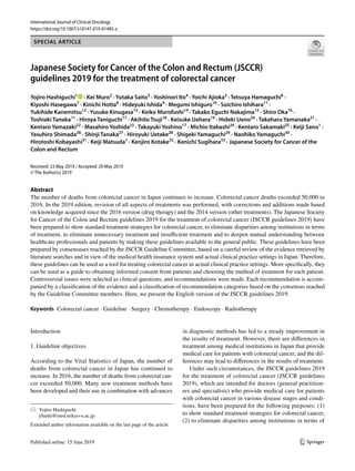
![International Journal of Clinical Oncology
1 3
treatment, (3) to eliminate unnecessary treatment and insuf‑
ficient treatment, and (4) to deepen mutual understanding
between healthcare professionals and patients by making
these guidelines available to the general public [1].
The following are expected to be achieved with these
guidelines: (1) improvement of the treatment of colorectal
cancer in Japan; (2) improvement of the results of treatment;
(3) reduction of the human and financial burden; and (4)
increased benefits for patients.
2. How to use these guidelines
These guidelines were prepared by consensuses reached by
the Guideline Committee of the Japanese Society for Can‑
cer of the Colon and Rectum, based on a careful review of
the evidence retrieved by the literature searches and in view
of the medical health insurance system and actual clinical
practice settings in Japan and, therefore, these guidelines
can be used as a tool for treating colorectal cancer in actual
clinical practice settings. More specifically, they can be used
as a guide to obtaining informed consent from patients and
choosing the method of treatment for each patient. How‑
ever, these guidelines provide only general recommendations
for choosing treatment strategies for colorectal cancer, and
they do not control or limit treatment strategies or treat‑
ment methods that are not described herein. They can also
be used as a document to explain the rationale for selecting
treatment strategies and treatment methods that differ from
those described therein.
The Japanese Society for Cancer of the Colon and Rec‑
tum (JSCCR) is responsible for the statements in these
guidelines. However, the personnel directly in charge of
treatment, not the JSCCR or the Guideline Committee, are
responsible for the outcome of treatment.
3. Users
The users of these guidelines are mainly clinical doctors
engaged in all aspects of the medical treatment of colorectal
cancer.
4. How to develop these guidelines
(1) Recording methods
We adopted the concept from the first edition, in which the
treatment policy algorithm was disclosed, a simple expla‑
nation thereof recorded, and added further comments with
regard to categories requiring additional explanation. Since
the 2009 edition, areas of debate have been raised as clini‑
cal questions (CQs) and included with recommendations
added. In the 2016 edition, systemic therapy was the only
treatment to be revised. In the 2019 edition, all aspects of
the treatments were revised, with corrections and additions
made to the CQs based on knowledge acquired since the
2016 version (systemic therapy) and the 2014 version (other
treatments).
Efforts were made to make the expression of the CQs
clear and unambiguous. When comparing multiple inter‑
ventions, we did not stick to ranking everything, and kept
the expression flexible to ensure that it is useful in clinical
practice. The clinicopathological terms conformed to those
described in the “Japanese Classification of Colorectal,
Appendiceal, and Anal Carcinoma, third English edition [2].
(2) Evidence level/strength of recommendations of CQs
The recommendations added to CQs included the evidence
level and strength of recommendations determined using the
following direction.
(2-1) Evidence level
Papers relating to the CQs were comprehensively collected,
and the evidence indicated by individual papers relating to
the critical outcomes included within the CQs was divided
into groups by study design [3]. The literature level and a
body of evidence (Table 1) were evaluated in reference to
the GRADE* System [4–26], before determining the final
CQ evidence level (Table 2).
*GRADE: The Grading of Recommendations Assess‑
ment, Development and Evaluation
(2-2) Strength of recommendations
Draft recommendation statements and the strength of the
recommendations were directed based on the outcomes and
the level of evidence obtained from the process described
above and were evaluated at a consensus meeting of the
Guideline Committee. In the CQ text, the recommendations
that were decided have been directly expressed, and ambigu‑
ous expressions were excluded.
The draft recommendations were evaluated from four cat‑
egories (① Quality of evidence, ② Patients’ views and prefer‑
ences, ③ Benefits and harms, and ④ Cost effectiveness). The
strength of recommendation (Table 3) was determined by
vote, based on the GRADE Grid method [11].
Method
1. We selected one of the following five options and voted.
① Strong “For” intervention
② A Weak “For” intervention
③ Weak “Against” intervention
④ Strong “Against” intervention
⑤ Not graded](data:image/gif;base64,R0lGODlhAQABAIAAAAAAAP///yH5BAEAAAAALAAAAAABAAEAAAIBRAA7)