
Recommended
More Related Content
What's hot
Similar to ppt xeropthalmia.pptx
Similar to ppt xeropthalmia.pptx (20)
Recently uploaded
Recently uploaded (20)
ppt xeropthalmia.pptx
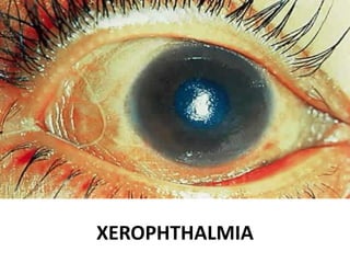
- 2. INTRODUCTION The term xerophthalmia ( xeros- dry, ophthalmia- eye) refers to the eye disease specifically caused by vitamin A deficiency. Xerophthalmia means dryness of the eye in which eye fails to produce tears. Xerophthalmia is most preventable cause of blindness in children at the age of 3 to 6 years.
- 3. The term xerophthalmia is reserved ( by a joint WHO AND USAID COMMITTEE, 1976) to cover all the ocular manifestation of vitamin A deficiency, including not only the structural changes affecting the conjunctiva cornea and occasionally retina, but also the biophysical disorder of retinal rods and cones functions.
- 4. CAUSES • The most common cause of xerophthalmia is vitamin A or lupus and rheumatoid arthritis. • Medication such as nasal decongestants, tranquilizers, antihistamines • Some chemical burn • Thickness and dryness caused due to trauma, local disease, diarrhea, and infection and from previous injury of cornea.
- 5. WHO classification • XN Night blindness • XIA conjunctival xerosis • XIB Bitot’s spot • X2 corneal xerosis
- 6. • X3A corneal ulceration/ keratomalacia affecting less than one- third corneal surface • X3B corneal ulceration/ keratomalacia affecting more than one-third corneal surface • XS corneal scar due to xerophthalmia
- 7. CLINICAL FEATURES Night blindness : inability to see in dim light due to impairment in dark adaptation. Conjunctival xerosis: First clinical sign Dryness or lack of luster Loss of ability to retain moisture no matter whether tears are present or absent loss of transparency, thickening
- 8. Clinical features contd…… Corneal xerosis The corneal surface has a rough, dull and dry fine pebbly appearance and lacks luster. Later, corneal ulceration may develop and healing corneal scar may affect vision Bitot’s spot • Triangular, pearly-white or yellowish, foamy spots on the bulbar conjunctiva • Are bilateral and on either side or cornea
- 9. Clinical features contd…… Keratomalacia • Consists of characteristic softening of the entire thickness of a part or, more often, the whole of the cornea • Leading to deformation or destruction of the eyeball • The process is a rapid one, the corneal structure melting into a cloudy gelatinous mass. • Vision is lost • Major cause of blindness
- 10. Clinical features contd…… Corneal scar • Healing of stromal defects results in corneal scars of different densities and sizes which may or may not cover the pupillary area. Xerophthalmic fundus • It is characterized by typical seed-like, raised, whitish lesions scattered uniformly over the part of the fundus at the level of optic disc.
- 12. Bitot’s spot
- 13. Keratomalacia
- 14. TREATMENT • Local ocular therapy : for conjunctival xerosis; artificial tears ( 0.7% hydroxypropyl methyl cellulose or 0.3% hypromellose) should be instilled every 3-4 hours. • Vitamin A therapy: treatment schedules apply to all stages of active xeropthalmia. • Treatment of underlying conditions: such as PEM and other nutritional disorder, diarrhea, dehydration and electrolyte imbalance, infection and parasitic condition should be considered simultaneously.
- 15. Prophylaxis WHO recommended schedule, which is universally recommended is as follows; • Infants 6-12 months old and older children weighing less than 8 kg – 100, 000 IU orally every 3-6 months. • Children over 1 year and under 6 years of age- 200,000 IU orally every 6 months. • Infants less than 6 months old, who are not being breastfed- 50,000 IU orally should be given before they attain every 6 months.
- 16. Precaution from xerophthalmia • The prevention of xerophthalmia intake of vitamin by fortification of food items such as in cooking oil, addition of vitamin A, skimmed milk etc. • Eat dark green leafy vegetables, pork, fish liver, beef, mango, papaya, eggs, butter, broccoli and apricots and drink lot of plenty water.
- 17. References • Sharma M, Paudel K, Gautam R.Comprehensive Text book of Medical Surgical Nursing.3rd ed. Ghattekulo: Samiksha publication; 2020. • Mandal GN. Textbook of medical surgical nursing(adult nursing).7th ed. kathmandu : Safal publication;2077. • Khurana A K. Comprehensive ophthalmology.7th edition. Jaypee Brothers medical publishers (p)Ltd; 2015