Cardiactumors 100910174533-phpapp02
•Download as PPT, PDF•
2 likes•262 views

Cardiactumors

Recommended
More Related Content
What's hot
What's hot (20)
Similar to Cardiactumors 100910174533-phpapp02
Similar to Cardiactumors 100910174533-phpapp02 (20)
More from Bhargav Kiran
More from Bhargav Kiran (20)
Recently uploaded
Recently uploaded (20)
Cardiactumors 100910174533-phpapp02
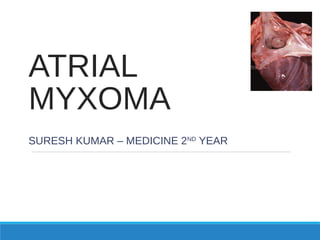
- 1. ATRIAL MYXOMA SURESH KUMAR – MEDICINE 2ND YEAR
- 2. HISTORY A series of six atrial tumors, with characteristics we now recognize as myxoma, was published in 1845 by King. The first echocardiographic diagnosis of an intracardiac tumor was made in 1959. An intracardiac myxoma was diagnosed by angiography in 1952 by Goldberg, but attempts at surgical removal were unsuccessful.
- 3. Treatment of cardiac tumors was profoundly influenced by two events: 1. The introduction of cardiopulmonary bypass in 1953 by John Gibbon, which allowed a safe and reproducible approach to the cardiac chambers 2. The introduction of cardiac echocardiography, which allowed safe and non invasive diagnosis of an intracardiac mass.
- 4. A large right atrial myxoma was removed by Bhanson in 1952 using caval inflow occlusion, but the patient died 24 days later Crafoord in Sweden first successfully removed a left atrial myxoma in 1954 using cardiopulmonary bypass, and Kay in Los Angeles first removed a left ventricular myxoma in 1959 By 1964, 60 atrial myxomas had been removed successfully, with improved results owing to the increasing safety of cardiopulmonary bypass and use of echocardiography for detection
- 5. INTRODUCTION Primary tumor of the heart Myxomas are one of the most benign neoplasm which comprise 50% out of which 15% myxomas are observed in children 5% of myxoma patients show a familial pattern of tumor development based on autosomal dominant inheritance. These patients and 20% of those with sporadic myxoma have an abnormal DNA genotype chromosomal pattern.
- 6. Familial patients are more likely to be younger, equally likely to be male or female, and more often (22%) have multicentric tumors originating from either the atrium or ventricle Approximately 20% of familial patients have associated conditions such as adrenocortical nodule hyperplasia, Sertoli cell tumors of the testes, pituitary tumors, multiple myxoid breast fibroadenomas, cutaneous myomas, and facial or labial pigmented spots. These conditions often are described as complex myxomas within the group of familial myxoma.
- 7. A familial syndrome with autosomal X-linked inheritance characterized by primary pigmented nodular adrenocortical disease with hypercortisolism, cutaneous pigmentous lentigines, and cardiac myxoma is referred to as Carney’s complex
- 8. Relative incidence of benign heart tumors
- 9. Relative incidence of primary malignant heart tumors
- 10. PATHOLOGY Myxomas are composed of stellate, frequently multinucleated myxoma cells, admixed with cells showing, endothelial, smooth muscle and or fibroblastic differentiation The cells are embedded in an abundant acid mucopolysacharide ground substance Hemmorhage, poorly organising thrombus & mononuclear inflammation also are usually present
- 12. Clinical Presentation Cardiac and non-cardiac manifestations Location and size of tumor are the major determinants of specific signs and symptoms Signs & symptoms similar to all form of heart disease: Chest pain Syncope Heart failure Murmurs Arrhythmias Conduction disturbance Pericardial effusion or tamponade
- 13. Myxoma Most common type of primary cardiac tumor (1/3 to ½ of all cases) Most commonly in 3rd – 6th decade; female > male Sporadic vs familial Majority sporadic; some are familial (autosomal dominant transmission) or part of a syndrome 1. Carney complex – spotty skin pigmentation, myxomas, endocrine overactivity, schwannomas 2. NAME syndrome – nevi, atrial myxoma, myxoid neurofibroma, ephelides 3. LAMB syndrome – lentigines, atrial myxoma, blue nevi
- 14. Myxoma Sporadic Familial or Syndrome Myxoma • Solitary • More common • Usually located in left atria • Arise from inter-atrial septum in vicinity of fossa ovalis • May also occur in the ventricles or multiple locations • Younger individual • Often multiple location • Less common (10%) • Autosomal dominant pattern of transmission • Associated with freckling, non- cardiac tumors, endocrine neoplasms • Recurrent after surgery
- 15. These benign masses are most often attached to the atrial wall, but can arise on a valve or in a ventricle They can produce a "ball valve" effect by intermittently occluding the atrioventricular valve orifice. Embolization of fragments of tumor may also occur Myxomas are easily diagnosed by echocardiography
- 16. Myxoma – Symptoms and Signs Symptoms Incidence (%) Dyspnea on exertion Paroxysmal dyspnea Fever Weight loss Severe dizziness/syncope Sudden death Hemoptysis >75 ~25 ~50 ~25 ~20 ~15 ~15
- 17. Myxoma – Symptoms and Signs Signs Incidence (%) Mitral diastolic murmur Mitral systolic murmur Pulmonary hypertension Right heart failure Pulmonary emboli Anemia Elevated ESR Third heart sound (tumor plop) Atrial fibrillation Elevated globulins Clubbing Raynaud’s phenomenon ~75 ~50 ~70 ~70 ~25 >33 >33 >33 ~15 ~10 ~5 <5
- 18. CLINICAL PRESENTATION Systemic or cardiovascular findings Cardiovascular findings: 1. Atrial resemble mitral valve disease most common clinical presentation Stenosis – tumor prolapse into the mitral orifice during diastole Regurgitation – injury to the valve by tumor-induced trauma 2. Ventricular – outflow obstruction syncope
- 19. DIAGNOSIS 1. Two-dimensional transthoracic or trans-esophageal echocardiography • Determine site of tumor attachment and tumor size • Screening of 1st degree relatives for familial or syndrome myxoma 2. CT scan and MRI • Tumor size, shape, composition, and surface characteristics 3. Cardiac catheterization • Risk of tumor emboli for suspected CAD
- 20. Myxoma TEE showing a large mass (M), in the left atrium with attachment to interatrial septum and prolapsing through the mitral valve into the left ventricular cavity in diastole. (M = myxoma).
- 23. SURGICAL MANAGEMENT Surgical resection is the only effective therapeutic option for patients with cardiac myxoma and should not be delayed because death from obstruction to flow within the heart or embolization may occur in as many as 8% of patients awaiting operation A median sternotomy approach with ascending aortic and bicaval cannulation usually is employed Manipulation of the heart before initiation of cardiopulmonary bypass is minimized in deference to the known friability and embolic tendency of myxomas CPB management
- 24. THANK YOU