SlideShare a Scribd company logo
1 of 12
Valvular Hear Disease
Valvular disease may resultin stenosis,insufficiency (regurgitation or incompetence), or both.
• Stenosis is the failureof a valveto open completely, obstructingforward flow.
• Insufficiency results from failureof a valve to close completely, thereby allowingregurgitation
(backflow) of blood.
Degenerative Valvedisease
Term used to describechanges that affect the integrity the valve ECM.
 Calcification- typically theaortic then the mitral valve.
 Alterations in ECM- increaseproteoglycan and diminished fibrillar collagen and elastin
(myxomatosis degeneration).
 Changes in the production of matrix metalloproteinaseor their inhibitors.
 Age
Calcific Aortic Stenosis
 Most common casuseof aortic stenosis
 Asympomatic
Morphology
 Hallmark is heaped-up calcified masses on te outflow sideof the cusps
 These protrude into the sinuses of valsalvaand mechanically impedevalve opening
 Commissural fusion notcommon but secodnary fibrosis and thinkeningo fthe cusp ma y occur
Myxomatous Mitral Valve
Morphology
Gross
 Myxomatous degeneration of the mitral valve
 one or both mitral valveleaflets arefloppy and prolapse
 Mitral valves balloon back into the left atriumduringsystole
 Women more than men
 Affected leaflets are enlarged,redundant, thick and rubbery
 The tendinous cords tend to be elongated thinned and occasionally rupture
Histology
 Thinningof the valvelayer know as tge fibrosa layer (structural integrity depends on that layer )
 Middlelayer the spongiosa is increased becauseof the deposition of myoxmatous material.
Rheumatic valve disease
The valvular disease principally takes the form of deforming fibrotic mitral stenosis; indeed
rheumatic heart disease is essentially the only cause of acquired mitral stenosis.
Morphology
Acute rheumatic fever
 characterized by discreteinflammatory foci within a variety of tissue(aschoff bodies)
 Aschoff bodies arepathognomonic for rheumatic fever
 Aschoff bodies composed of:
1. Collections of lymphocytes (primarily T cells),
2. Scattered plasma cells,
3. Plump activated macrophages called Anitschkow cells associated with zones of fibrinoid
necrosis.
 TheAnitschkow cells abundantcytoplasm,nuclei with chromatin that is centrally condensed into
a slender, wavy ribbon (so-called “caterpillar cells”).
 Duringacute rheumatic fever, Aschoff bodies can be found in any of the three layers of the
heart—pericardium,myocardium,or endocardium (includingvalves).
 Rheumatic fever is said to causepancarditis,with the followingsalientfeatures:
1. Pericardium may exhibita fibrinous exudate,which generally resolves without sequelae.
2. Myocardium-myocarditis—takes the form of scattered Aschoff bodies within the interstitial
connective tissue.
3. Valve -fibrinoid necrosisand fibrin deposition alongthelines of closure(Fig.11.19A) forming1-
to 2-mm vegetations—verrucae—that causelittledisturbancein cardiac function.
Chronic rheumatic
Gross
characterizedby
 Organization ofacute inflammation
 Subsequent scarring
 Aschoff bodies arereplaced by fibrous scar
 Valve cusps and leaflets becomes perminently thickened and retracted
 Clasically themitral valves exhitis:
1. Leaflet thickening
2. commissural fusion
3. shortening, and thickeningand fusion of the chordaetendineae (Fig. 11.19C to E).
 Fibrous bridging across thevalvular commissuresand calcification createfish mouth or
buttonhole stenoses
Histology
Neovascularization (visibility evidentin and diffuse fibrosisthatobliterates the normal leaflet
architecture
N. B:The most important functional consequenceof rheumatic heart diseaseis valvularstenosisand
regurgitation.
Infective Endocarditis
Infective endocarditis (IE) is a microbial infection of the heart valves or the mural endocardiumthat
leads to the formation of vegetations composed of thrombotic debris and organisms,often associated
with destruction of the underlyingcardiac tissues
Two forms.
• Acute endocarditis refers to tumultuous, destructiveinfections,frequently involvinga highly
virulentorganismattackinga previously normal valve.Itis associated with of substantial morbidity
and mortality, even with appropriateantibiotic therapy and/or surgery.
• Subacute endocarditis refers to infections by organisms of lowvirulenceaffectinga previously
abnormal heart, especially scarred or deformed valves.The diseasetypically appearsinsidiously and—
even if untreated— follows a protracted courseof weeks to months; most patients recover after
appropriateantibiotic therapy.
Morphology
In both forms:
 Friable
 bulky,
 and potentially destructive vegetations containingfibrin,inflammatory cells,and
microorganisms arepresenton the heart valves (Figs.11.20 and 11.21).
N.B:The aortic and mitral valves arethe most common sites of infection, although the tricuspid valve
is a frequent target in the setting of intravenous drugabuse.
N. B: Most consistent sign of infective endocardits is fever.
Non infective endocarditis
Non bacterial ThromboticEndocarditis (NBTE)
Nonbacterial thromboticendocarditis(NBTE) is characterized by thedepositionof sterilethrombi on
cardiac valves,typically inthosewith anunderlyinghypercoagulablestate.
The vegetations in NBTEaretypically small (1 to 5 mmin diameter) andvalvular damageisnota
prerequisite.Indeed,theconditionusually occurson previously normal valves.Rather,hypercoagulable
states aretheusual precursorto NBTE; such conditionsincludechronicdisseminated intravascular
coagulation.
Endocarditis in Systemic Lupus Erythematosus: Libman-Sacks Endocarditis
Libman-Sacks endocarditisis characterized by the presence of sterile vegetations on the valves of
patients with systemic lupus erythematosus.
Cardiomyopathies
Cardiac diseases dueto intrinsic myocardial dysfunction aretermed cardiomyopathies (literally,
“heart musclediseases”);these can be primary—that is,principally confined to the myocardium—or
secondary presenting as the cardiacmanifestation of a systemic disorder.
However, three timehonored clinical,functional,and pathologic patterns arerecognized (Fig. 11.23
and Table 11.5):
• Dilated cardiomyopathy (DCM) (including arrhythmogenic right ventricular cardiomyopathy)
• Hypertrophic cardiomyopathy (HCM)
• Restrictive cardiomyopathy
Of the three major patterns, DCM is most common (90% of cases),and restrictivecardiomyopathy is
the least frequent.
Dilated Cardiomyopathy
DCM characterized by:
 Progressivecardiac dilation
 Contractile(systolic) dysfunction
 Usually with concurrenthypertrophy
Morphology
Gross
 Enlarged (up to two to three times the normal weight )
 Flabby
 With dilation in all chambers
 Because of the wall thinning thataccompanies the dilation ,the ventricular thickness may be
less than,equal or greater than normal .
 Mural thrombi can also be present
Histology
 Most myoctes exhibithypertrophy with enlarged nuclei
 Myoctes may also beattenuated,stretched and irregular.
 Variable interstitial and endocardial fibrosis fibrosis with scattered areas of replacement fibrosis.
 In DCM caused by iron overload itis marked by accumulation of intramyocardial hemosiderin by
prussian bluestain.
Causes
Most cases of Dilated Cardiomyopathy are idiopathic.
Some common causes includes:
 Genetics
 Infections and other toxic exposure
 Alcohol
 Iron overload
 Peripartumcardiomyopathy
DCM(Arrhythmogen Right ventricular cardiopulmonary)
Is an autosomal dominantdisorder classically manifestwith rightsided heart failureand rhythm
disturbances which can causesudden cardiacdeath.
Morphology
 The right ventricular wall is severely thinned owing to myocytes
 Replacement by fatty infiltration and lesser amounts of fibrosis.
Hypertrophic cardiomyopathy
HCM characterized by:
 Myochardial hypertrophy
 Defective diastolicfilling
 In 1/3 of the cases ventricular outflowobstruction

Morphology
Gross
 Marked by massivemyocardial hypertrophy without ventricular dilation
 Asymmetric septal hypertrophy - Disproportionatethickeningof the ventricular septumrelate to
the left ventricle free wall
 In 10% of cases concentric hypertrophy is seen
 On longitudinal section the ventricular cavity loses itround - ovoid shapeand acquirea banana
likeconfiguration.
 Mitrialvalveis affected
Histology
 Marked by myocyte hypertrophy, haphazard myocyte(and myofiber ) dissarry
 Interstitial fibrosis
N.B: HCM is an important causeof sudden cardiac death.In almost1/3 of cases of of sudden cardiac
death in athletes younger than 35 years of age the underlyingcauseis HCM
RestrictiveCardiomyopathy
RC is characterized by :
Primary decreasein ventricular compliance
Resultingin impaired ventricular fillingduringdiastole(simply putthe wall becomes stiffer).
Morphology
Gross
 The ventricles arenormal approximately normal sizeor slightly enlarged
 The cavities arenot dilated
 The myocrium is firm
 Both atria aretypically dilated as a consequnce of restricted ventricular fillingand pressure
overloads
Histology
 Variabledegrees of interstitial fibrosis
N.B: Although the gross morphology is similarfor restrictivecardiomoptahy of disoarartecauses
endomyocardial biopsy often can reveal a specific etiology.
Types of restrictive cardiomyopathy
This form of cardiomyopathy may be idiopathicor may be associated with systemic diseases that
affect the myocardium, for example, radiation fibrosis,amyloidosis,sarcoidosis,or products of inborn
errors of metabolism.
Three forms of restrictivecardiomyopathy merit brief mention.
Amyloidosis is caused by the deposition of extracellular proteins with te dredilection from forming
insoluableBpleted sheets.
Endomyocardial fibrosis-is principally a diseaseof children and young adults in Africa and other
tropical areas.Itis characterized by dense diffusefibrosis of the ventricular endocardiumand
subendocardium,often involvingthe tricuspid and mitral valves.
Loeffler endomyocarditis also exhibits endocardial fibrosis,typically associated with ormation of large
mural thrombi. It has no geographic or population predilec tion.It is characterized by peripheral
hypereosinophilia and eosinophilictissueinfiltrates;releaseof eosinophil granulecontents,
especially major basic protein,prob ably engenders endocardial and myocardial necrosis, followed by
scarring,layeringof the endocardium by thrombus, and finally thrombus organization.
Cardiovascular system path docx

More Related Content

Similar to Cardiovascular system path docx

Thrombosis and Embolism
Thrombosis and EmbolismThrombosis and Embolism
Thrombosis and EmbolismHijab Siddiqi
 
RHEUMATIC FEVER & RHEUMATIC HEART DISEASE
RHEUMATIC FEVER & RHEUMATIC HEART DISEASERHEUMATIC FEVER & RHEUMATIC HEART DISEASE
RHEUMATIC FEVER & RHEUMATIC HEART DISEASESAMOEINESH
 
Pericardial dis.&cardiactumors 5
Pericardial dis.&cardiactumors 5Pericardial dis.&cardiactumors 5
Pericardial dis.&cardiactumors 5Forensic Pathology
 
Cardiomyopathies
CardiomyopathiesCardiomyopathies
CardiomyopathiesMilDoc
 
Cardiovascular system pathology
Cardiovascular system pathologyCardiovascular system pathology
Cardiovascular system pathologyRemix education
 
Aneurysms arterial swelling abnormality.pptx
Aneurysms arterial swelling abnormality.pptxAneurysms arterial swelling abnormality.pptx
Aneurysms arterial swelling abnormality.pptxAkilanN5
 
4. CARDIOMYOPATHIES.pptx
4. CARDIOMYOPATHIES.pptx4. CARDIOMYOPATHIES.pptx
4. CARDIOMYOPATHIES.pptxmariaidrees3
 
ARTERIOSCLEROSIS.pptx
ARTERIOSCLEROSIS.pptxARTERIOSCLEROSIS.pptx
ARTERIOSCLEROSIS.pptxSAMOEINESH
 
Myocardial infarction
Myocardial infarctionMyocardial infarction
Myocardial infarctionutsav parmar
 
Embolism - Causes, Symptoms, Treatment, Diagnosis
Embolism - Causes, Symptoms, Treatment, DiagnosisEmbolism - Causes, Symptoms, Treatment, Diagnosis
Embolism - Causes, Symptoms, Treatment, Diagnosissainavlefusion
 
Pathology of hemodynamic disorders part 2 nov 2017 Dr. Sufia Husain
Pathology of hemodynamic disorders part 2 nov 2017 Dr. Sufia HusainPathology of hemodynamic disorders part 2 nov 2017 Dr. Sufia Husain
Pathology of hemodynamic disorders part 2 nov 2017 Dr. Sufia HusainSufia Husain
 
416456384-6-Thrombosis-ppt.ppt
416456384-6-Thrombosis-ppt.ppt416456384-6-Thrombosis-ppt.ppt
416456384-6-Thrombosis-ppt.pptAxmedAbdiHasen
 
Ischemic Heart Disease.pptx
Ischemic Heart Disease.pptxIschemic Heart Disease.pptx
Ischemic Heart Disease.pptxAmalRashid10
 
Cerebrovascular diseases.pptx
Cerebrovascular diseases.pptxCerebrovascular diseases.pptx
Cerebrovascular diseases.pptxtesa10
 
3. ARRHYTHMIAS.pptx
3. ARRHYTHMIAS.pptx3. ARRHYTHMIAS.pptx
3. ARRHYTHMIAS.pptxmariaidrees3
 

Similar to Cardiovascular system path docx (20)

Thrombosis and Embolism
Thrombosis and EmbolismThrombosis and Embolism
Thrombosis and Embolism
 
Cardiovascular System
Cardiovascular SystemCardiovascular System
Cardiovascular System
 
RHEUMATIC FEVER & RHEUMATIC HEART DISEASE
RHEUMATIC FEVER & RHEUMATIC HEART DISEASERHEUMATIC FEVER & RHEUMATIC HEART DISEASE
RHEUMATIC FEVER & RHEUMATIC HEART DISEASE
 
Pericardial dis.&cardiactumors 5
Pericardial dis.&cardiactumors 5Pericardial dis.&cardiactumors 5
Pericardial dis.&cardiactumors 5
 
Cardiomyopathies
CardiomyopathiesCardiomyopathies
Cardiomyopathies
 
Cardiovascular system pathology
Cardiovascular system pathologyCardiovascular system pathology
Cardiovascular system pathology
 
Aneurysms arterial swelling abnormality.pptx
Aneurysms arterial swelling abnormality.pptxAneurysms arterial swelling abnormality.pptx
Aneurysms arterial swelling abnormality.pptx
 
IHD - TA L2.pptx
IHD - TA L2.pptxIHD - TA L2.pptx
IHD - TA L2.pptx
 
4. CARDIOMYOPATHIES.pptx
4. CARDIOMYOPATHIES.pptx4. CARDIOMYOPATHIES.pptx
4. CARDIOMYOPATHIES.pptx
 
ARTERIOSCLEROSIS.pptx
ARTERIOSCLEROSIS.pptxARTERIOSCLEROSIS.pptx
ARTERIOSCLEROSIS.pptx
 
Myocardial infarction
Myocardial infarctionMyocardial infarction
Myocardial infarction
 
Embolism - Causes, Symptoms, Treatment, Diagnosis
Embolism - Causes, Symptoms, Treatment, DiagnosisEmbolism - Causes, Symptoms, Treatment, Diagnosis
Embolism - Causes, Symptoms, Treatment, Diagnosis
 
Pathology of hemodynamic disorders part 2 nov 2017 Dr. Sufia Husain
Pathology of hemodynamic disorders part 2 nov 2017 Dr. Sufia HusainPathology of hemodynamic disorders part 2 nov 2017 Dr. Sufia Husain
Pathology of hemodynamic disorders part 2 nov 2017 Dr. Sufia Husain
 
416456384-6-Thrombosis-ppt.ppt
416456384-6-Thrombosis-ppt.ppt416456384-6-Thrombosis-ppt.ppt
416456384-6-Thrombosis-ppt.ppt
 
Ischemic Heart Disease.pptx
Ischemic Heart Disease.pptxIschemic Heart Disease.pptx
Ischemic Heart Disease.pptx
 
ATHEROSCLEROSIS & IHD.ppt
ATHEROSCLEROSIS & IHD.pptATHEROSCLEROSIS & IHD.ppt
ATHEROSCLEROSIS & IHD.ppt
 
Cerebrovascular diseases.pptx
Cerebrovascular diseases.pptxCerebrovascular diseases.pptx
Cerebrovascular diseases.pptx
 
Ischemic heart diseases 2
Ischemic heart diseases 2Ischemic heart diseases 2
Ischemic heart diseases 2
 
3. ARRHYTHMIAS.pptx
3. ARRHYTHMIAS.pptx3. ARRHYTHMIAS.pptx
3. ARRHYTHMIAS.pptx
 
Hemodynamic Disorders
Hemodynamic DisordersHemodynamic Disorders
Hemodynamic Disorders
 

Recently uploaded

Heredity: Inheritance and Variation of Traits
Heredity: Inheritance and Variation of TraitsHeredity: Inheritance and Variation of Traits
Heredity: Inheritance and Variation of TraitsCharlene Llagas
 
Grafana in space: Monitoring Japan's SLIM moon lander in real time
Grafana in space: Monitoring Japan's SLIM moon lander  in real timeGrafana in space: Monitoring Japan's SLIM moon lander  in real time
Grafana in space: Monitoring Japan's SLIM moon lander in real timeSatoshi NAKAHIRA
 
‏‏VIRUS - 123455555555555555555555555555555555555555
‏‏VIRUS -  123455555555555555555555555555555555555555‏‏VIRUS -  123455555555555555555555555555555555555555
‏‏VIRUS - 123455555555555555555555555555555555555555kikilily0909
 
Cytokinin, mechanism and its application.pptx
Cytokinin, mechanism and its application.pptxCytokinin, mechanism and its application.pptx
Cytokinin, mechanism and its application.pptxVarshiniMK
 
RESPIRATORY ADAPTATIONS TO HYPOXIA IN HUMNAS.pptx
RESPIRATORY ADAPTATIONS TO HYPOXIA IN HUMNAS.pptxRESPIRATORY ADAPTATIONS TO HYPOXIA IN HUMNAS.pptx
RESPIRATORY ADAPTATIONS TO HYPOXIA IN HUMNAS.pptxFarihaAbdulRasheed
 
TOPIC 8 Temperature and Heat.pdf physics
TOPIC 8 Temperature and Heat.pdf physicsTOPIC 8 Temperature and Heat.pdf physics
TOPIC 8 Temperature and Heat.pdf physicsssuserddc89b
 
Behavioral Disorder: Schizophrenia & it's Case Study.pdf
Behavioral Disorder: Schizophrenia & it's Case Study.pdfBehavioral Disorder: Schizophrenia & it's Case Study.pdf
Behavioral Disorder: Schizophrenia & it's Case Study.pdfSELF-EXPLANATORY
 
Dashanga agada a formulation of Agada tantra dealt in 3 Rd year bams agada tanta
Dashanga agada a formulation of Agada tantra dealt in 3 Rd year bams agada tantaDashanga agada a formulation of Agada tantra dealt in 3 Rd year bams agada tanta
Dashanga agada a formulation of Agada tantra dealt in 3 Rd year bams agada tantaPraksha3
 
Transposable elements in prokaryotes.ppt
Transposable elements in prokaryotes.pptTransposable elements in prokaryotes.ppt
Transposable elements in prokaryotes.pptArshadWarsi13
 
Best Call Girls In Sector 29 Gurgaon❤️8860477959 EscorTs Service In 24/7 Delh...
Best Call Girls In Sector 29 Gurgaon❤️8860477959 EscorTs Service In 24/7 Delh...Best Call Girls In Sector 29 Gurgaon❤️8860477959 EscorTs Service In 24/7 Delh...
Best Call Girls In Sector 29 Gurgaon❤️8860477959 EscorTs Service In 24/7 Delh...lizamodels9
 
Call Girls in Aiims Metro Delhi 💯Call Us 🔝9953322196🔝 💯Escort.
Call Girls in Aiims Metro Delhi 💯Call Us 🔝9953322196🔝 💯Escort.Call Girls in Aiims Metro Delhi 💯Call Us 🔝9953322196🔝 💯Escort.
Call Girls in Aiims Metro Delhi 💯Call Us 🔝9953322196🔝 💯Escort.aasikanpl
 
Twin's paradox experiment is a meassurement of the extra dimensions.pptx
Twin's paradox experiment is a meassurement of the extra dimensions.pptxTwin's paradox experiment is a meassurement of the extra dimensions.pptx
Twin's paradox experiment is a meassurement of the extra dimensions.pptxEran Akiva Sinbar
 
zoogeography of pakistan.pptx fauna of Pakistan
zoogeography of pakistan.pptx fauna of Pakistanzoogeography of pakistan.pptx fauna of Pakistan
zoogeography of pakistan.pptx fauna of Pakistanzohaibmir069
 
Speech, hearing, noise, intelligibility.pptx
Speech, hearing, noise, intelligibility.pptxSpeech, hearing, noise, intelligibility.pptx
Speech, hearing, noise, intelligibility.pptxpriyankatabhane
 
Gas_Laws_powerpoint_notes.ppt for grade 10
Gas_Laws_powerpoint_notes.ppt for grade 10Gas_Laws_powerpoint_notes.ppt for grade 10
Gas_Laws_powerpoint_notes.ppt for grade 10ROLANARIBATO3
 
Bentham & Hooker's Classification. along with the merits and demerits of the ...
Bentham & Hooker's Classification. along with the merits and demerits of the ...Bentham & Hooker's Classification. along with the merits and demerits of the ...
Bentham & Hooker's Classification. along with the merits and demerits of the ...Nistarini College, Purulia (W.B) India
 
Is RISC-V ready for HPC workload? Maybe?
Is RISC-V ready for HPC workload? Maybe?Is RISC-V ready for HPC workload? Maybe?
Is RISC-V ready for HPC workload? Maybe?Patrick Diehl
 
Vision and reflection on Mining Software Repositories research in 2024
Vision and reflection on Mining Software Repositories research in 2024Vision and reflection on Mining Software Repositories research in 2024
Vision and reflection on Mining Software Repositories research in 2024AyushiRastogi48
 

Recently uploaded (20)

Heredity: Inheritance and Variation of Traits
Heredity: Inheritance and Variation of TraitsHeredity: Inheritance and Variation of Traits
Heredity: Inheritance and Variation of Traits
 
Grafana in space: Monitoring Japan's SLIM moon lander in real time
Grafana in space: Monitoring Japan's SLIM moon lander  in real timeGrafana in space: Monitoring Japan's SLIM moon lander  in real time
Grafana in space: Monitoring Japan's SLIM moon lander in real time
 
‏‏VIRUS - 123455555555555555555555555555555555555555
‏‏VIRUS -  123455555555555555555555555555555555555555‏‏VIRUS -  123455555555555555555555555555555555555555
‏‏VIRUS - 123455555555555555555555555555555555555555
 
Cytokinin, mechanism and its application.pptx
Cytokinin, mechanism and its application.pptxCytokinin, mechanism and its application.pptx
Cytokinin, mechanism and its application.pptx
 
RESPIRATORY ADAPTATIONS TO HYPOXIA IN HUMNAS.pptx
RESPIRATORY ADAPTATIONS TO HYPOXIA IN HUMNAS.pptxRESPIRATORY ADAPTATIONS TO HYPOXIA IN HUMNAS.pptx
RESPIRATORY ADAPTATIONS TO HYPOXIA IN HUMNAS.pptx
 
TOPIC 8 Temperature and Heat.pdf physics
TOPIC 8 Temperature and Heat.pdf physicsTOPIC 8 Temperature and Heat.pdf physics
TOPIC 8 Temperature and Heat.pdf physics
 
Behavioral Disorder: Schizophrenia & it's Case Study.pdf
Behavioral Disorder: Schizophrenia & it's Case Study.pdfBehavioral Disorder: Schizophrenia & it's Case Study.pdf
Behavioral Disorder: Schizophrenia & it's Case Study.pdf
 
Dashanga agada a formulation of Agada tantra dealt in 3 Rd year bams agada tanta
Dashanga agada a formulation of Agada tantra dealt in 3 Rd year bams agada tantaDashanga agada a formulation of Agada tantra dealt in 3 Rd year bams agada tanta
Dashanga agada a formulation of Agada tantra dealt in 3 Rd year bams agada tanta
 
Transposable elements in prokaryotes.ppt
Transposable elements in prokaryotes.pptTransposable elements in prokaryotes.ppt
Transposable elements in prokaryotes.ppt
 
Hot Sexy call girls in Moti Nagar,🔝 9953056974 🔝 escort Service
Hot Sexy call girls in  Moti Nagar,🔝 9953056974 🔝 escort ServiceHot Sexy call girls in  Moti Nagar,🔝 9953056974 🔝 escort Service
Hot Sexy call girls in Moti Nagar,🔝 9953056974 🔝 escort Service
 
Best Call Girls In Sector 29 Gurgaon❤️8860477959 EscorTs Service In 24/7 Delh...
Best Call Girls In Sector 29 Gurgaon❤️8860477959 EscorTs Service In 24/7 Delh...Best Call Girls In Sector 29 Gurgaon❤️8860477959 EscorTs Service In 24/7 Delh...
Best Call Girls In Sector 29 Gurgaon❤️8860477959 EscorTs Service In 24/7 Delh...
 
Call Girls in Aiims Metro Delhi 💯Call Us 🔝9953322196🔝 💯Escort.
Call Girls in Aiims Metro Delhi 💯Call Us 🔝9953322196🔝 💯Escort.Call Girls in Aiims Metro Delhi 💯Call Us 🔝9953322196🔝 💯Escort.
Call Girls in Aiims Metro Delhi 💯Call Us 🔝9953322196🔝 💯Escort.
 
Twin's paradox experiment is a meassurement of the extra dimensions.pptx
Twin's paradox experiment is a meassurement of the extra dimensions.pptxTwin's paradox experiment is a meassurement of the extra dimensions.pptx
Twin's paradox experiment is a meassurement of the extra dimensions.pptx
 
zoogeography of pakistan.pptx fauna of Pakistan
zoogeography of pakistan.pptx fauna of Pakistanzoogeography of pakistan.pptx fauna of Pakistan
zoogeography of pakistan.pptx fauna of Pakistan
 
Speech, hearing, noise, intelligibility.pptx
Speech, hearing, noise, intelligibility.pptxSpeech, hearing, noise, intelligibility.pptx
Speech, hearing, noise, intelligibility.pptx
 
Gas_Laws_powerpoint_notes.ppt for grade 10
Gas_Laws_powerpoint_notes.ppt for grade 10Gas_Laws_powerpoint_notes.ppt for grade 10
Gas_Laws_powerpoint_notes.ppt for grade 10
 
Bentham & Hooker's Classification. along with the merits and demerits of the ...
Bentham & Hooker's Classification. along with the merits and demerits of the ...Bentham & Hooker's Classification. along with the merits and demerits of the ...
Bentham & Hooker's Classification. along with the merits and demerits of the ...
 
Is RISC-V ready for HPC workload? Maybe?
Is RISC-V ready for HPC workload? Maybe?Is RISC-V ready for HPC workload? Maybe?
Is RISC-V ready for HPC workload? Maybe?
 
Volatile Oils Pharmacognosy And Phytochemistry -I
Volatile Oils Pharmacognosy And Phytochemistry -IVolatile Oils Pharmacognosy And Phytochemistry -I
Volatile Oils Pharmacognosy And Phytochemistry -I
 
Vision and reflection on Mining Software Repositories research in 2024
Vision and reflection on Mining Software Repositories research in 2024Vision and reflection on Mining Software Repositories research in 2024
Vision and reflection on Mining Software Repositories research in 2024
 

Cardiovascular system path docx

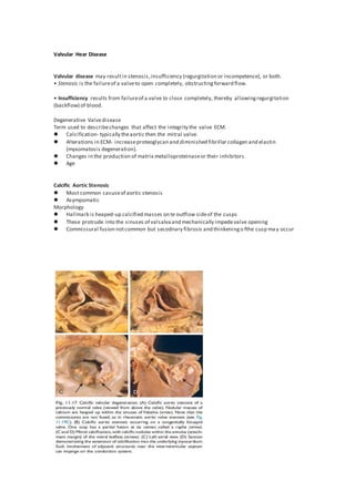
  • 1. Valvular Hear Disease Valvular disease may resultin stenosis,insufficiency (regurgitation or incompetence), or both. • Stenosis is the failureof a valveto open completely, obstructingforward flow. • Insufficiency results from failureof a valve to close completely, thereby allowingregurgitation (backflow) of blood. Degenerative Valvedisease Term used to describechanges that affect the integrity the valve ECM.  Calcification- typically theaortic then the mitral valve.  Alterations in ECM- increaseproteoglycan and diminished fibrillar collagen and elastin (myxomatosis degeneration).  Changes in the production of matrix metalloproteinaseor their inhibitors.  Age Calcific Aortic Stenosis  Most common casuseof aortic stenosis  Asympomatic Morphology  Hallmark is heaped-up calcified masses on te outflow sideof the cusps  These protrude into the sinuses of valsalvaand mechanically impedevalve opening  Commissural fusion notcommon but secodnary fibrosis and thinkeningo fthe cusp ma y occur
  • 2. Myxomatous Mitral Valve Morphology Gross  Myxomatous degeneration of the mitral valve  one or both mitral valveleaflets arefloppy and prolapse  Mitral valves balloon back into the left atriumduringsystole  Women more than men  Affected leaflets are enlarged,redundant, thick and rubbery  The tendinous cords tend to be elongated thinned and occasionally rupture Histology  Thinningof the valvelayer know as tge fibrosa layer (structural integrity depends on that layer )  Middlelayer the spongiosa is increased becauseof the deposition of myoxmatous material. Rheumatic valve disease The valvular disease principally takes the form of deforming fibrotic mitral stenosis; indeed rheumatic heart disease is essentially the only cause of acquired mitral stenosis. Morphology Acute rheumatic fever  characterized by discreteinflammatory foci within a variety of tissue(aschoff bodies)  Aschoff bodies arepathognomonic for rheumatic fever  Aschoff bodies composed of: 1. Collections of lymphocytes (primarily T cells), 2. Scattered plasma cells, 3. Plump activated macrophages called Anitschkow cells associated with zones of fibrinoid necrosis.  TheAnitschkow cells abundantcytoplasm,nuclei with chromatin that is centrally condensed into a slender, wavy ribbon (so-called “caterpillar cells”).  Duringacute rheumatic fever, Aschoff bodies can be found in any of the three layers of the heart—pericardium,myocardium,or endocardium (includingvalves).  Rheumatic fever is said to causepancarditis,with the followingsalientfeatures: 1. Pericardium may exhibita fibrinous exudate,which generally resolves without sequelae. 2. Myocardium-myocarditis—takes the form of scattered Aschoff bodies within the interstitial connective tissue. 3. Valve -fibrinoid necrosisand fibrin deposition alongthelines of closure(Fig.11.19A) forming1- to 2-mm vegetations—verrucae—that causelittledisturbancein cardiac function. Chronic rheumatic Gross characterizedby  Organization ofacute inflammation  Subsequent scarring  Aschoff bodies arereplaced by fibrous scar  Valve cusps and leaflets becomes perminently thickened and retracted  Clasically themitral valves exhitis:
  • 3. 1. Leaflet thickening 2. commissural fusion 3. shortening, and thickeningand fusion of the chordaetendineae (Fig. 11.19C to E).  Fibrous bridging across thevalvular commissuresand calcification createfish mouth or buttonhole stenoses Histology Neovascularization (visibility evidentin and diffuse fibrosisthatobliterates the normal leaflet architecture N. B:The most important functional consequenceof rheumatic heart diseaseis valvularstenosisand regurgitation.
  • 4. Infective Endocarditis Infective endocarditis (IE) is a microbial infection of the heart valves or the mural endocardiumthat leads to the formation of vegetations composed of thrombotic debris and organisms,often associated with destruction of the underlyingcardiac tissues Two forms. • Acute endocarditis refers to tumultuous, destructiveinfections,frequently involvinga highly virulentorganismattackinga previously normal valve.Itis associated with of substantial morbidity and mortality, even with appropriateantibiotic therapy and/or surgery. • Subacute endocarditis refers to infections by organisms of lowvirulenceaffectinga previously abnormal heart, especially scarred or deformed valves.The diseasetypically appearsinsidiously and— even if untreated— follows a protracted courseof weeks to months; most patients recover after appropriateantibiotic therapy. Morphology In both forms:  Friable  bulky,  and potentially destructive vegetations containingfibrin,inflammatory cells,and microorganisms arepresenton the heart valves (Figs.11.20 and 11.21). N.B:The aortic and mitral valves arethe most common sites of infection, although the tricuspid valve is a frequent target in the setting of intravenous drugabuse. N. B: Most consistent sign of infective endocardits is fever.
  • 5. Non infective endocarditis Non bacterial ThromboticEndocarditis (NBTE) Nonbacterial thromboticendocarditis(NBTE) is characterized by thedepositionof sterilethrombi on cardiac valves,typically inthosewith anunderlyinghypercoagulablestate. The vegetations in NBTEaretypically small (1 to 5 mmin diameter) andvalvular damageisnota prerequisite.Indeed,theconditionusually occurson previously normal valves.Rather,hypercoagulable states aretheusual precursorto NBTE; such conditionsincludechronicdisseminated intravascular coagulation.
  • 6. Endocarditis in Systemic Lupus Erythematosus: Libman-Sacks Endocarditis Libman-Sacks endocarditisis characterized by the presence of sterile vegetations on the valves of patients with systemic lupus erythematosus. Cardiomyopathies Cardiac diseases dueto intrinsic myocardial dysfunction aretermed cardiomyopathies (literally, “heart musclediseases”);these can be primary—that is,principally confined to the myocardium—or secondary presenting as the cardiacmanifestation of a systemic disorder.
  • 7. However, three timehonored clinical,functional,and pathologic patterns arerecognized (Fig. 11.23 and Table 11.5): • Dilated cardiomyopathy (DCM) (including arrhythmogenic right ventricular cardiomyopathy) • Hypertrophic cardiomyopathy (HCM) • Restrictive cardiomyopathy Of the three major patterns, DCM is most common (90% of cases),and restrictivecardiomyopathy is the least frequent. Dilated Cardiomyopathy DCM characterized by:  Progressivecardiac dilation  Contractile(systolic) dysfunction  Usually with concurrenthypertrophy Morphology Gross  Enlarged (up to two to three times the normal weight )  Flabby
  • 8.  With dilation in all chambers  Because of the wall thinning thataccompanies the dilation ,the ventricular thickness may be less than,equal or greater than normal .  Mural thrombi can also be present Histology  Most myoctes exhibithypertrophy with enlarged nuclei  Myoctes may also beattenuated,stretched and irregular.  Variable interstitial and endocardial fibrosis fibrosis with scattered areas of replacement fibrosis.  In DCM caused by iron overload itis marked by accumulation of intramyocardial hemosiderin by prussian bluestain. Causes Most cases of Dilated Cardiomyopathy are idiopathic. Some common causes includes:  Genetics  Infections and other toxic exposure  Alcohol  Iron overload  Peripartumcardiomyopathy DCM(Arrhythmogen Right ventricular cardiopulmonary) Is an autosomal dominantdisorder classically manifestwith rightsided heart failureand rhythm disturbances which can causesudden cardiacdeath. Morphology  The right ventricular wall is severely thinned owing to myocytes  Replacement by fatty infiltration and lesser amounts of fibrosis.
  • 9. Hypertrophic cardiomyopathy HCM characterized by:  Myochardial hypertrophy  Defective diastolicfilling  In 1/3 of the cases ventricular outflowobstruction  Morphology Gross  Marked by massivemyocardial hypertrophy without ventricular dilation  Asymmetric septal hypertrophy - Disproportionatethickeningof the ventricular septumrelate to the left ventricle free wall  In 10% of cases concentric hypertrophy is seen  On longitudinal section the ventricular cavity loses itround - ovoid shapeand acquirea banana likeconfiguration.  Mitrialvalveis affected Histology  Marked by myocyte hypertrophy, haphazard myocyte(and myofiber ) dissarry  Interstitial fibrosis N.B: HCM is an important causeof sudden cardiac death.In almost1/3 of cases of of sudden cardiac death in athletes younger than 35 years of age the underlyingcauseis HCM
  • 10. RestrictiveCardiomyopathy RC is characterized by : Primary decreasein ventricular compliance Resultingin impaired ventricular fillingduringdiastole(simply putthe wall becomes stiffer). Morphology Gross  The ventricles arenormal approximately normal sizeor slightly enlarged  The cavities arenot dilated  The myocrium is firm  Both atria aretypically dilated as a consequnce of restricted ventricular fillingand pressure overloads Histology  Variabledegrees of interstitial fibrosis N.B: Although the gross morphology is similarfor restrictivecardiomoptahy of disoarartecauses endomyocardial biopsy often can reveal a specific etiology. Types of restrictive cardiomyopathy This form of cardiomyopathy may be idiopathicor may be associated with systemic diseases that affect the myocardium, for example, radiation fibrosis,amyloidosis,sarcoidosis,or products of inborn errors of metabolism. Three forms of restrictivecardiomyopathy merit brief mention.
  • 11. Amyloidosis is caused by the deposition of extracellular proteins with te dredilection from forming insoluableBpleted sheets. Endomyocardial fibrosis-is principally a diseaseof children and young adults in Africa and other tropical areas.Itis characterized by dense diffusefibrosis of the ventricular endocardiumand subendocardium,often involvingthe tricuspid and mitral valves. Loeffler endomyocarditis also exhibits endocardial fibrosis,typically associated with ormation of large mural thrombi. It has no geographic or population predilec tion.It is characterized by peripheral hypereosinophilia and eosinophilictissueinfiltrates;releaseof eosinophil granulecontents, especially major basic protein,prob ably engenders endocardial and myocardial necrosis, followed by scarring,layeringof the endocardium by thrombus, and finally thrombus organization.