Spinal Cord Anatomy and Tracts
•Download as PPTX, PDF•
1 like•222 views

The document summarizes the anatomy and organization of the spinal cord. It describes that the spinal cord lies within the vertebral canal and extends from the foramen magnum to the lumbar vertebrae. It is covered by meninges and contains gray matter with nuclei and laminae containing different types of neurons, and white matter divided into anterior, lateral, and posterior columns. The document also outlines the ascending and descending tracts within the spinal cord that transmit sensory and motor signals to and from the brain.

Recommended
More Related Content
What's hot
What's hot (20)
Similar to Spinal Cord Anatomy and Tracts
Similar to Spinal Cord Anatomy and Tracts (20)
More from Ayub Abdi
More from Ayub Abdi (20)
Recently uploaded
Recently uploaded (20)
Spinal Cord Anatomy and Tracts
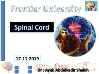
- 1. 17-11-2019
- 2. • Spinal cord lies loosely in the vertebral canal. • It extends from foramen magnum to first lumbar vertebra below. • Covered by meninges. • Cylindrical in shape. • Length of the spinal cord is about 45 cm in males and about 43 cm in females. • Cervical and lumbar enlargements. • Conus medullaris – filum terminale
- 5. • anterior (ventral) root and a posterior (dorsal) root. • Neural substance of spinal cord is divided into inner gray matter and outer white matter.
- 8. GRAY MATTER OF SPINAL CORD: • Neurons in Gray Matter of Spinal Cord: 1. Golgi type I neurons (Anterior horn). 2. Golgi type II neurons (posterior horn). Organization of Neurons in Gray Matter: 1. Nuclei or columns (Clusters of neurons). 2. Laminae or layers (neurons of different size and shape).
- 9. A) Nuclei in Posterior Gray Horn: 1. Marginal nucleus. 2. Substantia gelatinosa of Rolando. 3. Chief sensory nucleus or nucleus proprius. 4. Dorsal nucleus of Clarke.
- 10. B) Nuclei in Lateral Gray Horn: 1. Intermediolateral nucleus (T1 – L2). C) Nuclei in Anterior Gray Horn: 1. Alpha motor neurons (extrafusal fibers). 2. Gamma motor neurons (intrafusal fibers). 3. Renshaw cells (synaptic inhibition at the spinal cord).
- 11. A) Laminae in Posterior Gray Horn (sensory functions): 1. Marginal nucleus : Lamina I 2. Substantial gelatinosa : Laminae II and III of Rolando 3. Chief sensory nucleus : Laminae III, IV and V 4. Dorsal nucleus of Clarke : Lamina VI B) Lamina in Lateral Gray Horn: 1. Intermediolateral nucleus : Lamina VII
- 12. C) Laminae in Anterior Gray Horn (Motor function): 1. Motor internuncial neurons which are also called interneurons, : Lamina VIII 2. Motor neurons : Lamina IX D) Lamina Around Central Canal (Neuroglia): 1. lamina X.
- 13. WHITE MATTER OF SPINAL CORD: I. Anterior or Ventral White Column (anterior or ventral funiculus). II. Lateral White Column (lateral funiculus). III. Posterior or Dorsal White Column (posterior or dorsal funiculus).
- 14. TRACTS IN SPINAL CORD
- 15. • Tracts of the spinal cord are Groups of nerve fibers passing through spinal cord. • The spinal tracts are divided into two main groups. They are: 1. Short tracts (Fibers of the short tracts connect different parts of spinal cord itself). a) Association or intrinsic tracts – same segment. b) Commissural tracts – opposite halves. 2. Long tracts (connect the spinal cord with other parts of central nervous system). c) Ascending tracts - which carry sensory impulses from the spinal cord to brain. d) Descending tracts - which carry motor impulses from brain to the spinal cord.
- 16. ASCENDING TRACTS OF SPINAL CORD: • Ascending tracts of spinal cord carry the impulses of various sensations to the brain. • Pathway for each sensation is formed by two or three groups of neurons, which are: 1. First order neurons – spinal cord. 2. Second order neurons – subcortical area. 3. Third order neurons – cerebral cortex.
- 19. 1. ANTERIOR SPINOTHALAMIC TRACT: • Second order neurons. • Crude touch sensation. • Crossed fibers. 2. LATERAL SPINOTHALAMIC TRACT: • Second order neurons. • Pain and temperature. • Crossed fibers
- 20. 3. VENTRAL SPINOCEREBELLAR TRACT: • Second order neurons. • Subconscious kinesthetic sensation. • Crossed and uncrossed fibers. 4. DORSAL SPINOCEREBELLAR TRACT: • Second order neurons. • Subconscious kinesthetic sensation. • uncrossed.
- 21. 5. SPINOTECTAL TRACT: • second order neurons. • spinovisual reflex. • crossed fibers. 6. FASCICULUS DORSOLATERALIS: • first order neurons. • pain and thermal sensations. • uncrossed fibers.
- 22. 8. SPINOOLIVARY TRACT: • Second order neurons. • ascending reticular activating system and are concerned with consciousness and awareness. • crossed and uncrossed. 7. SPINORETICULAR TRACT: • is not specific. • the fibers terminate in olivary nucleus of medulla oblongata. • proprioception.
- 23. 9. SPINOVESTIBULAR TRACT: • arise from all the segments of spinal cord. • proprioception. 10. FASCICULUS GRACILIS (TRACT OF GOLL) AND 11. FASCICULUS CUNEATUS (TRACT OF BURDACH): • first order neurons.
- 24. • i. Fine (epicritic) tactile sensation ii. Tactile localization (ability to locate the area of skin where the tactile stimulus is applied with closed eyes) iii. Tactile discrimination or two point discrimination (ability to recognize the two stimuli applied over the skin simultaneously with closed eyes) iv. Sensation of vibration (ability to perceive the vibrations from a vibrating tuning fork placed over bony prominence conducted to deep tissues through skin). It is the synthetic sense produced by combination of touch and pressure sensations. v. Conscious kinesthetic sensation (sensation or awareness of various muscular activities in different parts of the body) vi. Stereognosis (ability to recognize the known objects by touch with closed eyes). It is also a synthetic sense produced by combination of touch and pressure sensations.
- 25. 12. COMMA TRACT OF SCHULTZE: • Between tracts of goll and burdach. • The short descending fibers. • Intersegmental communications and to form short reflex arc.
- 26. DESCENDING TRACTS OF SPINAL CORD: • Descending tracts of the spinal cord are formed by motor nerve fibers arising from brain and descend into the spinal cord. • Descending tracts of spinal cord are of two types: A. Pyramidal tracts. B. Extrapyramidal tracts.
- 28. • First tracts to be found in man. • The descending tracts concerned with voluntary motor activities of the body. • Also known as Corticospinal tracts. • 2 type: anterior corticospinal tract and lateral corticospinal tract. • These two tracts give the appearance of a pyramid. • Large fibers of pyramidal tracts have the tendency to disappear at old age – automatic shivering movements.
- 31. • Descending tracts of spinal cord other than pyramidal tracts are called extrapyramidal tracts.
- 32. 1. MEDIAL LONGITUDINAL FASCICULUS: • Anterior white column of the spinal cord. • Position of head and body. 2. ANTERIOR VESTIBULOSPINAL TRACT: • Anterior white column of the spinal cord. • Coordination of reflex ocular movements and the integration of ocular and neck movements.
- 33. • Lateral white column of the spinal cord. • Position of head and body. 3. LATERAL VESTIBULOSPINAL TRACT: • Anterior white column of the spinal cord. • Position of head and body. 4. RETICULOSPINAL TRACT:
- 34. • Anterior white column of the spinal cord. • Movement of head in response to visual and auditory stimuli. 5. TECTOSPINAL TRACT: • Lateral white column of the spinal cord. • Facilitatory influence upon flexor muscle tone. 6. RUBROSPINAL TRACT:
- 35. 7. OLIVOSPINAL TRACT: • Lateral white column. • Reflex movements arising from the proprioceptors.
Editor's Notes
- 2.5.15.16.28.29