SlideShare a Scribd company logo
1 of 18
Download to read offline
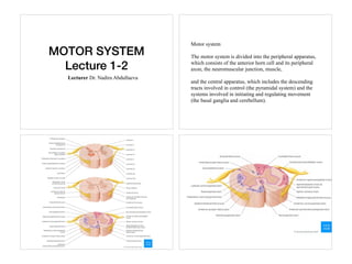
MOTOR SYSTEM
Lecture 1-2
Lecturer Dr. Nadira Abdullaeva
Motor system
The motor system is divided into the peripheral apparatus,
which consists of the anterior horn cell and its peripheral
axon, the neuromuscular junction, muscle,
and the central apparatus, which includes the descending
tracts involved in control (the pyramidal system) and the
systems involved in initiating and regulating movement
(the basal ganglia and cerebellum).
Descending pathways (motor tracts)
• Pyramidal tracts
• They are responsible for the voluntary control of the
musculature of the body and face.
• Extrapyramidal tracts
• They are responsible for the involuntary and automatic
control of all musculature, such as muscle tone, balance,
posture and locomotion
Pyramidal Tracts
The pyramidal tracts derive their name from the medullary
pyramids of the medulla oblongata, which they pass through.
These pathways are responsible for the voluntary control of the
musculature of the body and face.
• Corticospinal tracts – supplies the musculature of the body.
• Corticobulbar tracts – supplies the musculature of the head
and neck.
Corticospinal Tracts
The corticospinal tracts begin in the cerebral cortex of frontal
lobe, precentral gyrus, V layer of cortex where Betz cells present
They receive a range of inputs:
• Primary motor cortex
• Premotor cortex
• Supplementary motor area
After originating from the cortex(UMN or I order neurons-
Betz cells are in 5th layer of cortex, precentral gyrus of
frontal lobe), the neurons converge, and descend through
the internal capsule (a white matter pathway, located
between the thalamus and the basal ganglia).
After the internal capsule, the neurones pass through the crus
cerebri of the midbrain, the pons and into the medulla.
In the caudal part of the medulla, the tract divides into two:
Lateral corticospinal tract (90% of axons)-descends contralateral
after decussation and Anterior corticospinal tract(10% of axons)-
remain ipsilateral, and decussates when it reaches spinal cord.
They then descend into the spinal cord, terminating in the ventral
horn (at all segmental levels).
From the ventral horn, the lower motor neurons(II order neurons
are in ventral horns) go on to supply the muscles of the body.
The anterior corticospinal tract remains ipsilateral,
descending into the spinal cord. They then decussate and
terminate in the ventral horn of the cervical and
upper thoracic segmental levels.
Function. The corticospinal tract (along with the corticobulbar
tract) is the primary pathway that carries the motor commands
that underlie voluntary movement. The lateral corticospinal
tract is responsible for the control of the distal musculature and
fine control of the digits of the hand.
Anterior corticospinal tract is responsible for the control of
the proximal musculature.
The corticobulbar tracts arise from the lateral aspect of the
primary motor cortex. They receive the same inputs as the
corticospinal tracts. The fibres converge and pass through
the internal capsule to the brainstem.
The neurons terminate on the motor nuclei of the cranial
nerves. Here, they synapse with lower motor neurones,
which carry the motor signals to the muscles of the face
and neck.
Corticonuclear (Corticobulbar) Tract
Some of the fibers of the pyramidal tract branch off from
the main mass of the tract as it passes through the midbrain
and goes to the motor cranial nerve nuclei.
The nuclei receiving pyramidal tract input are the ones that
mediate voluntary movements of the cranial musculature
through cranial nerves V (the trigeminal nerve), VII (the
facial nerve), IX, X, and XI (the glossopharyngeal, vagus,
and accessory nerves), and XII (the hypoglossal nerve).
THE CORTICOBULBAR TRACT
A. Definitions
1.Bulb: the medulla + pons + mesencephalon
2.The corticobulbar fibers are similar to corticospinal fibers except
instead of terminating in the ventral horn of the cord, they end in
cranial nerve motor nuclei (EXCEPT THE EXTRAOCULAR
NUCLEI III, IV, and VI to be discussed later). The terms upper
motoneuron and pyramidal tract are often used collectively as a term
for both corticospinal and corticobulbar axons. Hence we have upper
motor neurons that end on cranial motor neurons.
B. Origin -- Similar to that of the corticospinal axons except face region
of cortex.
C. Course
1. Internal capsule -- posterior limb (some books say genu)
2. Cerebral peduncle -- the axons either leave the corticospinal fibers at this
point or at a slightly more caudal level and make their way to the appropriate
cranial nerve nuclei. Others travel more diffusely in the tegmentum. There is
no visible corticobulbar tract as there is for the corticospinal tract.
D. Termination: Examples.
1.Most muscles act together such as the pharynx, larynx. They get input
from
both hemispheres.
5.Termination on the hypoglossal nuclei motoneurons is mostly crossed and
can be useful in localizing lesions in the acute state. Signs may disappear
after a few days.
6.Muscles of facial expression
E. Corticobulbar tract to the facial nucleus lower motor
neurons.
1. The lower motor neurons that innervate muscles of the
lower face receive
only crossed corticobulbar axons from the cortex of the
opposite side.
2. The motor neurons that innervate muscle of the upper
face receive both crossed and uncrossed corticobulbar
axons. That is that both hemispheres send cortical fibers to
the nuclei on both sides of the brainstem.
Motor neurons are innervated bilaterally.
There are a few exceptions to this rule:
• Upper motor neurons for the facial nerve (CN VII)
have a contralateral innervation. This only affects the
muscles in the lower quadrant of the face – below the
eyes.
• Upper motor neurons for the hypoglossal (CN XII)
nerve only provide contralateral innervation.
Key points
• Corticospinal tract(Pyramidal) originates from 5th layer of cortex, precentral gyrus of frontal
lobe(UMN)
• Descends through posterior third of internal capsule
• Decussates in caudal medulla
• After decussation there are two tracts: Lateral and anterior corticospinal tracts
• Lateral contains (90% of axons) and anterior (10%)
• Anterior corticospinal tract remains ipsilateral until it reaches spinal cord where it decussates
• The second order neurons(LMN) are in anterior(ventral) horns of spinal cord, here axons of
UMN synapse with them
• Axons of LMN then go to innervate skeletal muscles (effector organs)
• Function of Corticospinal tract is innervation of skeletal musculature of body and extremities
Extrapyramidal Tracts
The extrapyramidal tracts originate in the brainstem,
carrying motor fibres to the spinal cord. They are
responsible for the involuntary and automatic control of
all musculature, such as muscle tone, balance, posture and
locomotion.
There are four tracts in total. The vestibulospinal and
reticulospinal tracts do not decussate, providing ipsilateral
innervation. The rubrospinal and tectospinal tracts
decussate
The motor tracts in the spinal cord are anatomically and
functionally divided into two groups: a lateral group: the
corticospinal and rubrospinal tracts, and a medial group,
comprising the reticulospinal, vestibulospinal, and
tectospinal tracts.
The lateral tracts mainly project to the distal musculature
(especially in the upper limbs).
Their function:voluntary movements of the forearms and
hands, for precise, highly differentiated, fine motor control.
The medial tracts innervate motor neurons lying more
medially in the anterior horn.
Function:They are primarily responsible for movements of
the trunk and lower limbs (stance and gait)
Extrapyramidal system
The EPS serves an essential function in maintaining posture and
regulating involuntary motor functions. In particular, the EPS provides:
• Postural tone adjustment
• Preparation of predisposing tonic attitudes for involuntary
movements
• Performing movements that make voluntary movements more natural
and correct
• Control of automatic modifications of tone and movements
• Control of the reflexes that accompany the responses to affective and
attentive situations (reactions)
• Control of the movements originally voluntary then become
automatic through exercise and learning (e.g., in writing)
• Inhibition of involuntary movements (hyperkinesias), which are
particularly evident in extrapyramidal diseases.
Vestibulospinal Tracts
There are two vestibulospinal pathways; medial and lateral.
They arise from the vestibular nuclei, which receive input
from the organs of balance. The tracts convey this balance
information to the spinal cord, where it remains ipsilateral.
Fibres in this pathway control balance and posture by
innervating the ‘anti-gravity’ muscles (flexors of the arm,
and extensors of the leg).
Vestibulospinal tracts.
The lateral vestibulospinal tract originates in the lateral
vestibular nucleus(Deiters’ nucleus lower medulla). It
courses through the brainstem and through the anterior
funiculus of the spinal cord on the ipsilateral side, before
exiting ipsilaterally at all levels of the spinal cord.
The medial vestibulospinal tract originates in the medial
vestibular nucleus( Schwalbe's nucleus) splits immediately
and courses bilaterally through the brainstem via the
medial longitudinal fasciculus (MLF) and through the
anterior funiculus of the spinal cord, to the ventral horns
and synapses with alfa and gamma motoneurons.
Function:
• Medial Vestibulospinal tract- extensor muscles and anti-
gravity muscles of neck, head
• Lateral VST -extensor muscles and anti-gravity muscles
of limbs and trunk
Reticulospinal Tracts
The two recticulospinal tracts have differing functions:
The medial reticulospinal tract arises from the pons. It
facilitates voluntary movements, and increases muscle
tone.
The lateral reticulospinal tract arises from the
medulla. It inhibits voluntary movements, and reduces
muscle tone.
Rubrospinal Tract
The rubrospinal tract originates from the red nucleus, a
midbrain structure. As the fibres emerge, they decussate,
and descend into the spinal cord.
They have a contralateral innervation.
It is thought to play a role in the fine control of hand
movements
Tectospinal Tracts
This pathway begins at the superior colliculus of the
midbrain. The superior colliculus is a structure that receives
input from the optic nerves. The neurones then quickly
decussate, and enter the spinal cord. They terminate at the
cervical levels of the spinal cord.
The tectospinal tract coordinates movements of the head in
relation to vision stimuli.
"Deep tendon" (muscle stretch; myotatic) reflexes
Deep tendon reflexes include:
the biceps, triceps, brachioradialis (radial periosteal),
quadriceps, hamstring and calf muscles (Achilles).
Upper Motor Neuron Syndrome
Damage to any part of the motor system hierarchy above
the level of alpha motor neurons results in a set of
symptoms termed the upper motor neuron syndrome.
Typically causes: stroke, tumors, and blunt trauma.
Signs of UMN damage
• Hyperreflexia
• Hypertonia(increased tone of muscles)
• Babinski +
• Spasticity
• Signs of LMN damage
• Hypo or areflexia
• Hypo or atonia of muscles
• Babinski “—“
• Fasciculations
• Fibrillations of muscles
Deep tendon (muscle stretch) reflex testing evaluates
afferent nerves, synaptic connections within the spinal cord,
motor nerves, and descending motor pathways.
Lower motor neuron lesions (affecting the anterior horn
cell, spinal root, or peripheral nerve) depress reflexes;
Reflexes tested include the following:
• Biceps (innervated by C5 and C6)
• Radial brachialis (by C6)
• Triceps (by C7)
• Distal finger flexors (by C8)
• Quadriceps knee jerk (by L4)
• Ankle jerk (by S1)
• Jaw jerk (by the 5th cranial nerve)
Pathologic reflexes (Babinski, Chaddock, Oppenheim,
snout, rooting, grasp) are reversions to primitive responses
and indicate loss of cortical inhibition.
Babinski, Chaddock, and Oppenheim reflexes all
evaluate the plantar response.
The normal reflex response is flexion of the great toe. An
abnormal response is slower and consists of extension of
the great toe with fanning of the other toes and often knee
and hip flexion.
For Babinski reflex, the lateral sole of the foot is firmly
stroked from the heel to the ball of the foot with an end of a
reflex hammer.
For Chaddock reflex, the lateral foot, from lateral malleolus
to small toe, is stroked with a blunt instrument.
For the Oppenheim reflex, the anterior tibia, from just below the patella to
the foot, is firmly stroked with a knuckle.
The Oppenheim test may be used with the Babinski test or the Chaddock
test to make withdrawal less likely.
The snout reflex is present if tapping a tongue blade across the lips causes
pursing of the lips.
The rooting reflex is present if stroking the lateral upper lip causes
movement of the mouth toward the stimulus.
The grasp reflex is present if gently stroking the palm of the patient’s
hand causes the fingers to flex and grasp the examiner’s finger.
The palmomental reflex is present if stroking the palm of
the hand causes contraction of the ipsilateral mentalis
muscle of the lower lip.
For the glabellar sign, the forehead is tapped to induce
blinking; normally, each of the first 5 taps induces a single
blink, then the reflex fatigues. Blinking persists in patients
with diffuse cerebral dysfunction.
This grading system.
• 0 No evidence of contraction
• 1+ Decreased, but still present (hypo-reflexic).
Hyporeflexia is generally associated with a lower motor
neuron deficit
• 2+ Normal
• 3+ Super-normal (hyper-reflexic)
• 4+ Clonus: Repetitive shortening of the muscle after a
single stimulation
General Clinical Features
The alterations of the extrapyramidal system can group into
hypokinesia and hyperkinesias manifestations.
Hypokinesias
The extrapyramidal involvement leads to hypokinesia and
akinesia.
Several voluntary acts such as walking, writing, and speaking
slow down.
Concerning involuntary movements, a reduction, or loss, of the
associated movements of pendulation of the upper limbs during
walking, or mimic and expressive movements can be observed.
Slowness in the execution of voluntary movements, especially
at the beginning of the movement or when this is about to
complete, can be present. This condition is termed bradykinesia.
Hyperkinesias
They concern abnormal involuntary movements that can
present in extrapyramidal diseases. These movements
become distinguished in:
• Choreic movements: sudden, irregular, incomplete,
aimless, variable movements;
• Athetotic: arrhythmic, slow, exaggerated, tentacular
movements;
• Hemiballism: the movements are similar to choreic, but
much more intense and persist during sleep;
• Rapid muscle contractions, which reproduce a
stereotyped movement, repeated obsessively; they can
become voluntarily inhibited, even if with effort;
• Tremors: the extrapyramidal tremor (typically the tremor
of PD) is rhythmic, at a slow pace (4 to 5 oscillations per
second), not very wide, uniform, more pronounced in
rest, attenuates during voluntary movements and in those
passive. It disappears in sleep; the fingers reproduce
movements such as counting coins
• Spasms: involuntary movements of a tonic type (intense
and lasting contraction, but transient) or clonic (series of
rhythmic contractions of short duration, separated by
periods of rest);
• Myoclonus: rapid, sudden contractions involving isolated
muscles or bundles of muscle fibers - usually do not
cause motor effects.
• Thank you for attention!

More Related Content

Similar to Motor system. Extrapyramydal system. (1,2,3 lectures).pdf

Anatomy of Spinal Cord.pdf
Anatomy of Spinal Cord.pdfAnatomy of Spinal Cord.pdf
Anatomy of Spinal Cord.pdfMaiGaber4
 
TRACTS OF THE SPINAL CORD.pptx
TRACTS OF THE SPINAL CORD.pptxTRACTS OF THE SPINAL CORD.pptx
TRACTS OF THE SPINAL CORD.pptxOlaniyiEmmanuel5
 
Tracts of spinal cord (1)
Tracts of spinal cord (1)Tracts of spinal cord (1)
Tracts of spinal cord (1)Zulcaif Ahmad
 
Neuroscience : Neuroanatomy of Ascending & Descending Tracts
Neuroscience : Neuroanatomy of Ascending & Descending TractsNeuroscience : Neuroanatomy of Ascending & Descending Tracts
Neuroscience : Neuroanatomy of Ascending & Descending TractsSado Anatomist
 
Descending tracts of spinal cord.pptx
Descending tracts of spinal cord.pptxDescending tracts of spinal cord.pptx
Descending tracts of spinal cord.pptxgopal8694
 
Pyramidal Tract
Pyramidal TractPyramidal Tract
Pyramidal TractM S
 
Spinalcorddisorders 170123051811 (1)
Spinalcorddisorders 170123051811 (1)Spinalcorddisorders 170123051811 (1)
Spinalcorddisorders 170123051811 (1)Abhishek Thakur
 
Spinalcorddisorders 170123051811 (1)
Spinalcorddisorders 170123051811 (1)Spinalcorddisorders 170123051811 (1)
Spinalcorddisorders 170123051811 (1)Abhishek Thakur
 
PATHWAYS_OF_THE_BRAIN_AND_SPINAL_CORD.pptx
PATHWAYS_OF_THE_BRAIN_AND_SPINAL_CORD.pptxPATHWAYS_OF_THE_BRAIN_AND_SPINAL_CORD.pptx
PATHWAYS_OF_THE_BRAIN_AND_SPINAL_CORD.pptxAparnaAnil20
 
DESCENDING MOTOR TRACTs.pptx
DESCENDING MOTOR TRACTs.pptxDESCENDING MOTOR TRACTs.pptx
DESCENDING MOTOR TRACTs.pptxAaron Paul Baliga
 
Corticospinal tract (Pyramidal tract)
Corticospinal tract (Pyramidal tract)Corticospinal tract (Pyramidal tract)
Corticospinal tract (Pyramidal tract)Ajith lolita
 
Pyramidal tract by Sunita.M.Tiwale,Prof. Dept of physiology,D.Y.Patil Medical...
Pyramidal tract by Sunita.M.Tiwale,Prof. Dept of physiology,D.Y.Patil Medical...Pyramidal tract by Sunita.M.Tiwale,Prof. Dept of physiology,D.Y.Patil Medical...
Pyramidal tract by Sunita.M.Tiwale,Prof. Dept of physiology,D.Y.Patil Medical...Physiology Dept
 
Physiology of equilibrium & balance
Physiology of equilibrium & balance Physiology of equilibrium & balance
Physiology of equilibrium & balance farhan_aq91
 

Similar to Motor system. Extrapyramydal system. (1,2,3 lectures).pdf (20)

Pyramidal tract
Pyramidal tractPyramidal tract
Pyramidal tract
 
Anatomy of Spinal Cord.pdf
Anatomy of Spinal Cord.pdfAnatomy of Spinal Cord.pdf
Anatomy of Spinal Cord.pdf
 
TRACTS OF THE SPINAL CORD.pptx
TRACTS OF THE SPINAL CORD.pptxTRACTS OF THE SPINAL CORD.pptx
TRACTS OF THE SPINAL CORD.pptx
 
Tracts of spinal cord (1)
Tracts of spinal cord (1)Tracts of spinal cord (1)
Tracts of spinal cord (1)
 
Neuroscience : Neuroanatomy of Ascending & Descending Tracts
Neuroscience : Neuroanatomy of Ascending & Descending TractsNeuroscience : Neuroanatomy of Ascending & Descending Tracts
Neuroscience : Neuroanatomy of Ascending & Descending Tracts
 
Descending tracts of spinal cord.pptx
Descending tracts of spinal cord.pptxDescending tracts of spinal cord.pptx
Descending tracts of spinal cord.pptx
 
Pyramidal Tract
Pyramidal TractPyramidal Tract
Pyramidal Tract
 
Spinalcorddisorders 170123051811 (1)
Spinalcorddisorders 170123051811 (1)Spinalcorddisorders 170123051811 (1)
Spinalcorddisorders 170123051811 (1)
 
Spinalcorddisorders 170123051811 (1)
Spinalcorddisorders 170123051811 (1)Spinalcorddisorders 170123051811 (1)
Spinalcorddisorders 170123051811 (1)
 
Ch11 eoc
Ch11 eocCh11 eoc
Ch11 eoc
 
PATHWAYS_OF_THE_BRAIN_AND_SPINAL_CORD.pptx
PATHWAYS_OF_THE_BRAIN_AND_SPINAL_CORD.pptxPATHWAYS_OF_THE_BRAIN_AND_SPINAL_CORD.pptx
PATHWAYS_OF_THE_BRAIN_AND_SPINAL_CORD.pptx
 
Medulla oblongata
Medulla oblongata Medulla oblongata
Medulla oblongata
 
SPINAL CORD.2.pptx
SPINAL CORD.2.pptxSPINAL CORD.2.pptx
SPINAL CORD.2.pptx
 
Spinal cord
Spinal cordSpinal cord
Spinal cord
 
DESCENDING MOTOR TRACTs.pptx
DESCENDING MOTOR TRACTs.pptxDESCENDING MOTOR TRACTs.pptx
DESCENDING MOTOR TRACTs.pptx
 
Corticospinal tract (Pyramidal tract)
Corticospinal tract (Pyramidal tract)Corticospinal tract (Pyramidal tract)
Corticospinal tract (Pyramidal tract)
 
Pyramidal tract by Sunita.M.Tiwale,Prof. Dept of physiology,D.Y.Patil Medical...
Pyramidal tract by Sunita.M.Tiwale,Prof. Dept of physiology,D.Y.Patil Medical...Pyramidal tract by Sunita.M.Tiwale,Prof. Dept of physiology,D.Y.Patil Medical...
Pyramidal tract by Sunita.M.Tiwale,Prof. Dept of physiology,D.Y.Patil Medical...
 
11. Descending tracts.pptx
11. Descending tracts.pptx11. Descending tracts.pptx
11. Descending tracts.pptx
 
Anatomy of Cerebellum(Part- 2)
Anatomy of Cerebellum(Part- 2)Anatomy of Cerebellum(Part- 2)
Anatomy of Cerebellum(Part- 2)
 
Physiology of equilibrium & balance
Physiology of equilibrium & balance Physiology of equilibrium & balance
Physiology of equilibrium & balance
 

More from Saicharitha15

Lec 5.pdf tuberculosis miliary tuberculosis meningitis
Lec 5.pdf tuberculosis miliary tuberculosis meningitisLec 5.pdf tuberculosis miliary tuberculosis meningitis
Lec 5.pdf tuberculosis miliary tuberculosis meningitisSaicharitha15
 
Еsophageal cancer-7.pdf oncology subject
Еsophageal cancer-7.pdf oncology subjectЕsophageal cancer-7.pdf oncology subject
Еsophageal cancer-7.pdf oncology subjectSaicharitha15
 
Oral candidiasis and stomatitis. name - seelam Sai charitha .pptx
Oral candidiasis and stomatitis. name - seelam Sai charitha .pptxOral candidiasis and stomatitis. name - seelam Sai charitha .pptx
Oral candidiasis and stomatitis. name - seelam Sai charitha .pptxSaicharitha15
 
Epilepsy.pdf neurology education information
Epilepsy.pdf neurology education  informationEpilepsy.pdf neurology education  information
Epilepsy.pdf neurology education informationSaicharitha15
 
lecture 4.ppt.pdf opthamology education ism
lecture 4.ppt.pdf opthamology education ismlecture 4.ppt.pdf opthamology education ism
lecture 4.ppt.pdf opthamology education ismSaicharitha15
 
1skin structure.pptx education dermatology
1skin structure.pptx education dermatology1skin structure.pptx education dermatology
1skin structure.pptx education dermatologySaicharitha15
 
Complex Regional Pain Syndrome.ppt education
Complex Regional Pain Syndrome.ppt educationComplex Regional Pain Syndrome.ppt education
Complex Regional Pain Syndrome.ppt educationSaicharitha15
 
lech. introduction — копия 3.ppt
lech. introduction — копия 3.pptlech. introduction — копия 3.ppt
lech. introduction — копия 3.pptSaicharitha15
 
Orthopedics Maheshwari 5th edition.pdf
Orthopedics Maheshwari 5th edition.pdfOrthopedics Maheshwari 5th edition.pdf
Orthopedics Maheshwari 5th edition.pdfSaicharitha15
 
Biochemical testing of renal function (1).pdf
Biochemical testing of renal function (1).pdfBiochemical testing of renal function (1).pdf
Biochemical testing of renal function (1).pdfSaicharitha15
 
Sensory system L3-4.pdf
Sensory system L3-4.pdfSensory system L3-4.pdf
Sensory system L3-4.pdfSaicharitha15
 
Acute-Diarrhoeal-Diseases.pdf
Acute-Diarrhoeal-Diseases.pdfAcute-Diarrhoeal-Diseases.pdf
Acute-Diarrhoeal-Diseases.pdfSaicharitha15
 
oesophages stomach.pptx
oesophages stomach.pptxoesophages stomach.pptx
oesophages stomach.pptxSaicharitha15
 
CVS HYPERTENSION.pdf
CVS HYPERTENSION.pdfCVS HYPERTENSION.pdf
CVS HYPERTENSION.pdfSaicharitha15
 
Normal anatomy of MSS part 1.pdf
Normal anatomy of MSS part 1.pdfNormal anatomy of MSS part 1.pdf
Normal anatomy of MSS part 1.pdfSaicharitha15
 
anatomy of MMS 2.pdf
anatomy of MMS 2.pdfanatomy of MMS 2.pdf
anatomy of MMS 2.pdfSaicharitha15
 
4 Different Economic Systems.ppt
4 Different Economic Systems.ppt4 Different Economic Systems.ppt
4 Different Economic Systems.pptSaicharitha15
 

More from Saicharitha15 (20)

Lec 5.pdf tuberculosis miliary tuberculosis meningitis
Lec 5.pdf tuberculosis miliary tuberculosis meningitisLec 5.pdf tuberculosis miliary tuberculosis meningitis
Lec 5.pdf tuberculosis miliary tuberculosis meningitis
 
Еsophageal cancer-7.pdf oncology subject
Еsophageal cancer-7.pdf oncology subjectЕsophageal cancer-7.pdf oncology subject
Еsophageal cancer-7.pdf oncology subject
 
Oral candidiasis and stomatitis. name - seelam Sai charitha .pptx
Oral candidiasis and stomatitis. name - seelam Sai charitha .pptxOral candidiasis and stomatitis. name - seelam Sai charitha .pptx
Oral candidiasis and stomatitis. name - seelam Sai charitha .pptx
 
Epilepsy.pdf neurology education information
Epilepsy.pdf neurology education  informationEpilepsy.pdf neurology education  information
Epilepsy.pdf neurology education information
 
lecture 4.ppt.pdf opthamology education ism
lecture 4.ppt.pdf opthamology education ismlecture 4.ppt.pdf opthamology education ism
lecture 4.ppt.pdf opthamology education ism
 
1skin structure.pptx education dermatology
1skin structure.pptx education dermatology1skin structure.pptx education dermatology
1skin structure.pptx education dermatology
 
Complex Regional Pain Syndrome.ppt education
Complex Regional Pain Syndrome.ppt educationComplex Regional Pain Syndrome.ppt education
Complex Regional Pain Syndrome.ppt education
 
Central vision.docx
Central vision.docxCentral vision.docx
Central vision.docx
 
lech. introduction — копия 3.ppt
lech. introduction — копия 3.pptlech. introduction — копия 3.ppt
lech. introduction — копия 3.ppt
 
Orthopedics Maheshwari 5th edition.pdf
Orthopedics Maheshwari 5th edition.pdfOrthopedics Maheshwari 5th edition.pdf
Orthopedics Maheshwari 5th edition.pdf
 
gastritis 16.ppt
gastritis 16.pptgastritis 16.ppt
gastritis 16.ppt
 
Biochemical testing of renal function (1).pdf
Biochemical testing of renal function (1).pdfBiochemical testing of renal function (1).pdf
Biochemical testing of renal function (1).pdf
 
Sensory system L3-4.pdf
Sensory system L3-4.pdfSensory system L3-4.pdf
Sensory system L3-4.pdf
 
Acute-Diarrhoeal-Diseases.pdf
Acute-Diarrhoeal-Diseases.pdfAcute-Diarrhoeal-Diseases.pdf
Acute-Diarrhoeal-Diseases.pdf
 
Radiology GIT.ppt
Radiology GIT.pptRadiology GIT.ppt
Radiology GIT.ppt
 
oesophages stomach.pptx
oesophages stomach.pptxoesophages stomach.pptx
oesophages stomach.pptx
 
CVS HYPERTENSION.pdf
CVS HYPERTENSION.pdfCVS HYPERTENSION.pdf
CVS HYPERTENSION.pdf
 
Normal anatomy of MSS part 1.pdf
Normal anatomy of MSS part 1.pdfNormal anatomy of MSS part 1.pdf
Normal anatomy of MSS part 1.pdf
 
anatomy of MMS 2.pdf
anatomy of MMS 2.pdfanatomy of MMS 2.pdf
anatomy of MMS 2.pdf
 
4 Different Economic Systems.ppt
4 Different Economic Systems.ppt4 Different Economic Systems.ppt
4 Different Economic Systems.ppt
 

Recently uploaded

CELL CYCLE Division Science 8 quarter IV.pptx
CELL CYCLE Division Science 8 quarter IV.pptxCELL CYCLE Division Science 8 quarter IV.pptx
CELL CYCLE Division Science 8 quarter IV.pptxJiesonDelaCerna
 
Capitol Tech U Doctoral Presentation - April 2024.pptx
Capitol Tech U Doctoral Presentation - April 2024.pptxCapitol Tech U Doctoral Presentation - April 2024.pptx
Capitol Tech U Doctoral Presentation - April 2024.pptxCapitolTechU
 
DATA STRUCTURE AND ALGORITHM for beginners
DATA STRUCTURE AND ALGORITHM for beginnersDATA STRUCTURE AND ALGORITHM for beginners
DATA STRUCTURE AND ALGORITHM for beginnersSabitha Banu
 
Roles & Responsibilities in Pharmacovigilance
Roles & Responsibilities in PharmacovigilanceRoles & Responsibilities in Pharmacovigilance
Roles & Responsibilities in PharmacovigilanceSamikshaHamane
 
ECONOMIC CONTEXT - PAPER 1 Q3: NEWSPAPERS.pptx
ECONOMIC CONTEXT - PAPER 1 Q3: NEWSPAPERS.pptxECONOMIC CONTEXT - PAPER 1 Q3: NEWSPAPERS.pptx
ECONOMIC CONTEXT - PAPER 1 Q3: NEWSPAPERS.pptxiammrhaywood
 
Biting mechanism of poisonous snakes.pdf
Biting mechanism of poisonous snakes.pdfBiting mechanism of poisonous snakes.pdf
Biting mechanism of poisonous snakes.pdfadityarao40181
 
Framing an Appropriate Research Question 6b9b26d93da94caf993c038d9efcdedb.pdf
Framing an Appropriate Research Question 6b9b26d93da94caf993c038d9efcdedb.pdfFraming an Appropriate Research Question 6b9b26d93da94caf993c038d9efcdedb.pdf
Framing an Appropriate Research Question 6b9b26d93da94caf993c038d9efcdedb.pdfUjwalaBharambe
 
Introduction to ArtificiaI Intelligence in Higher Education
Introduction to ArtificiaI Intelligence in Higher EducationIntroduction to ArtificiaI Intelligence in Higher Education
Introduction to ArtificiaI Intelligence in Higher Educationpboyjonauth
 
Earth Day Presentation wow hello nice great
Earth Day Presentation wow hello nice greatEarth Day Presentation wow hello nice great
Earth Day Presentation wow hello nice greatYousafMalik24
 
Employee wellbeing at the workplace.pptx
Employee wellbeing at the workplace.pptxEmployee wellbeing at the workplace.pptx
Employee wellbeing at the workplace.pptxNirmalaLoungPoorunde1
 
CARE OF CHILD IN INCUBATOR..........pptx
CARE OF CHILD IN INCUBATOR..........pptxCARE OF CHILD IN INCUBATOR..........pptx
CARE OF CHILD IN INCUBATOR..........pptxGaneshChakor2
 
Alper Gobel In Media Res Media Component
Alper Gobel In Media Res Media ComponentAlper Gobel In Media Res Media Component
Alper Gobel In Media Res Media ComponentInMediaRes1
 
Painted Grey Ware.pptx, PGW Culture of India
Painted Grey Ware.pptx, PGW Culture of IndiaPainted Grey Ware.pptx, PGW Culture of India
Painted Grey Ware.pptx, PGW Culture of IndiaVirag Sontakke
 
Presiding Officer Training module 2024 lok sabha elections
Presiding Officer Training module 2024 lok sabha electionsPresiding Officer Training module 2024 lok sabha elections
Presiding Officer Training module 2024 lok sabha electionsanshu789521
 
POINT- BIOCHEMISTRY SEM 2 ENZYMES UNIT 5.pptx
POINT- BIOCHEMISTRY SEM 2 ENZYMES UNIT 5.pptxPOINT- BIOCHEMISTRY SEM 2 ENZYMES UNIT 5.pptx
POINT- BIOCHEMISTRY SEM 2 ENZYMES UNIT 5.pptxSayali Powar
 
Meghan Sutherland In Media Res Media Component
Meghan Sutherland In Media Res Media ComponentMeghan Sutherland In Media Res Media Component
Meghan Sutherland In Media Res Media ComponentInMediaRes1
 

Recently uploaded (20)

CELL CYCLE Division Science 8 quarter IV.pptx
CELL CYCLE Division Science 8 quarter IV.pptxCELL CYCLE Division Science 8 quarter IV.pptx
CELL CYCLE Division Science 8 quarter IV.pptx
 
Capitol Tech U Doctoral Presentation - April 2024.pptx
Capitol Tech U Doctoral Presentation - April 2024.pptxCapitol Tech U Doctoral Presentation - April 2024.pptx
Capitol Tech U Doctoral Presentation - April 2024.pptx
 
DATA STRUCTURE AND ALGORITHM for beginners
DATA STRUCTURE AND ALGORITHM for beginnersDATA STRUCTURE AND ALGORITHM for beginners
DATA STRUCTURE AND ALGORITHM for beginners
 
Roles & Responsibilities in Pharmacovigilance
Roles & Responsibilities in PharmacovigilanceRoles & Responsibilities in Pharmacovigilance
Roles & Responsibilities in Pharmacovigilance
 
ECONOMIC CONTEXT - PAPER 1 Q3: NEWSPAPERS.pptx
ECONOMIC CONTEXT - PAPER 1 Q3: NEWSPAPERS.pptxECONOMIC CONTEXT - PAPER 1 Q3: NEWSPAPERS.pptx
ECONOMIC CONTEXT - PAPER 1 Q3: NEWSPAPERS.pptx
 
Biting mechanism of poisonous snakes.pdf
Biting mechanism of poisonous snakes.pdfBiting mechanism of poisonous snakes.pdf
Biting mechanism of poisonous snakes.pdf
 
ESSENTIAL of (CS/IT/IS) class 06 (database)
ESSENTIAL of (CS/IT/IS) class 06 (database)ESSENTIAL of (CS/IT/IS) class 06 (database)
ESSENTIAL of (CS/IT/IS) class 06 (database)
 
Framing an Appropriate Research Question 6b9b26d93da94caf993c038d9efcdedb.pdf
Framing an Appropriate Research Question 6b9b26d93da94caf993c038d9efcdedb.pdfFraming an Appropriate Research Question 6b9b26d93da94caf993c038d9efcdedb.pdf
Framing an Appropriate Research Question 6b9b26d93da94caf993c038d9efcdedb.pdf
 
OS-operating systems- ch04 (Threads) ...
OS-operating systems- ch04 (Threads) ...OS-operating systems- ch04 (Threads) ...
OS-operating systems- ch04 (Threads) ...
 
Introduction to ArtificiaI Intelligence in Higher Education
Introduction to ArtificiaI Intelligence in Higher EducationIntroduction to ArtificiaI Intelligence in Higher Education
Introduction to ArtificiaI Intelligence in Higher Education
 
Earth Day Presentation wow hello nice great
Earth Day Presentation wow hello nice greatEarth Day Presentation wow hello nice great
Earth Day Presentation wow hello nice great
 
Employee wellbeing at the workplace.pptx
Employee wellbeing at the workplace.pptxEmployee wellbeing at the workplace.pptx
Employee wellbeing at the workplace.pptx
 
CARE OF CHILD IN INCUBATOR..........pptx
CARE OF CHILD IN INCUBATOR..........pptxCARE OF CHILD IN INCUBATOR..........pptx
CARE OF CHILD IN INCUBATOR..........pptx
 
Model Call Girl in Tilak Nagar Delhi reach out to us at 🔝9953056974🔝
Model Call Girl in Tilak Nagar Delhi reach out to us at 🔝9953056974🔝Model Call Girl in Tilak Nagar Delhi reach out to us at 🔝9953056974🔝
Model Call Girl in Tilak Nagar Delhi reach out to us at 🔝9953056974🔝
 
Alper Gobel In Media Res Media Component
Alper Gobel In Media Res Media ComponentAlper Gobel In Media Res Media Component
Alper Gobel In Media Res Media Component
 
Painted Grey Ware.pptx, PGW Culture of India
Painted Grey Ware.pptx, PGW Culture of IndiaPainted Grey Ware.pptx, PGW Culture of India
Painted Grey Ware.pptx, PGW Culture of India
 
Model Call Girl in Bikash Puri Delhi reach out to us at 🔝9953056974🔝
Model Call Girl in Bikash Puri  Delhi reach out to us at 🔝9953056974🔝Model Call Girl in Bikash Puri  Delhi reach out to us at 🔝9953056974🔝
Model Call Girl in Bikash Puri Delhi reach out to us at 🔝9953056974🔝
 
Presiding Officer Training module 2024 lok sabha elections
Presiding Officer Training module 2024 lok sabha electionsPresiding Officer Training module 2024 lok sabha elections
Presiding Officer Training module 2024 lok sabha elections
 
POINT- BIOCHEMISTRY SEM 2 ENZYMES UNIT 5.pptx
POINT- BIOCHEMISTRY SEM 2 ENZYMES UNIT 5.pptxPOINT- BIOCHEMISTRY SEM 2 ENZYMES UNIT 5.pptx
POINT- BIOCHEMISTRY SEM 2 ENZYMES UNIT 5.pptx
 
Meghan Sutherland In Media Res Media Component
Meghan Sutherland In Media Res Media ComponentMeghan Sutherland In Media Res Media Component
Meghan Sutherland In Media Res Media Component
 

Motor system. Extrapyramydal system. (1,2,3 lectures).pdf

  • 1. MOTOR SYSTEM Lecture 1-2 Lecturer Dr. Nadira Abdullaeva Motor system The motor system is divided into the peripheral apparatus, which consists of the anterior horn cell and its peripheral axon, the neuromuscular junction, muscle, and the central apparatus, which includes the descending tracts involved in control (the pyramidal system) and the systems involved in initiating and regulating movement (the basal ganglia and cerebellum).
  • 2. Descending pathways (motor tracts) • Pyramidal tracts • They are responsible for the voluntary control of the musculature of the body and face. • Extrapyramidal tracts • They are responsible for the involuntary and automatic control of all musculature, such as muscle tone, balance, posture and locomotion Pyramidal Tracts The pyramidal tracts derive their name from the medullary pyramids of the medulla oblongata, which they pass through. These pathways are responsible for the voluntary control of the musculature of the body and face. • Corticospinal tracts – supplies the musculature of the body. • Corticobulbar tracts – supplies the musculature of the head and neck. Corticospinal Tracts The corticospinal tracts begin in the cerebral cortex of frontal lobe, precentral gyrus, V layer of cortex where Betz cells present They receive a range of inputs: • Primary motor cortex • Premotor cortex • Supplementary motor area
  • 3.
  • 4.
  • 5. After originating from the cortex(UMN or I order neurons- Betz cells are in 5th layer of cortex, precentral gyrus of frontal lobe), the neurons converge, and descend through the internal capsule (a white matter pathway, located between the thalamus and the basal ganglia). After the internal capsule, the neurones pass through the crus cerebri of the midbrain, the pons and into the medulla. In the caudal part of the medulla, the tract divides into two: Lateral corticospinal tract (90% of axons)-descends contralateral after decussation and Anterior corticospinal tract(10% of axons)- remain ipsilateral, and decussates when it reaches spinal cord. They then descend into the spinal cord, terminating in the ventral horn (at all segmental levels). From the ventral horn, the lower motor neurons(II order neurons are in ventral horns) go on to supply the muscles of the body. The anterior corticospinal tract remains ipsilateral, descending into the spinal cord. They then decussate and terminate in the ventral horn of the cervical and upper thoracic segmental levels. Function. The corticospinal tract (along with the corticobulbar tract) is the primary pathway that carries the motor commands that underlie voluntary movement. The lateral corticospinal tract is responsible for the control of the distal musculature and fine control of the digits of the hand. Anterior corticospinal tract is responsible for the control of the proximal musculature.
  • 6. The corticobulbar tracts arise from the lateral aspect of the primary motor cortex. They receive the same inputs as the corticospinal tracts. The fibres converge and pass through the internal capsule to the brainstem. The neurons terminate on the motor nuclei of the cranial nerves. Here, they synapse with lower motor neurones, which carry the motor signals to the muscles of the face and neck. Corticonuclear (Corticobulbar) Tract Some of the fibers of the pyramidal tract branch off from the main mass of the tract as it passes through the midbrain and goes to the motor cranial nerve nuclei. The nuclei receiving pyramidal tract input are the ones that mediate voluntary movements of the cranial musculature through cranial nerves V (the trigeminal nerve), VII (the facial nerve), IX, X, and XI (the glossopharyngeal, vagus, and accessory nerves), and XII (the hypoglossal nerve). THE CORTICOBULBAR TRACT A. Definitions 1.Bulb: the medulla + pons + mesencephalon 2.The corticobulbar fibers are similar to corticospinal fibers except instead of terminating in the ventral horn of the cord, they end in cranial nerve motor nuclei (EXCEPT THE EXTRAOCULAR NUCLEI III, IV, and VI to be discussed later). The terms upper motoneuron and pyramidal tract are often used collectively as a term for both corticospinal and corticobulbar axons. Hence we have upper motor neurons that end on cranial motor neurons. B. Origin -- Similar to that of the corticospinal axons except face region of cortex. C. Course 1. Internal capsule -- posterior limb (some books say genu) 2. Cerebral peduncle -- the axons either leave the corticospinal fibers at this point or at a slightly more caudal level and make their way to the appropriate cranial nerve nuclei. Others travel more diffusely in the tegmentum. There is no visible corticobulbar tract as there is for the corticospinal tract. D. Termination: Examples. 1.Most muscles act together such as the pharynx, larynx. They get input from both hemispheres. 5.Termination on the hypoglossal nuclei motoneurons is mostly crossed and can be useful in localizing lesions in the acute state. Signs may disappear after a few days. 6.Muscles of facial expression
  • 7. E. Corticobulbar tract to the facial nucleus lower motor neurons. 1. The lower motor neurons that innervate muscles of the lower face receive only crossed corticobulbar axons from the cortex of the opposite side. 2. The motor neurons that innervate muscle of the upper face receive both crossed and uncrossed corticobulbar axons. That is that both hemispheres send cortical fibers to the nuclei on both sides of the brainstem.
  • 8. Motor neurons are innervated bilaterally. There are a few exceptions to this rule: • Upper motor neurons for the facial nerve (CN VII) have a contralateral innervation. This only affects the muscles in the lower quadrant of the face – below the eyes. • Upper motor neurons for the hypoglossal (CN XII) nerve only provide contralateral innervation.
  • 9. Key points • Corticospinal tract(Pyramidal) originates from 5th layer of cortex, precentral gyrus of frontal lobe(UMN) • Descends through posterior third of internal capsule • Decussates in caudal medulla • After decussation there are two tracts: Lateral and anterior corticospinal tracts • Lateral contains (90% of axons) and anterior (10%) • Anterior corticospinal tract remains ipsilateral until it reaches spinal cord where it decussates • The second order neurons(LMN) are in anterior(ventral) horns of spinal cord, here axons of UMN synapse with them • Axons of LMN then go to innervate skeletal muscles (effector organs) • Function of Corticospinal tract is innervation of skeletal musculature of body and extremities Extrapyramidal Tracts The extrapyramidal tracts originate in the brainstem, carrying motor fibres to the spinal cord. They are responsible for the involuntary and automatic control of all musculature, such as muscle tone, balance, posture and locomotion. There are four tracts in total. The vestibulospinal and reticulospinal tracts do not decussate, providing ipsilateral innervation. The rubrospinal and tectospinal tracts decussate
  • 10. The motor tracts in the spinal cord are anatomically and functionally divided into two groups: a lateral group: the corticospinal and rubrospinal tracts, and a medial group, comprising the reticulospinal, vestibulospinal, and tectospinal tracts. The lateral tracts mainly project to the distal musculature (especially in the upper limbs). Their function:voluntary movements of the forearms and hands, for precise, highly differentiated, fine motor control. The medial tracts innervate motor neurons lying more medially in the anterior horn. Function:They are primarily responsible for movements of the trunk and lower limbs (stance and gait) Extrapyramidal system The EPS serves an essential function in maintaining posture and regulating involuntary motor functions. In particular, the EPS provides: • Postural tone adjustment • Preparation of predisposing tonic attitudes for involuntary movements • Performing movements that make voluntary movements more natural and correct • Control of automatic modifications of tone and movements • Control of the reflexes that accompany the responses to affective and attentive situations (reactions) • Control of the movements originally voluntary then become automatic through exercise and learning (e.g., in writing) • Inhibition of involuntary movements (hyperkinesias), which are particularly evident in extrapyramidal diseases.
  • 11. Vestibulospinal Tracts There are two vestibulospinal pathways; medial and lateral. They arise from the vestibular nuclei, which receive input from the organs of balance. The tracts convey this balance information to the spinal cord, where it remains ipsilateral. Fibres in this pathway control balance and posture by innervating the ‘anti-gravity’ muscles (flexors of the arm, and extensors of the leg). Vestibulospinal tracts. The lateral vestibulospinal tract originates in the lateral vestibular nucleus(Deiters’ nucleus lower medulla). It courses through the brainstem and through the anterior funiculus of the spinal cord on the ipsilateral side, before exiting ipsilaterally at all levels of the spinal cord. The medial vestibulospinal tract originates in the medial vestibular nucleus( Schwalbe's nucleus) splits immediately and courses bilaterally through the brainstem via the medial longitudinal fasciculus (MLF) and through the anterior funiculus of the spinal cord, to the ventral horns and synapses with alfa and gamma motoneurons. Function: • Medial Vestibulospinal tract- extensor muscles and anti- gravity muscles of neck, head • Lateral VST -extensor muscles and anti-gravity muscles of limbs and trunk
  • 12. Reticulospinal Tracts The two recticulospinal tracts have differing functions: The medial reticulospinal tract arises from the pons. It facilitates voluntary movements, and increases muscle tone. The lateral reticulospinal tract arises from the medulla. It inhibits voluntary movements, and reduces muscle tone.
  • 13. Rubrospinal Tract The rubrospinal tract originates from the red nucleus, a midbrain structure. As the fibres emerge, they decussate, and descend into the spinal cord. They have a contralateral innervation. It is thought to play a role in the fine control of hand movements
  • 14. Tectospinal Tracts This pathway begins at the superior colliculus of the midbrain. The superior colliculus is a structure that receives input from the optic nerves. The neurones then quickly decussate, and enter the spinal cord. They terminate at the cervical levels of the spinal cord. The tectospinal tract coordinates movements of the head in relation to vision stimuli. "Deep tendon" (muscle stretch; myotatic) reflexes Deep tendon reflexes include: the biceps, triceps, brachioradialis (radial periosteal), quadriceps, hamstring and calf muscles (Achilles).
  • 15. Upper Motor Neuron Syndrome Damage to any part of the motor system hierarchy above the level of alpha motor neurons results in a set of symptoms termed the upper motor neuron syndrome. Typically causes: stroke, tumors, and blunt trauma. Signs of UMN damage • Hyperreflexia • Hypertonia(increased tone of muscles) • Babinski + • Spasticity • Signs of LMN damage • Hypo or areflexia • Hypo or atonia of muscles • Babinski “—“ • Fasciculations • Fibrillations of muscles
  • 16. Deep tendon (muscle stretch) reflex testing evaluates afferent nerves, synaptic connections within the spinal cord, motor nerves, and descending motor pathways. Lower motor neuron lesions (affecting the anterior horn cell, spinal root, or peripheral nerve) depress reflexes; Reflexes tested include the following: • Biceps (innervated by C5 and C6) • Radial brachialis (by C6) • Triceps (by C7) • Distal finger flexors (by C8) • Quadriceps knee jerk (by L4) • Ankle jerk (by S1) • Jaw jerk (by the 5th cranial nerve) Pathologic reflexes (Babinski, Chaddock, Oppenheim, snout, rooting, grasp) are reversions to primitive responses and indicate loss of cortical inhibition. Babinski, Chaddock, and Oppenheim reflexes all evaluate the plantar response. The normal reflex response is flexion of the great toe. An abnormal response is slower and consists of extension of the great toe with fanning of the other toes and often knee and hip flexion.
  • 17. For Babinski reflex, the lateral sole of the foot is firmly stroked from the heel to the ball of the foot with an end of a reflex hammer. For Chaddock reflex, the lateral foot, from lateral malleolus to small toe, is stroked with a blunt instrument. For the Oppenheim reflex, the anterior tibia, from just below the patella to the foot, is firmly stroked with a knuckle. The Oppenheim test may be used with the Babinski test or the Chaddock test to make withdrawal less likely. The snout reflex is present if tapping a tongue blade across the lips causes pursing of the lips. The rooting reflex is present if stroking the lateral upper lip causes movement of the mouth toward the stimulus. The grasp reflex is present if gently stroking the palm of the patient’s hand causes the fingers to flex and grasp the examiner’s finger. The palmomental reflex is present if stroking the palm of the hand causes contraction of the ipsilateral mentalis muscle of the lower lip. For the glabellar sign, the forehead is tapped to induce blinking; normally, each of the first 5 taps induces a single blink, then the reflex fatigues. Blinking persists in patients with diffuse cerebral dysfunction. This grading system. • 0 No evidence of contraction • 1+ Decreased, but still present (hypo-reflexic). Hyporeflexia is generally associated with a lower motor neuron deficit • 2+ Normal • 3+ Super-normal (hyper-reflexic) • 4+ Clonus: Repetitive shortening of the muscle after a single stimulation
  • 18. General Clinical Features The alterations of the extrapyramidal system can group into hypokinesia and hyperkinesias manifestations. Hypokinesias The extrapyramidal involvement leads to hypokinesia and akinesia. Several voluntary acts such as walking, writing, and speaking slow down. Concerning involuntary movements, a reduction, or loss, of the associated movements of pendulation of the upper limbs during walking, or mimic and expressive movements can be observed. Slowness in the execution of voluntary movements, especially at the beginning of the movement or when this is about to complete, can be present. This condition is termed bradykinesia. Hyperkinesias They concern abnormal involuntary movements that can present in extrapyramidal diseases. These movements become distinguished in: • Choreic movements: sudden, irregular, incomplete, aimless, variable movements; • Athetotic: arrhythmic, slow, exaggerated, tentacular movements; • Hemiballism: the movements are similar to choreic, but much more intense and persist during sleep; • Rapid muscle contractions, which reproduce a stereotyped movement, repeated obsessively; they can become voluntarily inhibited, even if with effort; • Tremors: the extrapyramidal tremor (typically the tremor of PD) is rhythmic, at a slow pace (4 to 5 oscillations per second), not very wide, uniform, more pronounced in rest, attenuates during voluntary movements and in those passive. It disappears in sleep; the fingers reproduce movements such as counting coins • Spasms: involuntary movements of a tonic type (intense and lasting contraction, but transient) or clonic (series of rhythmic contractions of short duration, separated by periods of rest); • Myoclonus: rapid, sudden contractions involving isolated muscles or bundles of muscle fibers - usually do not cause motor effects. • Thank you for attention!