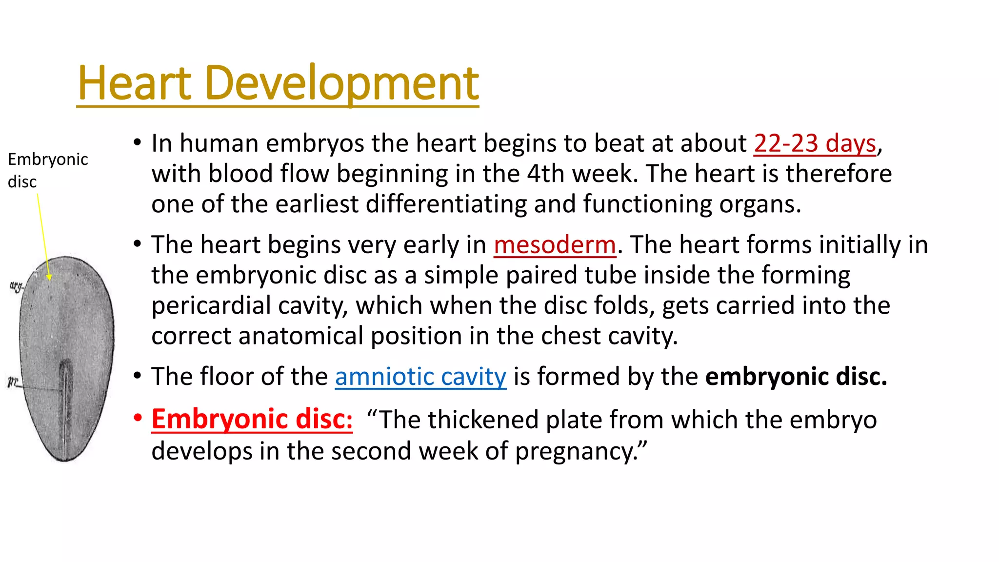
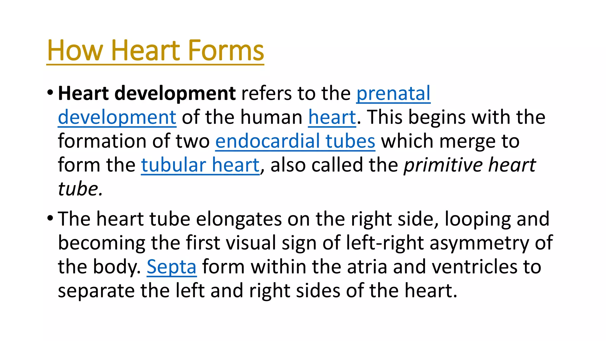
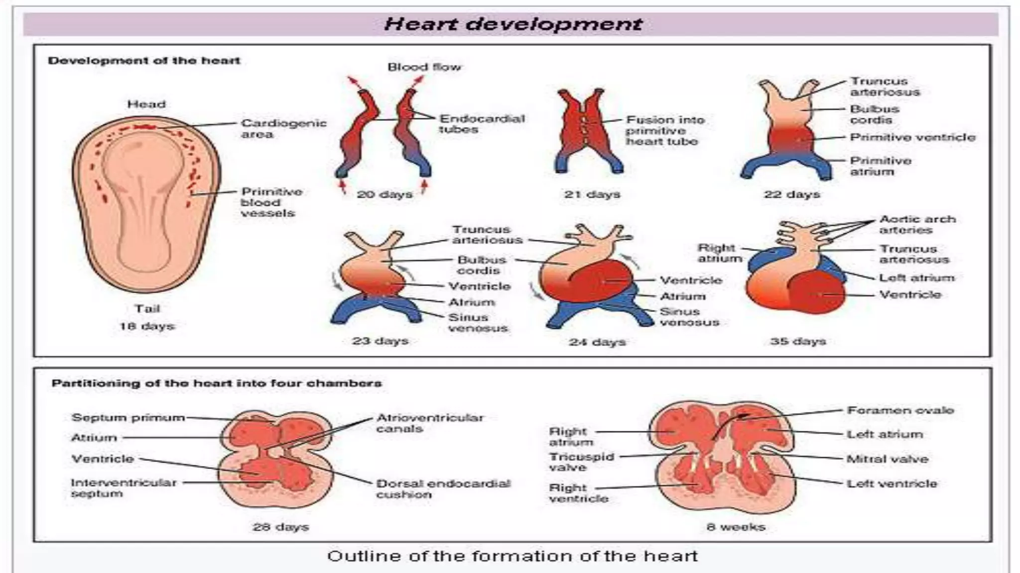
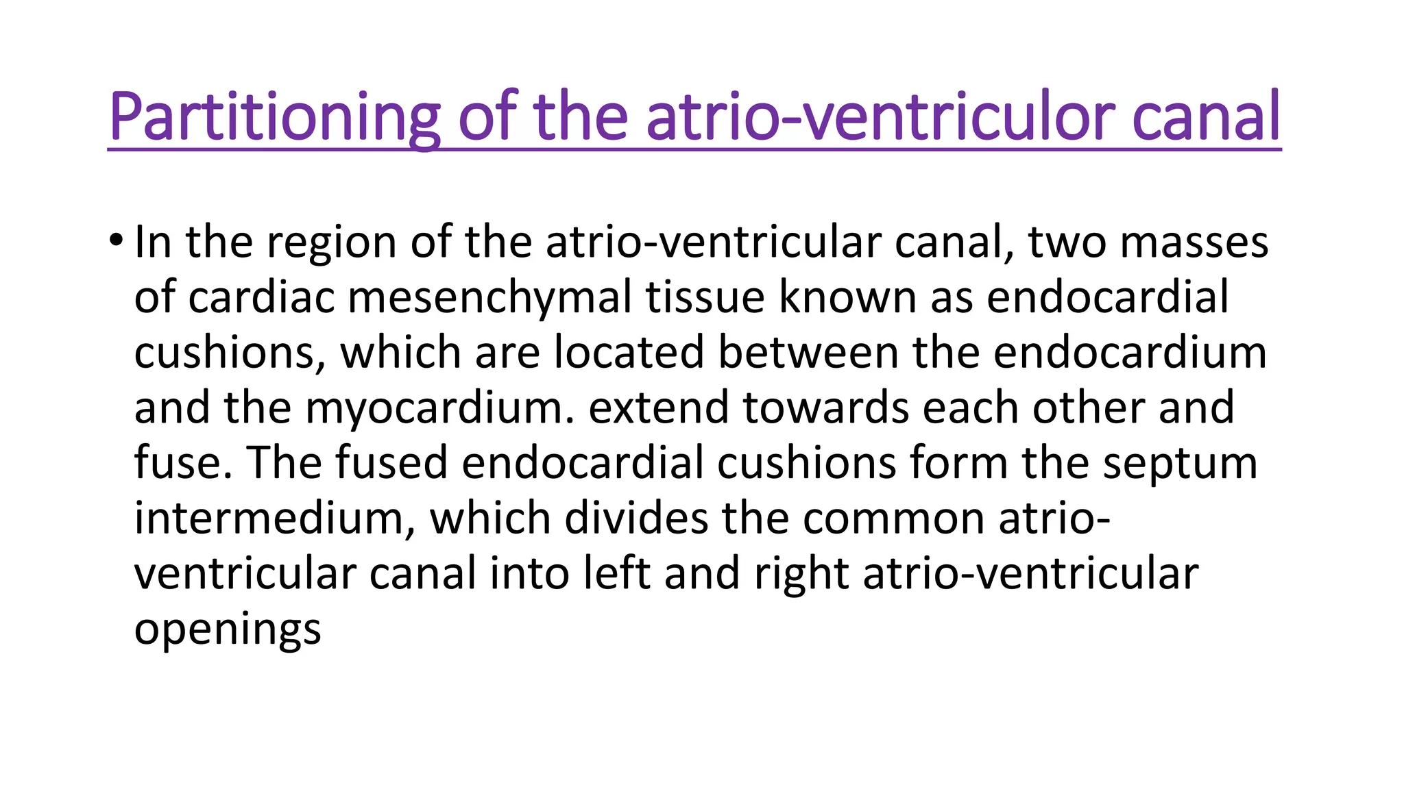
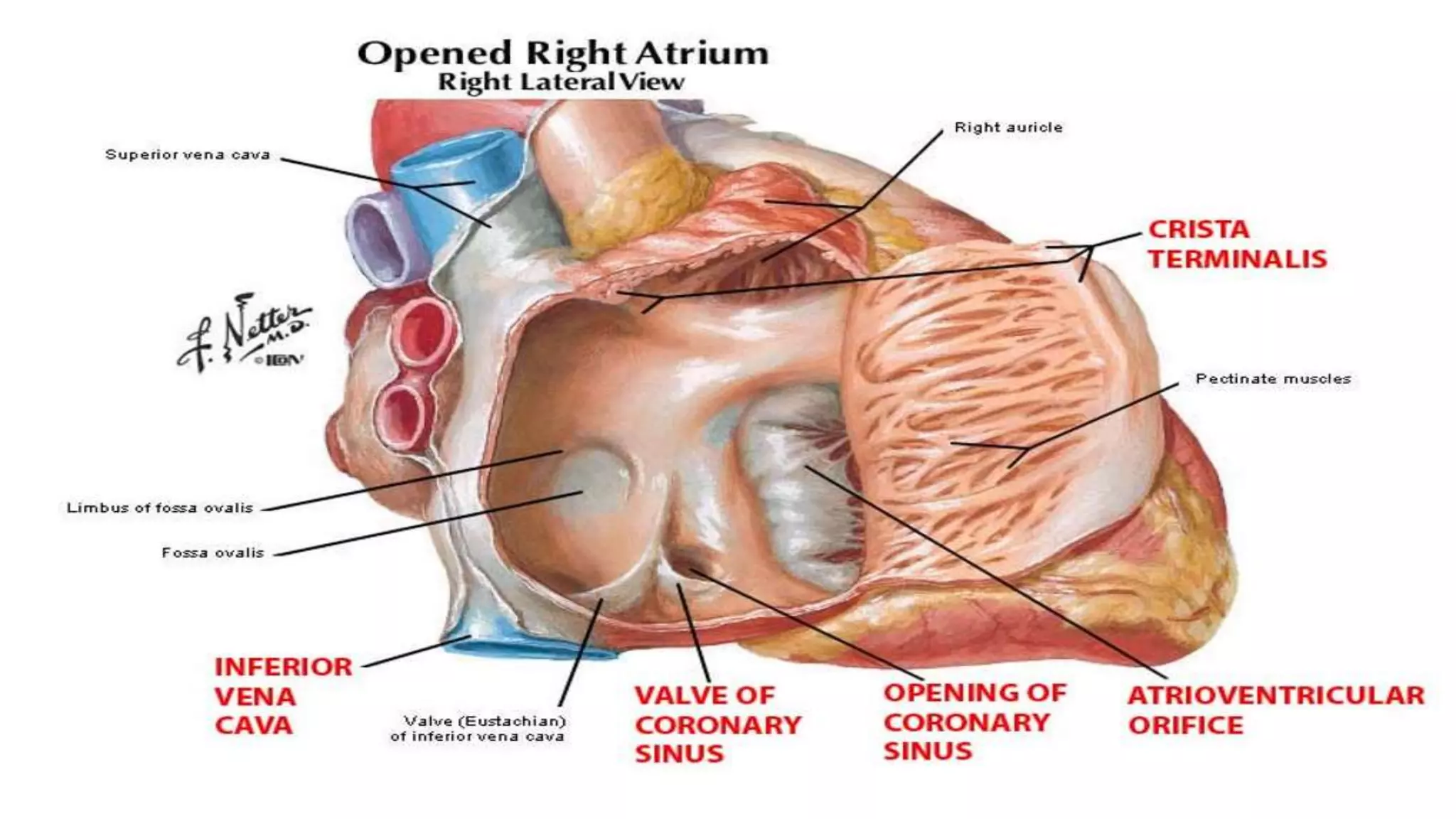
Embryonic Normal
Development of Heart
Submitted to :DR. RIAZ HUSSAIN PASHA
Submitted by:
ATTIQUE HASSAN
ZIKRIA MANZOOR
M.ABDULLAH
DVM 2nd semester(EVENING)
Some important terms
• Embryo :
“an unborn or unhatched offspring in the
process of development, in particular a human offspring
during the period from approximately the second to the
eighth week after fertilization (after which it is usually
termed a fetus).”
• Heart:
“ Heart is pumping organ. Which pumps and distribute
blood throughout the body.”
Importance of cardiac system
• In the early stages of embryological development, the
respiratory, excretory and nutritional requirements of
the embryo are provided through simple diffusion.
• As the mammalian embryo increases in size, diffusion is
inadequate for its nutritional, respiratory and excretory
needs. Because of its increasing size and complexity, the
developing embryo requires a system for delivering
oxygen and nutrients to its tissues, and for transporting
its waste products. These requirements are supplied by
the cardiovascular system.
Heart Development
• In human embryos the heart begins to beat at about 22-23 days,
with blood flow beginning in the 4th week. The heart is therefore
one of the earliest differentiating and functioning organs.
• The heart begins very early in mesoderm. The heart forms initially in
the embryonic disc as a simple paired tube inside the forming
pericardial cavity, which when the disc folds, gets carried into the
correct anatomical position in the chest cavity.
• The floor of the amniotic cavity is formed by the embryonic disc.
• Embryonic disc: “The thickened plate from which the embryo
develops in the second week of pregnancy.”
Embryonic
disc
How Heart Forms
• Heart development refers to the prenatal
development of the human heart. This begins with the
formation of two endocardial tubes which merge to
form the tubular heart, also called the primitive heart
tube.
• The heart tube elongates on the right side, looping and
becoming the first visual sign of left-right asymmetry of
the body. Septa form within the atria and ventricles to
separate the left and right sides of the heart.
Formation of heart chambers
• Partitions which form in the primordial mammalian
heart gradually convert the single pulsating cardiac tube
into a complex four-chambered organ. Although
formation of cardiac septa takes place at approximately
the same time, for descriptive purposes their formation
is described as if they were separate events. The foetal
heart continues to function effectively as these ongoing
major structural changes occur.
Partitioning of the atrio-ventriculor canal
• In the region of the atrio-ventricular canal, two masses
of cardiac mesenchymal tissue known as endocardial
cushions, which are located between the endocardium
and the myocardium. extend towards each other and
fuse. The fused endocardial cushions form the septum
intermedium, which divides the common atrio-
ventricular canal into left and right atrio-ventricular
openings
Final form of Right Atrium
• The terminal portion of the pectinate muscles
form crista terminalis.
• sulcus terminalis: A sulcus which externally
represent the crista terminalis.
• The regressing left horn of the sinus venosus
contributes to the formation of the coronary
venous sinus which opens into the right atrium.
• Venous Sinus also called as Sinus venorum.
Left Atrium
• During the embryonic development the common
pulmonary vein become incorporated into left atrium.
• Structures inside left atrium are similar to those of right
atrium.
Formation of the left and right ventricles
• The dilated portion of the bulbus cordis and the
embryonic ventricle form a common chamber.
•BULBUS CORDIS:
“In the embryo the segments of the
heart tube destined to be a partitioned and to form the
two ventricular outflow segments to the aorta on the
left to the pulmonary trunk on the right.”
Conti
• As the walls of the ventricle and bulbus cordis increase
in thickness diverticulation of their inner surfaces
imparts a trahecular appearance to the myocardium.
• At this stage, the embryonic ventricle can be considered
as the primitive left ventricle and the dilated bulbus
cordis as the primitive right ventricle.
• As the inter-ventricular sulcus deepens and the walls of
the expanding ventricles meet medially at the sulcus.
the walls become apposed and fuse, contributing to the
elongation of the intervenlricular septum.
Final form of right ventricle
• Conus arteriosum:
“ A doom shaped structure formed by
right ventricle at cranial border of heart.”
• Trabeculae carnea:
“Rough wall of ventricle which bears
muscular ridges and bands.”
• Moderate bands:
“Extend from septum to opposite wall.”
Final form of left ventricle
• Trabeculae carnea:
“Rough wall of ventricle which
bears muscular ridges and bands.”
• Moderate bands:
“Extend from septum to opposite
wall.”
• Ventricular septum:
“ It is a portioning which separate
the cavities of two ventricles.”
Formation of cardiac valves
• The aortic and pulmonary valves, which are necessary
for prevention of backflow of blood into the left and
right ventricles, arise from three swellings of sub-
endothelial mesenchymal tissue at the origins of the
aortic and pulmonary trunks.
• Mesenchymal tissue:
Type of connective tissue found
in embryonic life only. Composed of ground substance
And a few fibers/cells.
Heart Walls
• The main walls of the heart are formed between day 27 and 37
of the development of the early embryo.
• The myocardium thickens and secretes a thick layer of
rich extracellular matrix containing hyaluronic acid which separates
the endothelium. Then mesothelial cells form the pericardium and
migrate to form most of the epicardium. Then the heart tube is
formed by the endocardium, which is the inner endothelial lining of
the heart, and the myocardial muscle wall which is the epicardium
that covers the outside of the tube.
Embryonic normal development of heart

Embryonic normal development of heart

  • 1.
    Embryonic Normal Development ofHeart Submitted to :DR. RIAZ HUSSAIN PASHA
  • 2.
    Submitted by: ATTIQUE HASSAN ZIKRIAMANZOOR M.ABDULLAH DVM 2nd semester(EVENING)
  • 3.
    Some important terms •Embryo : “an unborn or unhatched offspring in the process of development, in particular a human offspring during the period from approximately the second to the eighth week after fertilization (after which it is usually termed a fetus).” • Heart: “ Heart is pumping organ. Which pumps and distribute blood throughout the body.”
  • 4.
    Importance of cardiacsystem • In the early stages of embryological development, the respiratory, excretory and nutritional requirements of the embryo are provided through simple diffusion. • As the mammalian embryo increases in size, diffusion is inadequate for its nutritional, respiratory and excretory needs. Because of its increasing size and complexity, the developing embryo requires a system for delivering oxygen and nutrients to its tissues, and for transporting its waste products. These requirements are supplied by the cardiovascular system.
  • 5.
    Heart Development • Inhuman embryos the heart begins to beat at about 22-23 days, with blood flow beginning in the 4th week. The heart is therefore one of the earliest differentiating and functioning organs. • The heart begins very early in mesoderm. The heart forms initially in the embryonic disc as a simple paired tube inside the forming pericardial cavity, which when the disc folds, gets carried into the correct anatomical position in the chest cavity. • The floor of the amniotic cavity is formed by the embryonic disc. • Embryonic disc: “The thickened plate from which the embryo develops in the second week of pregnancy.” Embryonic disc
  • 6.
    How Heart Forms •Heart development refers to the prenatal development of the human heart. This begins with the formation of two endocardial tubes which merge to form the tubular heart, also called the primitive heart tube. • The heart tube elongates on the right side, looping and becoming the first visual sign of left-right asymmetry of the body. Septa form within the atria and ventricles to separate the left and right sides of the heart.
  • 8.
    Formation of heartchambers • Partitions which form in the primordial mammalian heart gradually convert the single pulsating cardiac tube into a complex four-chambered organ. Although formation of cardiac septa takes place at approximately the same time, for descriptive purposes their formation is described as if they were separate events. The foetal heart continues to function effectively as these ongoing major structural changes occur.
  • 9.
    Partitioning of theatrio-ventriculor canal • In the region of the atrio-ventricular canal, two masses of cardiac mesenchymal tissue known as endocardial cushions, which are located between the endocardium and the myocardium. extend towards each other and fuse. The fused endocardial cushions form the septum intermedium, which divides the common atrio- ventricular canal into left and right atrio-ventricular openings
  • 11.
    Final form ofRight Atrium • The terminal portion of the pectinate muscles form crista terminalis. • sulcus terminalis: A sulcus which externally represent the crista terminalis. • The regressing left horn of the sinus venosus contributes to the formation of the coronary venous sinus which opens into the right atrium. • Venous Sinus also called as Sinus venorum.
  • 13.
    Left Atrium • Duringthe embryonic development the common pulmonary vein become incorporated into left atrium. • Structures inside left atrium are similar to those of right atrium.
  • 14.
    Formation of theleft and right ventricles • The dilated portion of the bulbus cordis and the embryonic ventricle form a common chamber. •BULBUS CORDIS: “In the embryo the segments of the heart tube destined to be a partitioned and to form the two ventricular outflow segments to the aorta on the left to the pulmonary trunk on the right.”
  • 15.
    Conti • As thewalls of the ventricle and bulbus cordis increase in thickness diverticulation of their inner surfaces imparts a trahecular appearance to the myocardium. • At this stage, the embryonic ventricle can be considered as the primitive left ventricle and the dilated bulbus cordis as the primitive right ventricle. • As the inter-ventricular sulcus deepens and the walls of the expanding ventricles meet medially at the sulcus. the walls become apposed and fuse, contributing to the elongation of the intervenlricular septum.
  • 16.
    Final form ofright ventricle • Conus arteriosum: “ A doom shaped structure formed by right ventricle at cranial border of heart.” • Trabeculae carnea: “Rough wall of ventricle which bears muscular ridges and bands.” • Moderate bands: “Extend from septum to opposite wall.”
  • 19.
    Final form ofleft ventricle • Trabeculae carnea: “Rough wall of ventricle which bears muscular ridges and bands.” • Moderate bands: “Extend from septum to opposite wall.” • Ventricular septum: “ It is a portioning which separate the cavities of two ventricles.”
  • 21.
    Formation of cardiacvalves • The aortic and pulmonary valves, which are necessary for prevention of backflow of blood into the left and right ventricles, arise from three swellings of sub- endothelial mesenchymal tissue at the origins of the aortic and pulmonary trunks. • Mesenchymal tissue: Type of connective tissue found in embryonic life only. Composed of ground substance And a few fibers/cells.
  • 23.
    Heart Walls • Themain walls of the heart are formed between day 27 and 37 of the development of the early embryo. • The myocardium thickens and secretes a thick layer of rich extracellular matrix containing hyaluronic acid which separates the endothelium. Then mesothelial cells form the pericardium and migrate to form most of the epicardium. Then the heart tube is formed by the endocardium, which is the inner endothelial lining of the heart, and the myocardial muscle wall which is the epicardium that covers the outside of the tube.