
Recommended
More Related Content
What's hot
What's hot (20)
Similar to Hemiplegic Gait Rehabilitation Techniques
Similar to Hemiplegic Gait Rehabilitation Techniques (20)
More from Ashik Dhakal
More from Ashik Dhakal (20)
Recently uploaded
Recently uploaded (20)
Hemiplegic Gait Rehabilitation Techniques
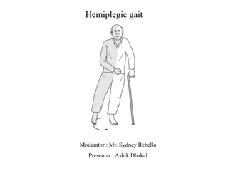
- 1. Hemiplegic gait Moderator : Mr. Sydney Rebello Presentar : Ashik Dhakal
- 2. Normal gait
- 3. Terminologies used in gait • Step length • Stride length • Step width • Cadence • Foot angle
- 4. • Step time • Stride time • Swing time • Double support time
- 6. Hemiplegia • Paralysis of one side of the body.
- 8. Gait deviations commonly seen following stroke Stance Phase Trunk/pelvis Unawareness of affected side: poor proprioception Forward trunk: Weak hip extension, Flexion contracture Hip Poor hip position Trendelenburg limp: weak abductors Scissoring: spastic adductors
- 9. Stance Phase Knee Increase knee flexion Flexion contracture, Weak hip and knee extensors Ankle dorsiflexion range past neutral Weakness in extension pattern or in selective motion of hip and knee extensors and plantarflexors
- 10. Stance Phase Knee Decreased knee flexion (or hyper extension) Insufficient active tension with knee flexors. Impaired proprioception: Knee wobbles or snaps back into recurvatum Severe spasticity in quadriceps Weak knee extensors: compensatory locking of knee in hyperextension
- 11. Stance Phase Ankle/foot Equinus gait (heel does not touch the ground: spasticity or contractures of gastrocnemius soleus Varus foot (patient bears weight on the lateral surface of the foot) Lack of dorsiflexion range on the affected side (approximately 10˙ is needed)
- 12. Swing Phase Trunk/pelvis Insufficient forward pelvic rotation (pelvic retraction): weak abdominal muscles Inclination to sound side for foot clearance: weakness of flexor muscles Hip Inadequate flexion: Weak hip flexors, Poor proprioception.
- 13. Swing Phase Knee Inadequate knee flexion Inadequate hip flexion and poor foot clearance Exaggerated but delayed knee flexion Strong flexor synergy Inadequate knee extension at weight acceptance Spastic hamstrings
- 14. Swing Phase Ankle/foot Persistent equinus and/or equinovarus: Plantarflexor contracture or spasticity, Weak dorsiflexors, Delayed contraction of dorsiflexors, Toe drags during midswing Exaggerated dorsiflexion: strong flexor synergy pattern
- 15. Video
- 17. Description of gait • Hip hike • Circumduction of the leg • Reduced hip and knee flexion • Poor dorsiflexion — foot drop — toe first or flat foot placement • Decreased weight shift towards affected side
- 18. 11 • Broader base of support • Increased double stance time • Shorter steps and stride length • Slower gait speed • Decreased walking efficacy • Poor endurance,
- 19. Weak or imbalanced muscles • Kinematic deviation occurs as a result of the inability to appropriately activate muscles, as well as, from adaptive muscle shortening. • Other than weakness, the person may experience clonus, spasticity, exaggerated deep tendon reflexes, and decreased endurance. • Weakened muscles include hip abductors, flexors, knee flexors, and weak ankle dorsiflexors.
- 20. Functional task affected • Difficulty maintaining balance due to limb weakness. • Inability to properly shift their body weight. • Staggering and stumbling. • Difficulty performing activities of daily living. (barthel)
- 21. Physical therapy intervention • Patient aimed at improving balance and restoring coordination. • Tx is focused on • Symmetrical weight bearing • Weight shifting • Step training • Heel strike • Single leg stance • Push off
- 22. Treatment approaches : • Conventional physiotherapy • Neuro physiological techniques • Functional electrical stimulation • Robot assisted • Brain computer interface • Motor imagery • Mirror therapy
- 23. 1. Neurophysiological techniques • Physiotherapist supports the patient's movement patterns, acting as an active participant and decision maker, so the patient acts as relatively passive recipient. • Most commonly used : • Bobath Method is the most widely accepted treatment. • This method consists on trying to inhibit increased muscle tone (spasticity) by passive mobilization associated with tactile and proprioceptive stimuli. • During exercise, pathologic synergies or reflex activities are not stimulated.
- 24. • Brunnstrom method is also well known but practiced less commonly. • It is different to the Bobath strategy and focus on raising pathologic synergies in order to achieve a normal movement pattern and promotes return of voluntary movement through reflex facilitation and sensory stimulation. • Rood technique uses peripheral input (sensory stimulation) to facilitate movement and postural responses in the same automatic way as they normally occur. • Johnson method assumes that damaged reflex mechanisms responsible for spasticity are the leading cause of posture and movement impairment. These pathological reflexes are controlled through positioning and splinting.
- 25. 2. Functional Electrical Stimulation • FES consists delivering an electric current through electrodes to the muscles. • The current elicits action potentials in the peripheral nerves of axonal branches and thus generates muscle contractions. • This technique is found effective to improve gait performance in hemiplegic subjects and thought to give better results if used along with BWS.
- 26. • Video
- 27. 3. Robotic devices • These devices provide safe, intensive and task-oriented rehabilitation with a minimal physical assistance required to walk — reduces health care costs. • It also provides kinematic and kinetic data in order to control intensity of practice, assess changes and measure motor impairments. • Robotic systems for gait recovery have been projected as simple electromechanical assistances for walking, such as the treadmill with body weight support (BWS) , as end- effectors, such as the Gait Trainer (Reha-Technologies, Germany, GT), lokomat.
- 29. 4. Motor imagery • Imagining of an action without its physical execution; using all of the senses. • It is an active process during which the representation of an action is internally reproduced within working memory without any overt (observable) output.
- 30. 5. Brain Computer Interfaces • BCIs establish a direct link between a brain and a computer without any use of peripheral nerves or muscles enabling communication and control without any motor output by the user. • The users brain activity extracts specific features from brain signals that reflect the intent of subjects and transform them into action.
- 31. • Video
- 32. 6. Mirror therapy • Type of motor imagery where the patient moves his/her unaffected limb while watching the movement in a mirror. • This in turn sends a visual stimulus to the brain to promote movement in the affected limb.
- 33. Outcome measures • Functional ambulation profile • Dynamic gait index • Stroke impact scale • Gait abnormality rating scale • Functional independence measures • Functional gait assessment • Timed up and go test • 6 min walk test • 2 min walk test • 10 meter walk test
- 39. Gait abnormality rating scale
- 44. Thank you