
Recommended
More Related Content
What's hot
What's hot (20)
Similar to CANCER OF THE ORAL CAVITY.pdf
Similar to CANCER OF THE ORAL CAVITY.pdf (20)
More from AnushriSrivastav
Recently uploaded
Recently uploaded (20)
CANCER OF THE ORAL CAVITY.pdf
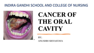
- 1. CANCER OF THE ORAL CAVITY BY; ANUSHRI SRIVASTAVA INDIRA GANDHI SCHOOL AND COLLEGE OF NURSING
- 2. INTRODUCTION Cancer of the oral cavity are associated with the use of tobacco and alcohol as they seems to have a synergistic carcinogenic effect. More common after the age of 35 years, with 65 years behind the average age of diagnosis. Oral cavity cancer is two times more common in men than in women. The common sites of oral malignant lesions are lower lip (mostly), lateral border and undersurface of tongue, labial commissure and buccal mucosa.
- 3. DEFINITION • According to NATIONAL CANCER INSTITUTE, ‘Oral cancer is defined as the cancer that forms in tissues of the oral cavity (the mouth) or the oropharynx (the part of the throat at the back of the mouth).’ • According to FDI World Dental Federation, ‘Oral cancer is a type of head and neck cancer and is any cancerous tissue growth located in the oral cavity.’ • Oral cancer is defined as the abnormal uncontrolled growth of cells in the oral cavity, characterized by lesions, thickened mass and dysphagia.
- 4. TYPES OF ORAL CANCER There are two types of oral cancer:- • Oral cavity cancer (cancer that starts in mouth) • Oropharyngeal cancer (cancer that starts in throat behind the mouth) Head and Neck Squamous Cell Carcinoma (HNSCC) is a term used for the cancers of oral cavity, pharynx and larynx, accounts 90% malignant tumors.
- 5. ETIOLOGY • The exact cause is unknown • Long term use of tobacco • History of frequent alcohol consumption • Prolong sunlight exposure may lead to lip cancer • Irritation from the pipe stem resting on the lip in Pipe smokers • HPV contributes 25% of oral cancer cases • Multiple oral sex partners • Low serum Vitamin A, C and E levels • Smoked meat ingestion • Poor oral hygiene • Recurrent herpetic lesion may lead to lip cancer • Immunosuppression • Syphilis • Chronic irritation (jagged tooth, ill fitting prosthesis, chemical or mechanical irritants)
- 6. TNM CLASSIFICATION OF ORAL CANCER • T- Primary tumor TX Primary tumor cannot be assessed T0 No evidence of primary tumor Tis Carcinoma in situ T1 Tumor 2 cm or less in greatest dimension T2 Tumor more than 2 cm but not more than 4 cm in greatest dimension T3 Tumor more than 4 cm in greatest dimension T4a Tumor invades through cortical bone, into deep/ extrinsic muscle of tongue, maxillary sinus, or skin of face T4b Tumor invades masticator space, pterygoid plates, or skull base, or encases internal carotid artery
- 7. TNM CLASSIFICATION OF ORAL CANCER • N- Regional Lymph nodes NX Regional lymph node cannot be assessed N0 No regional lymph node metastasis N1 Metastasis in a single ipsilateral lymph node, 3 cm or less in greatest dimension N2 Metastasis in lymph node, more than 3 cm but not more than 6 cm in greatest dimension N3 Metastasis in a lymph node more than 6 cm in greatest dimension • M- Distant Metastasis M0 No distant metastasis M1 Distant metastasis
- 8. CLINICAL MANIFESTATION Lip Cancer • Indurated • Painless ulcer Tongue Cancer • Ulcer or area of thickening • Soreness or pain • Increased salivation • Slurred speech • Dysphagia • Toothache • Earache(later sign) This Photo by Unknown Author is licensed under CC BY
- 9. Continued… Oral Cavity Cancer • Leukoplakia Also known as Smoker's patch, white patch on the mouth mucosa or tongue, often considered as a precancerous lesion and less than 15% chance to transform into malignant cells. The patch becomes keratinized (hard and leathery) and it sometimes described as hyperkeratosis. Leukoplakia results from chronic irritation, especially from smoking.
- 10. Continued… • Erythroplakia Also known as Erythroplasia, is a red velvety pouch on the mouth or tongue, also a precancerous lesion. More than 50% of cases of Erythroplakia may progress to squamous cell carcinoma.
- 11. Continued… . • Ulceration • Rough area pain • Enlarged cervical lymph node • Sore spot • Dysphagia • A lump or thickening in the cheek • Asymptomatic neck mass Nonspecific signs • Chronic sore throat • Sore mouth • Voice changes Late signs • A sore throat or a feeling that something is stuck • Difficulty chewing and speaking
- 12. DIAGNOSTIC EVALUATION History Collection Physical Examination Biopsy Oral Exfoliative cytology Toluidine Blue Test Computed Tomography scan Magnetic Resonance Imaging Positron Emission Test
- 13. Medical Management • Chemotherapy 5 FU, Methotrexate, Cisplatin, Carboplatin, Bleomycin, Cetuximab • Radiation therapy • Brachytherapy with implantation of radioactive seeds to the early stage of cancer • Nutritional therapy • Palliative treatment
- 14. SURGICAL MANAGEMENT Most effective on early stages and the type of surgery depend on location and extent of tumor. • Partial Mandibulectomy • Hemiglossectomy • Glossectomy • Resection of buccal mucosa and floor of mouth • Reconstructive surgery • Radial Neck Dissection It includes wide excision of the primary lesion with the removal of lymph nodes, the deep cervical lymph nodes, and their lymphatic channels and other structures may also be removed or transected depending on the extent of primary lesion.
- 15. Complication Mucositis Xerostomia Swelling Stomatitis Dysphagia
- 16. Nursing Management • Assess the patient condition • Provide comfortable position • Monitor vital signs • Monitor intake and output chart • Administer medication as prescribed by physician • Administer analgesic as prescribed by physician • Placement of Percutaneous Endoscopic Gastrostomy considered before radiation treatment or surgery • Parenteral Fluids are given for first 24 to 48 hours. After this time enteral nutrition via NG, Gastrostomy or Nasointestinal tube is provided. • Frequent suctioning of oral cavity at time when swallowing becomes difficult • Observe for feeding tolerance and adjust the amount, time and formula if nausea, vomiting, diarrhea or distension occurs • Give small amount of water when the patient can swallow • Observe for choking • Suctioning is also necessary to prevent aspiration
- 17. NURSING DIAGNOSIS • Imbalanced nutrition less than body requirement related to difficulty in swallowing as evidence by weight loss • Chronic pain related to tumor as evidence by patient facial expression • Ineffective health maintenance related to lack of knowledge of disease process as evidence by poor oral hygiene.
- 18. bibliography • Bucher, Heitkemper, Dirksen. A Textbook of Medical Surgical Nursing: Assessment and Management of Clinical Problems. Edition IX. 3251 Riverport Lane, St Loius, Missouri 63043.ELSEVIER publisher; Page number 929-31 • https://screening.iarc.fr/atlasoralclassiftnm.php • https://www.cancer.gov/search/results?swKeyword=Oral+cancer • https://www.fdiworlddental.org/oral- cancer#:~:text=It%20includes%20malignancy%20of%20the,highly%20 lethal%2C%20incapacitating%20and%20disfiguring
- 19. . thanking you!