SlideShare a Scribd company logo
1 of 243
Download to read offline
Video 13:
Introduction to the
Endomembrane System
Gergely, Z.R., Martinez, D.E., Donohoe, B.S. et al. J of Biol Res-Thessaloniki
25, 15 (2018). https://doi.org/10.1186/s40709-018-0086-2
The Endomembrane
System Derived from the
cytoplasmic membrane
and forming a
coordinated unit
Include the ER, Golgi,
endosomes,
lysosomes, and
vacuoles (NOT the
mitochondria and
chloroplasts)
Allow transportation of
proteins, lipids, and
complex
polysaccharides
Transport System
Transport vesicles
allow movement
between cellular
compartments
Travel along the
cytoskeleton mediated
by motor proteins
Sorting signals within
amino acid sequences
or oligosaccharides
allow targeting to
appropriate cellular
compartment
Transport Pathways
All begin with the the
biosynthetic pathway
Proteins produced in
RER
●
Vesicles released
from RER fuse with
the cis Golgi
complex,
undergoing further
modifications
●
Transport vesicles
released from the
trans Golgi
Karp’s Cell & Molecular Biology (2020) Fig 8.2
Transport Pathways
Secretory pathways
result in exocytosis and
may be constitutive
(continuous) or
regulated (require
signal for release)
Materials are also
imported through
endocytosis and
digested within the
lysosome
Karp’s Cell & Molecular Biology (2020) Fig 8.2
Pulse-Chase &
Autoradiography
Monitor movement of silver grains (in
red) through secretory pathway
Karp’s Cell & Molecular Biology (2020) Fig 8.3
(Green)
Fluorescent
Proteins
Karp’s Cell & Molecular Biology (2020) Fig 8.4
Temperature-sensitive mutation
prevents movement of VSVG from ER
at 40OC
Other Experimental
Approaches
Subcellular fractionation (differential centrifugation)
●
Idenification of fraction containing particular enzymes or
functions
●
Now extend cell-free systems to include synthetic
liposomes
Effects of genetic mutations
●
Introduction of mutations in identify function of particular
gene
●
Newer methods include RNA interference and CRISPR
Video 14:
The Endoplasmic
Reticulum
https://www.sigmaaldrich.com/life-science/cell-biology/cell-biology-
products.html?TablePage=106514528
The Endoplasmic
Reticulum
Likely from invagination
of cytoplasmic
membrane
Differentiated into rough
and smooth due
to presence
or absence of
bound ribosomes
on the
cytoplasmic
surface
ER Similarities
RER and SER are joined, having a
continual luminal space which
allows for free diffusion
Number of
proteins are
common to both
RER & SER
Both synthesize
some shared
lipids and
cholesterol
Karp’s Cell & Molecular Biology Fig. 8.10
Highly curved, tubular network with large numbers of
reticulons (cause bending of the membranes)
Karp’s Cell & Molecular Biology Fig. 8.10
Smooth Endoplasmic
Reticulum
Detoxification of drugs
such as ethanol and
barbituates in the liver
by oxygenases
Ca2+ sequestering in
muscle cells (called
sarcoplasmic reticulum)
Synthesis of steroidal hormones in
endocrine cells
Continuous with the outer nuclear membrane and
composed of flattened sacs (cisternae) connected by
helical “ramps”
Karp’s Cell & Molecular Biology Fig. 8.10
Rough Endoplasmic
Reticulum
Increased in secretory cells
(acinar cells of pancreas &
mucous cells in digestive
tract)
The site of synthesis of the
majority of phospholipids for
inclusion in cellular
membranes
Karp’s Cell & Molecular Biology Fig. 8.10
Rough Endoplasmic
Reticulum
Undergo co-translational
translocation from bound
ribosomes to ER lumen
Synthesizes carbohydrate
chains and initiates N-
linked glycosylation of
proteins
Site of synthesis of approx. 1/3 of cellular proteins: those
destined for secretion or inclusion in endomembrane
organelles, integral
membrane proteins
Membrane-bound or Free
Ribosome?
Remainder of proteins are synthesized by free
(cytoplasmic) ribosomes
●
Cytosolic proteins
●
Peripheral membrane proteins for cytosolic surface)
●
Nuclear proteins (contain nuclear localization signals)
●
Proteins for other organelles (lysosomes, mitochondria,
chloroplasts)
All translation is initiated on free ribosomes
Proteins which should go to ER contain an N-terminal
signal sequence at N-terminus: results in ribosome
binding to ER surface and co-translational translocation
Co-translational Translocation
SRP binds signal sequence and guides ribosome to
translocon channel protein by binding to the SRP
receptor
SRP = signal recognition particle
Karp’s Cell & Molecular Biology Fig. 8.13
Polypeptide binding to translocon opens
“plug” to allow polypeptide to enter ER
Signal removal
and folding
Synthesis of Integral Proteins
Hydrophobic “stop-transfer” sequence blocks
translocation into ER lumen
The next steps dependent upon location of positive and
negative charges at ends of the transmembrane region
Karp’s Cell & Molecular Biology Fig. 8.14
●
If N-terminus side is
negative, the lateral gate
of the translocon allows
the transmembrane
region to migrate into the
membrane
●
Results in C-terminus on
cytosolic side
Synthesis of Integral Proteins
●
If N-terminus side is positive, the translocon first
reorients the transmembrane region, which is then able
to migrate through the lateral gate
●
This results in the N-terminus being located on the
cytosolic side
●
Multi-pass polypeptides utilize translocon many times in
an antiparallel
manner
●
Some proteins
utilize
alternative
pathways
Karp’s Cell & Molecular Biology Fig. 8.14
Video 15:
Protein Processing
https://hms.harvard.edu/news/folding-revolution
Protein
Processing
Many secretory cells have a
polarized or directional structure
Reflects movement of
polypeptides from synthesis in the
RER, processing in the RER &
Golgi, and secretion via transport
vesicles or secretory granules
Processing is a highly regulated
multi-step process required for
the production of proteins
(biologically-active form)
Karp’s Cell & Molecular Biology Fig 8.12
Protein
Processing
Removal of signal peptide by
signal peptidase
Protein folding & molecular
chaperones
Formation and shuffling of
disulfide bonds by disulfide
isomerase
Addition of carbohydrates by
oligosaccharyltransferase
Protein Folding
Occurs spontaneously
during translation
Folding
directed by
amino acid
sequence and
the production
of favorable
interactions
Molecular Chaperones
Chaperones
redirect back to
proper folding
pathway
Membrane
Asymmetry
Cellular membranes “grow”
rather than being produced
through de novo synthesis
Protein and lipid
composition are altered as
membranes are moved from
one compartment to the next
Through the means of
membrane fusion, specific
asymmetry of membrane is
maintained
Karp’s Cell & Molecular Biology Fig 8.15
Altering Lipid
Composition
Karp’s Cell & Molecular Biology Fig 8.16
Enzymatic
conversion
Preferential
inclusion of
specific lipids
within vesicles
Movement from one
compartment to another by lipid
transfer proteins
Newly synthesized
lipids are inserted
in cytosolic leaflet,
but may be moved
by flippases
Glycosylation
Majority of proteins produced in the RER become
glycoproteins: starts in the RER & completed in the Golgi
Addition of sugars to oligosaccaride
catalyzed by specific
glycosyltransferases
●
Transfer a monosaccharide
from a nucleotide sugar
Oligosaccharide first synthesized
on dolichol phosphate
Transferred to protein by
oligosaccharyltransferase
enzyme
Glycosylation
Flippases
catalyze altered
orientation
Karp’s Cell & Molecular Biology Fig 8.17
Modification & Quality Control
Following
acquisition,
two
terminal
glucose
residues
removed
Bound by calnexin or
calreticulin (chaperone)
Final glucose removed
Karp’s Cell & Molecular Biology Fig 8.18
Modification & Quality Control
Conformation-sensing
enzyme (UGGT) determines
whether it is misfolded based
upon whether hydrophobic
regions are exposed
Karp’s Cell & Molecular Biology Fig 8.18
Modification & Quality Control
Eventually, a mannose is
removed & it is
transported to proteasome
for destruction
Karp’s Cell & Molecular Biology Fig 8.18
If it is
misfolded,
glucose
residue
again
added and
cycle
repeated
Transport from ER to Golgi
Special exit sites on edges of ER lack ribosomes and are
sites of vesicle budding
These small vesicles fuse together to form larger transport
vesicles and interconnected tubules
Vesicular-tubule carriers (VTCs) move towards the Golgi
complex and fuse with the cis-Golgi network
●
VTC transport occurs via microtubule tracks
The Golgi will allow for further modification of proteins and
sorting for final transport
Video 16:
Golgi Complex &
Transport
https://www.cancer.gov/about-nci/organization/dcb/research-programs
Golgi Complex
Tubule networks on cis and trans faces are involved in
sorting of proteins:
●
cis will send back to ER or on to the cisternae
●
trans will sort to plasma membrane or intracellular
organelles
Golgi Complex
Golgi cisternae are involved in the sequential modification
of the proteins synthesized by the ER
●
Differential composition in each compartment
Osmium
tetroxide
stains
unsaturated
fatty acids
in the Cis
Cisternae
Mannosidase II enzyme
in the Medial Cisternae
Nucleoside
diphosphatase
Enzyme in the
Trans
Cisternae
Golgi Complex
Alters the N-linked oligosaccharides added within the ER
as they progress through the cisternae
Assembles O-linked oligosaccarides
Majority of
complex
polysaccharide
assembly
Different
enzymes
found in each
compartment
Glycosylation
Enzymes involved in glycosylation are integral proteins
with active sites facing the interior of the Golgi
Those involved in adding sugars are transferases
Karp’s Cell and Molecular Biology (2020) Fig. 8.23
Vesicular Transport Model
Popular in the 1980s & 90s
Vesicles move from one
cisternae to the next, while
the cisternae themselves are
“static”
In vitro evidence of budding
and fusing vesicles
Believed to occur in vivo in
an anterograde direction
Karp’s Cell and Molecular Biology (2020)
Fig. 8.24
Cisternal Maturation Model
First & currently accepted
Cisternae contents change
over time and develop in an
anterograde direction
Vesicles travel in retrograde:
are they returning enzymes?
Could both potentially be
occurring?
Karp’s Cell and Molecular Biology (2020)
Fig. 8.24
Transport from ER
to Golgi:Revisited
ERGIC = ER Golgi intermediate
compartment
Comprised of the larger vesicles & tubules
formed through the fusion of transport
vesicles
3 kinds of coated transport vesicles:
●
COPI = posterograde from Golgi
●
COPII = anterograde from ER
●
Clathrin-coated = from plasma membrane
& trans Golgi network to other organelles
Karp’s Cell and Molecular Biology (2020)
Fig. 8.26
Coated Vesicles
Transport vesicles are
coated in proteins which
mediate
●
Membrane curvature
●
Selective uptake of cargo
●
Selective binding to target
compartment
Karp’s Cell and Molecular Biology (2020)
Fig. 8.25
COPII Vesicles
Karp’s Cell and Molecular Biology (2020)
Fig. 8.25
Form outer scaffold = framework
Remainder of
proteins are
inner layer of
adaptors
COPII Vesicles
Guanine exchange factor
catalyzes Sar1-GDP to
Sar1-GTP: implants into
cytoplasmic membrane of
RER: recruits Sec23-Sec24
Karp’s Cell and Molecular Biology (2020)
Fig. 8.26
Sec13-
Sec31
form outer
scaffold
Sec24 recruits
Golgi enzymes,
membrane
proteins for
docking/fusion,
and soluble
cargo receptors
Outer Vesicle
Coats
Karp’s Cell and Molecular Biology (2020) Fig. 8.28
Transport vesicle shapes and sizes
are dependent upon the external
coat proteins & their interactions
Disassemble &
released into
cytoplasm to allow
docking and fusion
For COPII, catalyzed
by hydrolysis of GTP
to GDP
COPI: Retrograde Transport
Karp’s Cell and Molecular Biology (2020) Fig. 8.29
●
Majority of ER-resident
proteins are retained and
unlikely to be included in
transport vesicles
●
Soluble ER-resident proteins
contain KDEL (lys-asp-glu-
leu) amino acid retrieval
sequence in C-terminus which
bind specific KDEL receptors
for return in COPI vesicle
●
Similarly KKXX signal used
for membrane proteins
●
Unique signals likely for
specific Golgi compartments
Video 17:
Trans-Golgi Transport
& Protein Sorting
http://www.nsf.gov/news/mmg/media/images/myosin_nucleus_h.jpg
Sorting to the Membrane
Both secretory proteins &
cytoplasmic membrane
proteins must be targeted
to the cytoplasmic
membrane.
Both may be transported
through secretory vesicles
in the constitutive
pathway
Some secretory proteins
also follow regulated
pathways: aggregates in
granules require hormone
or nervous stimulus
Karp’s Cell & Molecular Biology (2020) Fig. 8.2
Targeting Vesicles
4 Steps: targeted transport, tethering, docking & fusion of
membranes
Karp’s Cell & Molecular Biology (2020) Fig. 8.32
Tethering may be through
fibrous or multiprotein
complexes
●
Specificity mediated
by G proteins called
Rabs
●
Rabs specific to
particular
compartments
Rabs also
recruit
motor
proteins
for
transport
through
cytoplasm
Targeting Vesicles
Docking due to interaction of v-SNARE & t-SNARE
Karp’s Cell & Molecular Biology (2020) Fig. 8.32
v-SNARE
t-SNARE
Model of Synaptic Vesicle
Exocytosis
Karp’s Cell & Molecular Biology (2020) Fig. 8.33
Synaptobrevin (v-SNARE) & Syntaxin (t-SNARE) both
contain transmembrane domains that anchor them to the
vesicle & plasma
membrane
Including
SNAP-25
(t-SNARE),
4 helix bundle
forms through
hydrophobic
interactions
Model of Synaptic Vesicle
Exocytosis
Karp’s Cell & Molecular Biology (2020) Fig. 8.33
●
For regulated release,
may stay in interlocked
formation until
stimulus received
●
Leads to formation of
a water-filled pore as a
transition state
●
Transmembrane
domains become
embedded in single
bilayer to form the
fusion pore
Synaptic Vesicle Exocytosis
Karp’s Cell & Molecular Biology (8th
ed) Fig. 4.57
●
Binding of Ca2+
by v-SNARE triggers fusion and release
of neurotransmitters
Exocytosis
Karp’s Cell & Molecular Biology (2020) Fig. 8.33
Contacts between vesicle & membrane result in pore
formation on outside of an alveolus
Fusion Pore
Fusion of vesicle and
plasma membrane
results in release of
contents into the
extracellular
environment
Luminal part of vesicle is
now on the surface of
the plasma membrane
Plant Cell Central Vacuoles
Karp’s Cell & Molecular Biology (2020) Fig. 8.38
Used for storage of
many biomoleucles
including sugars,
polysaccharides, &
amino acids
Tonoplast (membrane)
site of active transport of
ions to ensure turgor
pressure
Site of cellular digestion,
in lieu of lysosomes:
contain hydrolases
Posttranslational Uptake
The proteins destined
for organelles which are
not a part of the
endomembrane system
are synthesized by free
ribosomes
Following translation,
they are transported to
the target organelle
Relies on presence of
amino acid localization
signal
Organelles with
posttranslational uptake:
●
Peroxisomes
●
Nucleus
●
Mitochondria
●
Chloroplasts
Interaction of specific
chaperones or receptors
allow directed transport
from cytoplasm
Pore proteins regulate
entry into the organelle
Mitochondrial
Uptake
Majority of polypeptides
synthesized in cytoplasm
and transported in as
unfolded polypeptides by
specific chaperones.
Different amino acid
sequences interact with
different chaperones and
protein complexes to
determine appropriate
localization
Chaperones
TOM Complex
TIM Complex
Karp’s Cell & Molecular Biology (2020) Fig. 8.49
Mitochondrial Uptake
Binding of chaperones involved in targeting to TOM
complex on outer mitochondrial membrane
TOM receptor recognizes mitochondrial protein and
channel complex allows its translocation
Karp’s Cell & Molecular Biology (2020) Fig. 8.49
Location of a
positively
charged
localization
signal
determines
compartment
Mitochondrial Uptake
Internal sequence results in transport through the TIM22
complex = inner mitochondrial membrane protein
Karp’s Cell & Molecular Biology (2020) Fig. 8.49
A terminal
sequence
(presequence)
results in transfer
through TIM23 =
matrix protein
●
Bound by
mitochondrial
chaperones,
folded and
presequence
removed
Chloroplast Uptake
Karp’s Cell & Molecular Biology (2020) Fig. 8.50
Sequential import
through Toc and
Tic complexes
for entry into
the stroma
Stroma domain
removed
Also imported in unfolded state and interact with
molecular chaperones
Targeted through presence of the N-terminal transit
peptide
Chloroplast Uptake
Karp’s Cell & Molecular Biology (2020) Fig. 8.50
Chaperones assist
folding of stromal
proteins
Thylakoid transfer
domain allows
transfer into
membrane or
lumen
Some thylakoid
membrane
proteins synthesized
for direct insertion
Video 18:
Lysosomes,
Endocytosis
& Phagocytosis
https://www.labroots.com/trending/immunology/5913/role-macrophages-molecular-housekeeping
Lysosomes
Are the digestive organelle
of the cell, containing
multiple acid hydrolases for
the digestion of:
●
Food particles
●
Extracellular debris
●
Pathogens
●
Biomolecule degradation
●
Autophagy
Proton transport used to
create acidic environment
Karp’s Cell & Molecular Biology (2020) Fig. 8.35
Autophagy
Digestion of cellular
organelles:
●
Turnover of worn out/
defective components
●
Source of biomolecules/
energy
●
Protection from
intracellular pathogens
●
Prevents accumulation
of specific cellular
components: protective
Karp’s Cell & Molecular Biology (2020) Fig. 8.37
Retained
indefinitely
Sorting of Lysosomal
Proteins
1)Mannose residue
phosphorylated in
the cis-Golgi
2)Incorporation into
clathrin-coated
vesicles
3)Mannose 6-
phosphate receptors
interact with
enzymes and
adaptors
Karp’s Cell & Molecular Biology (2020) Fig. 8.30
Clathrin-coated Vesicles
Mannose-6-P
acts as targeting
mechanism for
lysosomal
enzymes
Interaction of
receptor and/or
lysosomal
membrane
proteins with
adaptor
Adaptor also
required for
assembly of
clathrin coat
Karp’s Cell & Molecular Biology (2020) Fig. 8.30
Sorting of Lysosomal
Proteins
4) Clathrin coat &
receptors disassemble
5) Receptors return to
trans-Golgi
6) Develop into
endosomes, then
lysosomes
7) Secreted lysosomal
enzymes also
returned by mannose-
6-P receptors
Karp’s Cell & Molecular Biology (2020) Fig. 8.30
Cellular Importation
Endocytosis
involves the
importation of
extracellular
fluid, dissolved
solutes, &
suspended
macromolecules
●
May be bulk
phase (general)
or receptor-
mediated
(specific)
Karp’s Cell & Molecular Biology (2020) Fig. 8.39
Phagocytosis is the uptake of particulate matter
RECEPTOR-MEDIATED ENDOCYTOSIS
Clathrin Coat Assembly
Karp’s Cell & Molecular Biology (2020) Fig. 8.42
Assembly of clathrin coats
for both endocytosis and
lysosomal vesicles
involves interaction of
clathrin subunits and
adaptor proteins
Clathrin-coated Pits
Phosphorylated
phosphoinositol
(phosophoinositides)
recruit the AP2 adaptor
proteins to the plasma
membrane
Binding allows m-subunit
to interact with tails of
receptors
b-adaptin recruits clathrin
Results in concentration
of specific receptors within
the pit
Karp’s Cell & Molecular Biology (2020) Fig. 8.42
Vesicle
Formation
Dynamin proteins
polymerize to form ring
around the “stalk” of the
forming vesicle
GTP hydrolysis leads to
separation of the vesicle
from the membrane
Blocking of hydrolysis
with analog shows helical
association of dynamin
Karp’s Cell & Molecular Biology (2020) Fig. 8.42
Endocytic Pathway
Karp’s Cell & Molecular Biology (2020) Fig. 8.42
Fate of receptors &
ligands are dependent
on their purpose
Housekeeping
receptors (red)
Ligands include needed
substances (eg. iron
and cholesterol)
●
Receptors are recycled
to the plasma
membrane
●
Ligands to late
endosomes
Endocytic Pathway
Karp’s Cell & Molecular Biology (2020) Fig. 8.42
Signaling receptors
(green)
Ligands include insulin
and growth factors
●
Both receptors &
ligands to late
endosomes
Late endosomes fuse
with additional vesicles
containing lysosomal
enzymes & fuse with
lysosomes
Phagocytosis
Food acquisition (unicellular organisms), immune cell
engulfment of pathogens
Karp’s Cell & Molecular Biology (2020) Fig. 8.48
Yeast
cell
Leukocyte
Some pathogens
escape digestion:
●
M. tuberculosis
prevents fusion
with lysosome
●
L. monocytogenes
phospholipases
allow escape from
lysosome into
cytoplasm
Entrapment of particle,
followed by engulfment
to form a phagosome
Fuse with a lysosome for
digestion: usable
nutrients transferred to
cytoplasm
Residual body forms and
contents released
through exocytosis or
retained permanently as
lipofuscin pigment
granule
Phagocytosis
Karp’s Cell & Molecular Biology (2020) Fig. 8.48
Public Domain doi:10.7295/W9CIL240
Video 19:
Introduction &
Microtubule
Structure
The Cytoskeleton
Divided into
three classes:
microfilaments
(actin),
intermediate
filaments (IFs),
and
microtubules
(MT)
Noncovalently
bonded protein
polymers:
highly dynamic
Microfilaments Microtubules
Merge
Intermediate
filaments
Major Functions
All three classes play
various roles in providing
structure and support
Microtubules act as
“tracks” for the movement
of organelles and
materials
Also a framework for
organization of organelles
Karp’s Cell & Molecular Biology (2020) Fig 9.1
Major Functions
Microtubule network and peroxisome transport
Peroxisomes
Peroxisomes
Peroxisomes
Karp’s Cell & Molecular Biology (2020) Fig 9.2
Major Functions
Microfilaments involved in growth
of axons
Also form the force-generating
apparatus for cell motility
Karp’s Cell & Molecular Biology (2020) Fig 9.1
Major Functions
Movement of chromosomes
Contractile ring for cytokinesis
Formation of the mitotic spindle
Karp’s Cell & Molecular Biology (2020) Fig 9.1
Microtubules
Components of cytoskeleton, mitotic spindle, cilia & flagella
Hollow tubular structures of 25 nm outer diameter
Karp’s Cell & Molecular Biology (2020) Fig 9.3
13 protofilaments shown in cross-section
Microtubules
Karp’s Cell & Molecular Biology (2020) Fig 9.3
Plus End
Minus End
Protofilaments formed of
alternating a- and b-tubulin
subunits
Offset alignment
results in helical
structure
Plus end
(b-tubulin terminus)
and minus end
(a-tubulin terminus)
results in
microtubule
structural and
functional polarity
Microtubule-Associated
Proteins (MAPs)
Karp’s Cell & Molecular Biology (2020) Fig 9.4
Multiple other proteins typically associated with
microtubules
Roles in increasing
microtubule stability and
promoting assembly of
tubulin subunits
Many regulated by
phosphorylation
Alterations in Tau function linked
to various forms of dementia
Tau & Dementia
Debate: cause or effect of
Alzheimer’s?
Brain 2006 129(11):3035-3041
Hyperphosphorylated Tau protein incapable of binding
to microtubules: found in neurofibrillary tagles/plaques
Structural Support
Organization of microtubules determines the overall shape
of the cell:
●
Radial arrangement seen in cultured animal cells results
in round, flattened
shape
●
Basal to apical
arrangement in
columnar epithelial
cells
●
Lengthwise
arrangement along
axons
Karp’s Cell & Molecular Biology (2020) Fig 9.5
Structural Organization
Association with integral membrane proteins influences
organization
Cellulose synthase
association determines
location/orientation
of cellulose
microfibrils which
determines plant
cell shape and
manner of growth
Organelle localization
eg. Golgi apparatus
Karp’s Cell & Molecular Biology (2020) Fig 9.6
Cellulose synthase Microtubules
https://foglets.com/molecular-nanomachines-kinesin-proteins/
Video 20:
Microtubules
& Transport
Intracellular Motility
Microtubule networks act as “roads” through the cell,
allowing specific transport of materials to a given target
Karp’s Cell & Molecular Biology (2020) Fig 9.7
Microtubule tracks
within for transport of
organelles, transport
vesicles and
molecules such as
mRNA & ribosomes
Neurofilaments
support
structure of
axons
Motor Proteins
Large variety of motor proteins in a given cell, each
specialized for a specific type of cargo
Three broad classes: kinesins, dyneins, & myosins
●
Both kinesins & dyneins use microtubule pathways
●
Myosins use microfilaments
Movement based on binding and hydrolysis of ATP to
cause conformational changes allowing stepwise
movement from one subunit to the next
Kinesins
Heads are highly conserved motor region: binding and
hydrolysis of ATP for movement along the microtubule
Tail sequences very diverse bind the cargo specific for that
particular type of kinesin
Karp’s Cell & Molecular Biology (2020) Fig 9.11
Kinesins
Each “step” requires the binding and hydrolysis of one
molecule of ATP
Rate of movement is dependent upon ATP concentration
Karp’s Cell & Molecular Biology (2020) Fig 9.11
Kinesins
The neck region of the molecule plays an important role in
“transmitting” the conformational changes from one head
subunit to the other
Majority of kinesins move toward the plus end of the
microtubule, but some do move toward the minus end
●
Neck region also determines the direction of movement
along the microtubule
Small group of kinesins are involved in microtubule
depolymerization instead: called microtubule
depolymerases
●
Critical in cell division processes
Kinesins & Organelle Transport
Normal Kinesin mutant
Normal
Microtubu
les
Mitochond
ria
Karp’s Cell & Molecular Biology (2020) Fig 9.12
Cytoplasmic Dynein
Dynein first discovered as the motor protein associated
with cilia and flagella
Karp’s Cell & Molecular Biology (2020) Fig 9.13
Composed of two identical
heavy chains
Involved in positioning of
spindle & chromosome
movement
Transport towards
minus end of the microtubule:
positioning of centrosomes &
Golgi, and in various transport
Cellular
Transport
Model for use of
kinesins and dyneins
in cellular transport
based upon
microtubule polarity
Vesicles may have
both types bound at
one time: inactive vs
active forms, or a tug-
of-war?
Karp’s Cell & Molecular Biology (2020) Fig 9.13
https://foglets.com/molecular-nanomachines-kinesin-proteins/
Video 21:
Microtubule
Dynamics
Microtubule-Organizing
Centers (MTOCs)
Specialized structure where the initial formation (or
nucleation) of a small section of the microtubule occurs,
followed by its rapid elongation
MTOCs include:
●
Centrosomes
●
Basal body: associated with cilia/flagella & similar to
centrosomes
●
Plant MTOC: more dispersed on outer surface of
nuclear envelope
The Centrosome
Karp’s Cell & Molecular Biology (2020) Fig 9.14
Contains two barrel-shaped
centrioles surrounded by electron
dense pericentriolar material (PCM)
Major site of microtubule initiation in
animal cells & is the centre of the
cell’s microtubule network
Nucleation
Karp’s Cell & Molecular Biology (2020) Fig 9.15
Microtubules always assembled with the minus end
associated with the centrosome
Growth occurs
through the
addition of
tubulin dimers
to the plus end
May remain
associated or
severed and
anchored in
other regions
of the cell
Nucleation
Karp’s Cell & Molecular Biology (2020) Fig 9.16
Ring of 13 g-tubulin monomers in complex
with additional proteins at site of nucleation
g-tubulin, a/b-tubulin, DNA
Microtubule Dynamics
Karp’s Cell & Molecular Biology (2020) Fig 9.17
Noncovalent association of dimers allows rapid
reorganization or polymerization/depolymerization
as needed
Interphase
Preprophase
band: future
division plane
Mitotic
spindle
formation
Phragmoplast:
cell wall
formation
between cells
Role of GTP
Karp’s Cell & Molecular Biology (2020) Fig 9.21
b-tubulin is a GTPase and
must be bound to GTP for
polymerization to occur
GTP is hydrolyzed shortly
after polymerization
GDP-bound tubulin lacks
stability on its own, & will
rapidly depolymerize in the
absence of MAPs
Structural cap model
describes effect
Dynamic Instability
Karp’s Cell & Molecular Biology (2020) Fig 9.24
Plus-end tracking
proteins (+TIPs) bind to
ends of microtubules to
regulate rate of growth or
shrinkage
+TIPs mediate
attachment to structures
(eg. kinetochores, actin)
Growing & shrinking microtubules found within the same
region of the cell
Rapid shift between growing & shrinking phases
Video 22:
Cilia & Flagella
https://www.mdpi.com/2313-7673/3/2/5/htm
Cilia & Flagella
Hair-like cellular projections which are usually motile
Both are constructed from microtubules in the same
fashion
Both may be used for cellular motility by unicellular
organisms, and flagella present on animal gametes
Differentiation based upon cell type and type of motion
Cilia
Single, non-motile cilium
on almost all cells used
as sensory “antennae”
Used for cellular motility, but also found on
surface of non-motile cells for the purpose of
moving extracellular materials
Use a coordinated oar-like
motion
Karp’s Cell & Molecular Biology (2020) Fig 9.25
& 9.26
Flagella
Karp’s Cell & Molecular Biology (2020) Fig 9.27
Found in singles or pairs
for locomotion
Variety of patterns of
motions, including
assymetric waveforms and
symmetrical propellar-like
movements
Cilia/Flagella Structure
Karp’s Cell & Molecular Biology (2020) Fig 9.30
Membrane is continuous with the plasma membrane
Basal body is an
MTOC identical to
the centriole
Primary cilium
derived from
centriole, new basal
bodies in secondary
cilia
A and B tubules of
basal body form
doublets of the
axoneme
Karp’s Cell & Molecular Biology (2020) Fig 9.28
Axoneme is the core structure of
longitudinal microtubules and associated
proteins
Axoneme Structure
9 peripheral doublets
surrounding 2 central
single microtubules
(9 + 2 array)
Central sheath connects
to A tubules by radial
spoke proteins
Karp’s Cell & Molecular Biology (2020) Fig 9.28 & 9.29
Axoneme Structure
Nexin-dynein
regulatory
complex
(N-DRC)
Movement of outer & inner
axonemal dynein arms result in
bending motion of flagella/cilia
Karp’s Cell & Molecular Biology (2020) Fig 9.31
Intraflagellar
Transport
Growth occurs only at the
(+) end
Transport uses IFT
particles (protein
complexes) to carry
tubulin or other cargo
Kinesin transports
outward, and cytoplasmic
dynein inward
Karp’s Cell & Molecular Biology (2020) Fig 9.32 & 9.33
Mechanism
Dynein stem is
anchored to the
A tubule
Movement due to binding
& release of dynein heads
1) Heads bind to B tubule
2) Conformational change in dynein
causes sliding of anchored doublet
3) Heads release B tubule allowing
return to original position
4) Cycle repeats
Nexin bridges results in limited
movement and bending
Karp’s Cell & Molecular Biology (2020) Fig 9.34
Sliding Mechanism
Central position occurs when outer
doublets are all at the same level
Sliding of doublets in relation
to the position of its
neighbours will
determine the direction
of bending
Inner tubules slide towards base
of the cilium/flagellum
*Different positional relationships can
result in different stroke patterns
Video 23:
Intermediate Filaments
https://micro.magnet.fsu.edu/primer/techniques/fluorescence/gallery/cells/mdok/
mdokcellsexlarge8.html
Intermediate Filaments
Provide mechanical strength to the cells, particularly those which
undergo large amounts of stress (nerves, muscles, epithelia)
Only in animals so far, but other insoluble fibres of diverse
protein sequences found in other eukaryotes
Chemically heterogeneous, with approx. 70 different genes in
humans
●
All of similar structure: solid & unbranched with 10-12 nm
diameter
●
5 classes based on cell type of expression, and biochemical,
genetic, and immuonologic criteria
– Cytoplasmic filaments (Types I – IV)
– Lamins (Type V): support nuclear membrane
IF Structure All IFs share same basic
structure:
●
a-helical central fibrous
domain: homologous
sequences
●
Terminal globular domains
of variable size/sequence
●
Different N- and C-terminal
sequences and their
association together results
in polarity within the dimers
●
Anti-parallel association of
dimers into nonpolar
tetramer = basic unit
Karp’s Cell & Molecular Biology Fig 9.36
IF Structure ●
Subunits associate in
groups of 8 to form a unit
length
●
End-to-end association to
form the polymerized IF
●
Growth through
intercalation within
existing IF
●
Polymerization &
depolymerization
regulated through
phosphorylation
●
Only cytoskeletal fibres
that lack polarity
Karp’s Cell & Molecular Biology Fig 9.36
~60 nm
Cytoplasmic IFs
Include proteins
such as keratin
(epithelia),
desmin (muscle),
and the
neurofilaments
(CNS and PNS
nerves)
Interconnected to
other cytoskeletal
filaments by
plectin cross-
bridges
Karp’s Cell & Molecular Biology Fig. 9.35
Epithelial Cells IFs (Types I and II) are
composed of keratin
Found radiating
throughout the cell
Often terminate on
desmosomes
(communication points
between cells)
Karp’s Cell & Molecular Biology Fig. 9.43 & 7.27
At least three different
proteins (Type IV Ifs)
Unique in structure
due to presence of
sidearms : ensure
appropriate spacing
between parallel fibres
Increasing amounts of
IFs as neuron matures
Karp’s Cell & Molecular Biology Fig. 9.43 & 7.27
Neurofilaments
Lamins
Form a strong,
mesh-like grid on
the nucleoplasmic
side of the inner
nuclear membrane
called the nuclear
lamina
Disassembled
during prophase of
mitosis/meiosis
Cellular
IF
Organization
Video 24:
Microfilaments and
Myosin
By Howard Vindin - Own work, CC BY-SA 4.0, https://commons.wikimedia.org/w/index.php?
curid=39436133
Microfilaments
= Actin filaments = F-actin
Roles:
●
Cellular motility
●
Intracellular motile processes
(vesicle transport,
phagocytosis, cytokinesis)
●
Muscle contraction
Highly conserved, with different
isoforms for specialized roles
Karp’s Cell & Molecular Biology (2020) Fig 9.39
Microfilaments
Assembled from monomers into
polar fibre with “barbed” and
“pointed” ends
Organized into:
●
Ordered arrays
●
Highly branched networks
●
Tightly anchored bundles
Karp’s Cell & Molecular Biology (2020) Fig 9.39
(+) “barbed”
end
(-) “pointed”
end
Assembly &
Disassembly
Regulated through
presence of ATP/ADP with
hydrolysis after
incorporation
Critical concentration for
the addition of ATP-actin
to either end is different:
lower concentrations
required for addition to
(+) end
At 0.3 mM (-) end removal
= (+) end addition
Karp’s Cell & Molecular Biology (2020) Fig 9.41
Myosin
Karp’s Cell & Molecular Biology (2020) Fig 9.43
Superfamily of motor proteins which
associate with actin filaments
Minimum of 17 classes
identified, with about 40
different myosins in humans
Type II (conventional)
myosins first identified
in muscle
Actin subunit
comprised of 6
polypeptide
chains
ATP-binding
cleft & motor
function
associated with
the head
Myosin Type II Filaments
Karp’s Cell & Molecular Biology (2020) Fig 9.45
Tails associate with
heads on either end to
form a bipolar filament
Myosin moves towards
(+) end of the
microfilament
Bipolar arrangement
results in
microfilaments being
pulled towards one
another
Unconvensional Myosins
Karp’s Cell & Molecular Biology (2020) Fig 9.61
Do not form filaments
and generally operate
independently
Myosin I has a single
head and acts as
attachment point for
microfilaments to the
plasma membrane
●
Potential involvement
in the alteration of
membrane shape
Unconvensional
Myosins
Karp’s Cell & Molecular Biology (2020) Fig 9.46
Many involved in the transport of
vesicles and organelles, or as
organelle tethers
Myosin V shows movement
similar to kinesin, with one head
bound to actin at all times
Unconvensional Myosins
Karp’s Cell & Molecular Biology (2020) Fig 9.47
Believed that long distance
transport occurs on
microtubules, with
microfilaments used for local
movement
Video 25:
Muscle Contraction
https://www.oldschoollabs.com/what-is-hypertrophy/
Muscle
Organization
Karp’s Cell & Molecular Biology (2020) Fig 9.49
Skeletal muscle cell
(fiber) formed through
fusion of mononucleated
cells early in
development
Each myofibril is
composed of repeating
contractile units called
sarcomeres
Sarcomeres
Visible striations due to
the presence of thin and
thick filaments and their
regions of overlap
Z-lines are borders
between sarcomeres
Karp’s Cell & Molecular Biology (2020) Fig 9.49
Sarcomeres
Composed of actin (thin)
filaments and conventional
myosin (thick filaments)
Karp’s Cell & Molecular Biology (2020) Fig 9.50
Actin
Myosin
Sliding
Filament
Model
Contraction results
in decrease in width
of the I and H bands
as actin moves
inward, shortening
the sarcomere
Karp’s Cell & Molecular Biology
(2020) Fig 9.51
Composition & Organization
Thin filaments are the microfilaments and associated
proteins
●
Tropomyosin fits within the goove of the microfilament,
7 actin subunits in length
●
Troponin globular protein which interacts with the end
of tropomyosin and with actin
Karp’s Cell & Molecular Biology (2020) Fig 9.52
Composition & Organization
Thick filaments are composed of several hundred myosin
molecules with bipolar orientation
Titin is an elastic protein stretching from M line to Z line
●
Maintains overall structure, aids relaxation
Karp’s Cell & Molecular Biology (2020) Fig 9.53
Molecular Basis of
Contraction
Each myosin filament head undergoes conformational
change to move actin filament 10 nm.
Each filament interacts with approx 100 myosin
molecules, resulting in continuous contraction of several
hundred nm
Karp’s Cell & Molecular Biology (2020) Fig 9.52
Swinging Lever Mechanism
1) Myosin tightly bound in absence of ATP
2) ATP binding results in release
3) ATP hydrolysis results in conformational change
4) Rebinding to actin 5) ADP realeased and repeat
http://www.esrf.fr/UsersAndScience/Publications/Highlights/2003/MX/MX03/
Lever arm
swings
Excitation-Contraction
Coupling
Transverse tubules
transmit impulses
from action
potentials to interior
of cell
Sarcoplamic
reticulum stores
calcium ions
needed to induce
muscle contraction
Karp’s Cell & Molecular Biology (2020) Fig 9.52
Muscle fibers organized into motor
units stimulated by a single nerve
Excitation-Contraction
Coupling
https://schoolbag.info/biology/mcat/53.html
Action potential results in release of
calcium ions from SR
Binding of calcium to
troponin causes change in
conformation
Video 26:
Cellular Motility
http://entertoys.blogspot.com/2016/09/
Cellular Motility
Relies upon the presence of
actin networks in the cell
cortex (below the cell
membrane)
Processes include:
●
Endocytosis &
phagocytosis
●
Extension of processes
●
Cytokinesis
Organization/function
dependent on actin binding
proteins Karp’s Cell & Molecular Biology (2020) Fig 9.39
1) Nucleating proteins
●
Spire contains multiple actin-binding domains to form
cluster for nucleation
●
Formins used to create and lengthen unbranched
filaments at the (+) end
●
Arp 2/3 complex is
template for addition
of actin monomers to
add branches
Actin Binding Proteins
Karp’s Cell & Molecular Biology (2020)
Fig 9.66
Actin Binding Proteins
Karp’s Cell & Molecular Biology (2020) Fig 9.60
2) Monomer-sequestering proteins (thymosins)
●
Bind actin-ATP monomers to prevent
polymerization
●
Regulates rate of polymerization/depolymerization
3) End-blocking (capping)
proteins
●
Prevent addition or loss
of actin
●
May be associated with
the (+) or (-) end
Actin Binding Proteins
4) Monomer polymerizing proteins
●
Profilin binds to same site as thymosin
●
Promotes removal of ADP, allowing ATP to bind
and polymerization to occur
5) Depolymerizing proteins (cofilin family)
●
Bind to pointed end to cause depolymerization
7) Filament-severing proteins
●
Cause fragmentation (eg. cofilin and gelsolin)
●
May create additional free barbed ends for growth,
or may act as capping proteins
Actin Binding Proteins
Karp’s Cell & Molecular Biology
(2020) Fig 9.60
6) Cross-linking and bundling
●
Flexible rod-like proteins
for forming mesh-like
network for support
(eg. filamin)
●
Globular bundling proteins
for parallel groupings
as seen in microvilli
(eg. villin & fimbrin)
8) Membrane-binding proteins
●
Anchorage for changes in
membrane shape
Cell Locomotion: “Crawling”
Karp’s Cell & Molecular Biology (2020) Fig 9.63
Protrusion formed
on leading edge
Lower surface of
protrusion attaches
to substratum
Majority of cell is
pulled forward
Rear contacts
broken
Lamellipodia
Karp’s Cell & Molecular Biology (2020) Fig 9.64
Dynamic fan-like or “ruffled”
protrusion on leading edge
of motile cells
Directed Motility
Karp’s Cell & Molecular Biology (2020) Fig 9.65
Stimulus results in activation of filament nucleating
proteins (Arp 2/3 complex) by WASP/WAVE
Polymerization of actin-ATP mediated by profilin
Directed Motility
Karp’s Cell & Molecular Biology (2020) Fig 9.65
Capping of older filaments occurs,
while allowing new branches to
continue growing, resulting in
the outgrowth of the
lamellipodium
Directed Motility
Karp’s Cell & Molecular Biology (2020) Fig 9.65
Older filaments are
comprised of actin-
ADP
Depolymerization of
the (-) ends of
capped
microfilaments
mediated by cofilin
Profilin allows
release of ADP to
allow formation of
actin-ATP for further
use
Traction Forces
Karp’s Cell & Molecular Biology (2020) Fig 9.65
Focal adhesions are
formed between the
leading edge of the
lamellipodium and
the substratum
These focal
adhesions allow for
the bulk of the cell to
be moved forward
by myosin and actin
Vinculin staining at
focal adhesions
Axonal Outgrowth
Karp’s Cell & Molecular Biology (2020) Fig 9.69
Growth cone at tip resembles a highly motile fibroblast
●
Broad lamellipodium
●
Short microspikes towards edge of lamellipodia
●
Elongated filipodia
Actin
Tubulin
Axonal Outgrowth
Karp’s Cell & Molecular Biology (2020) Fig 9.70
Direction of growth is determined by physical and chemical
stimuli detected by receptors in the growth cone
Tubulin Ephrin
f
●
Response to
chemoattractants
(eg. netrin) which
diffuse through
extracellular
environment
●
Interacting with
membrane-bound
molecules (eg. ephrin)
to encourage growth in
that particular direction.
Video 27:
Video 27:
Introduction to
Introduction to
Cell Signalling
Cell Signalling
https://www.kurzweilai.net/using-cells-chemical-signaling-to-control-cancer-or-detect-toxins
Cell Signalling &
Communication
Cells receive signals from the environment, which results
in a particular response:
●
Alterations in cell behavior (eg. Chemotaxis)
●
Alterations in gene expression (eg. Gene expression)
Multicellular organisms need to coordinate their behavior
●
Embryonic development
●
Cell division
Vocabulary
Signal molecules: extracellular molecules which act as
messengers between cells
Signalling cell: the cell which produces the signal molecule
Target cell: the cell receiving the signal
Receptor protein: a protein in the target cell which
recognizes and responds specifically to that signal
Signal transduction: means by which the message is
translated into a response
Receptors may be found
on the cell surface or
within the cell
Hydrophilic signals bind
to surface receptors
Hydrophobic signals
bind to intracellular
receptors
Receptor
Proteins
Autocrine Signalling
Receptors on the surface of the cell allow it to respond to a
signalling molecule it produces itself
Karp’s Cell & Molecular Biology (2020) Fig 15.1
Paracrine Signalling
Karp’s Cell & Molecular Biology (2020) Fig 15.1
Messenger produced by signalling cell acts on nearby
target cells: signal travels short distance through
extracellular space
Limited due to
their stability,
digestion by
enzymes, or
through
interactions
with the
extracellular
matrix
Endocrine Signalling
Karp’s Cell & Molecular Biology (2020) Fig 15.1
Messenger produced for long
distance signalling: signal
travels through bloodstream
Many hormones are
produced in organs distant
from the site of the target
cell
GAP JUNCTIONS
●
Hydrophilic channels
allow communication
between cells that form
during development
CNX OpenStax / Wikimedia Commons
Other Forms
of Cell
Signalling
https://projects.ncsu.edu/project/bio183de/
Lab/cells_celldivision/cells_celldivision1.html
SYNAPTIC
Signalling Overview
Karp’s Cell & Molecular Biology (2020) Fig 15.2
Diverse signalling molecules ranging from small soluble
compounds and proteins to large cell surface bound
glycoproteins
Specific receptors on
the target cell will bind
to their ligand
(signalling molecule)
Diversity of Extracellular
Messengers
Amino acids & their derivatives (eg. Epinephrine &
dopamine) act as neurotransmitters and hormones
Gases (NO, CO)
Cholesterol-derived steroid hormones
Eicosanoids (derived from fatty acids) act in regulating
various systemic responses (inflammation, blood
pressure)
Polypeptides/proteins on membrane of signalling cell or
secreted into extracellular environment
Cell Signalling Receptors
G protein-coupled receptors (GPCR): binding of ligand
results in activation of GTP-binding protein
Receptor protein-tyrosine kinases (RTK): binding of
ligand results in activation of kinase activity and
phosphorylation of target proteins
Ligand-gated channels: alterations in ion concentrations
lead to altered cellular activities (neurotransmission,
muscle contraction, calcium as a second messenger)
Steroid hormone receptors: intracellular, act as
transcription factors
Signalling Overview
Ligand binding to receptor results in conformational
change: transmits signal across membrane
Effector enzyme
generates small molecule
second messenger Karp’s Cell & Molecular Biology (2020) Fig 15.2
Recruitment of
signalling proteins
OR
Signalling
Overview
Sequential
interaction of
proteins, resulting in
conformational
changes which affect
their activity
Final target protein is
activated leading to
alterations in cellular
responses
Karp’s Cell & Molecular Biology (2020) Fig 15.2
RESPONSE
The addition (kinases) or removal
(phosphatases) of phosphate
groups alter protein
conformation and
therefore activity
Termination of signalling
requires the destruction
or inactivation of
receptors, second
messengers, and
transduction proteins Karp’s Cell & Molecular Biology (2020) Fig 15.2
Signal Transduction
Pathways
Phosphorylation occurs
on serine, threonine
or tyrosine
Proteins are cytoplamic
or membrane-bound
Video 28:
G Protein-Coupled
Receptors
http://www.aureliabio.com/news/gpcrs-significance-pharmaceutical-world/
Superfamily of receptors that mediate signalling through
interaction with G proteins
G Protein-Coupled
Receptors (GPCR)
Karp’s Cell & Molecular Biology (2020) Fig. 15.5
G proteins are
trimeric proteins that
bind to GDP/GTP
Following activation,
G proteins interact
with specific
effectors to initiate
signalling
Largest group of proteins in animals, with highly varied
roles in the cell
G Protein-Coupled
Receptors
Karp’s Cell & Molecular Biology (2020) Table 15.1
G Protein-Coupled Receptor
Action
Karp’s Cell & Molecular Biology (2020) Fig. 15.5
Consist of 7 a-helical
transmembrane
domains (also called
7TM receptors)
Extracellular loops form
binding site for ligand
Ligand binding will
results in
conformational change
in transmembrane
domains
N-term
C-term
G Protein-Coupled Receptor
Action
Karp’s Cell & Molecular Biology (2020) Fig. 15.5
Conformational change
results in strong affinity for
the G protein
Heterotrimeric G proteins
consist of three subunits:
●
a (contains GDP/GTP
binding site), b, and γ
●
Binding to receptor
results in exchange of
GDP for GTP
N-term
C-term
a
GDP/GTP
binding site
GPCR
Action
Karp’s Cell & Molecular Biology (2020) Fig. 15.6
GTP binding results in
dissociation of a from
b/γ and its interaction
with the effector
molecule to initiate the
signalling cascade
Hydrolysis of GTP to
GDP results in a
dissociation and the
effector being “turned
off”
GPCR Action
GDP-bound a loses affinity
for effector molecule and
associates with b/γ again
4 different types of G
proteins with different a
subunits which interact with
different effectors:
● Gs activate adenyl cyclase
● Gi inhibit adenyl cyclase
● Gq with phospholipase C b
(PLCb)
● G12/13 not characterized
Karp’s Cell & Molecular Biology (2020) Fig. 15.6
b/γ also involved in
signalling and interact
with adenyl cyclase
PLCb, and K+ and Ca2+
channels
Regulation of G Protein
Activity
G proteins with GTP bound interact with the effector
molecule to activate them
G proteins are slow-acting GTPases, resulting in their
eventual hydrolysis of bound GTP to GDP + Pi
Acceleration of this process occurs through the action of
regulators of G protein signaling (RGSs)
Termination of Response
●
G protein-coupled
receptor kinase (GRK)
are serine-threonine
kinases which
phosphorylate activated
GPCR
●
Phosporylated residues
are bound by arrestin,
which compete with G
proteins for binding to
GPCR Karp’s Cell & Molecular Biology (2020) Fig. 15.6
Effectors are no longer active after dissociation of the a
subunit, but the receptors need to undergo desensitization
in order to deactivate them:
Arrestin & Internalization
●
Inclusion in
signalling
complexes &
activation of
transcription
factors (3)
●
Digestion (4)
●
Recycled to
cell surface
(5/6)
Karp’s Cell & Molecular Biology (2020) Fig. 15.6
Arrestins associate with AP2 in clathrin-coated pits, which
results in GPCR accumulation within the pits
Endocytosis occurs, resulting in 3 potential roles:
GPCRs and Bacterial Toxins
Some bacterial toxins target the functioning of GPCRs
through targeting the function of the a subunits
●
Cholera toxin modifies a subunits by inhibiting GTPase
activity, leading to continual activation of adenylate
cyclase.
– This is the cause of diarrhea due to altered water
retention by intestinal cells.
●
Pertussis toxin inhibits function of a subunits, interfering
with signalling required for immune defense
Video 29:
Second Messengers
Interact with multiple targets, resulting in a coordinated
rapid response to external stimuli
Most are soluble (except phosphoinositides & DAG) and
will rapidly diffuse through the cell
Include:
●
cyclic AMP/GMP (cAMP/cGMP)
●
phosphoinositides, diacylglycerol (DAG), inositol
triphosphate (IP3)
●
Ca2+
●
NO
Second Messengers
Cyclic AMP
Karp’s Cell & Molecular Biology (2020) Fig. 15.13
Production catalyzed by adenylyl cyclase
●
Interacts with GTP-bound G protein a
subunits
●
Recall that G proteins may either
activate adenylyl cyclase (as) or
inhibit its function (ai)
Each activated
adenylyl cyclase
allows a rapid
amplification of the
signal initiated through
ligand binding
Karp’s Cell & Molecular Biology (2020) Fig. 15.12
Reg
ulati
on
of
Bloo
d
Gluc
ose
In response to
glucagon (from
pancreatic a-cells)
or epinephrine
●
Through G protein-
coupled receptors
●
Also cause inhibition of
glycogen synthase
Activated by
insulin from
pancreatic b-cells
●
Mediated by a
receptor
protein-tyrosine
kinase
Glucose Mobilization
Karp’s Cell & Molecular Biology (2020) Fig. 15.14
2) Adenylyl cyclase in cell membrane
is activated & cAMP is produced
●
Downregulation may occur
through action of
phosphodiesterase
1) Ligand binding
results in G
protein activation
& mobilization of
the as subunit
3) cAMP then binds to &
activates protein kinase A
(PKA)
PKA
https://www.creative-enzymes.com/resource/protein-kinase-a_15.html
Inactive form is
tetramer of 2 catalytic
and 2 regulatory
subunits
Following binding of
cAMP, regulatory
subunits dissociate,
releasing 2 active PKA
PKA phosphorylates
targets within the cell,
regulating their activity
Glucose Mobilization
Karp’s Cell & Molecular Biology (2020) Fig. 15.14
4) Inactivates glycogen synthase
5) Activates phosphorylase
kinase
6) Phosphorylase kinase
activates glycogen
phosphorylase
7) Glycogen
phosphorylase
depolymerizes
glycogen
Phosphatases
involved in
downregulation
PKA
Glucose Mobilization
Karp’s Cell & Molecular Biology (2020) Fig. 15.14
9) PKA which enters the nucleus
phosphorylates the cAMP response
element-binding protein (CREB)
10) CREB binds response elements
within the promoter of genes to
upregulate their transcription
Regulation of PKA Targeting
PKA is known to
phosphoylate over 100
different substrates
Which responses occur
in a given cell type due
to:
●
Differential
expression of these
targets
●
Presence of PKA-
anchoring proteins
(AKAPs)
Karp’s Cell & Molecular Biology (2020) Fig. 15.15
Regulation of PKA Targeting
Different AKAPs found within specific membranes in
different cell types
Karp’s Cell & Molecular Biology (2020) Fig. 15.16
Sequester PKAs to certain cellular
regions to coordinate their
interactions with targets
Phosphatidylinositol (PI)
Numerous lipid derivatives function as second messengers
Enzymatically altered by phospholipases, phospholipid
kinases, and phospholipid phosphatases
Those derived from PI called phosphoinositides
Hbf878, CC0, via Wikimedia Commons
Phosphoinositides
Number of derivatives are created through the actions of
kinases which remain associated with the membrane
PI(4)P, PI(4,5)P2, and PIP3 (not shown) all act though
interaction of the phosphorylated inositol ring with lipid-
binding domain containing proteins
Karp’s Cell & Molecular Biology (2020) Fig. 15.10
Domain acts
to recruit
these
proteins to
the membrane
for protein-
protein
interactions
Phospholipase C (PLC)
Karp’s Cell & Molecular Biology (2020) Fig. 15.10
GPCR binding may also lead to
activation of phosphatidylinositol-
specific phospholipase C-b, which
cleaves PI(4,5)P2
Diacylglycerol
Inositol 1,4,5-
triphosphate
Diacylglycerol (DAG)
https://www.mdpi.com/2072-6694/6/2/860
DAG remains associated with the membrane and binds to
proteins containing the DAG-binding domain
Protein kinase C (PKC) associates with DAG and
phosphorylates various targets:
●
Extracellular
signal molecule
release
●
Cell growth &
differentiation
●
Metabolism
●
Apoptosis
●
Immunity
Inositol 1,4,5-triphosphate
Karp’s Cell & Molecular Biology (2020) Fig. 15.10
IP3
is a soluble second messenger which will rapidly
diffuse through the cell
IP3
receptor on SER is a calcium channel, which opens
upon binding
Ca2+
also a second
messenger,
interacting with
various targets:
●
Muscle
contraction
●
Exocytosis
●
Cell shape
Video 30: Receptor
Protein-Tyrosine Kinases
By Emw - Own work, CC BY-SA 3.0, https://commons.wikimedia.org/w/index.php?curid=8762948
Receptor Protein-Tyrosine
Kinases
Large number of kinases expressed by different cell types
Approximately 2/3 of tyrosine kinases are membrane-
bound receptors which are activated through ligand
binding and dimerization
●
Ligands inclued growth factors & hormones
Regulate diverse processes:
●
Cell growth & proliferation
●
Differentiation
●
Cellular motility
Receptor Protein-Tyrosine
Kinases
Karp’s Cell & Molecular Biology (2020) Fig. 15.17
Monomeric
receptors undergo
dimerization due to
ligand binding
Receptor Protein-Tyrosine
Kinases
Karp’s Cell & Molecular Biology (2020) Fig. 15.17
In the absence of
phosphorylation, the
ATP-binding active site
of the kinase domain is
blocked by the
activation loop
Phosphorylation results
in activation loop being
in stable conformation
away from the active
site, allowing trans-
autophosphorylation of
adjacent cytoplasmic
domains
Receptor Protein-Tyrosine
Kinases
Karp’s Cell & Molecular Biology (2020) Fig. 15.17
The phosphorylated
tyrosines and their
surrounding amino acid
sequences determine
the specificity of
interactions with
signalling proteins
Most characterized
domains:
●
Src-homology 2
(SH2)
●
Phosphotyrosine-
binding (PTB)
RTK Signal
Transduction
Karp’s Cell & Molecular Biology (2020) Fig. 15.19
May be bound by adaptor
or scaffolding proteins
which allow the
recruitment of other
proteins
Docking proteins may
themselves be
phosphorylated by the
receptor to allow
interaction with signalling
molecules
RTK Signal Transduction
Karp’s Cell & Molecular Biology (2020) Fig. 15.19
Many transcription
factors are able to bind
to activated RTKs
Binding results in
phosphorylation, leading
to dimerization
Following dimerization,
the transcription factor is
transported into the
nucleus, where it binds
specific DNA sequences
to allow transcription
RTK Signal Transduction
Karp’s Cell & Molecular Biology (2020) Fig. 15.19
Activation of enzymes
through:
●
Recruitment to membrane
& co-localization with
substrate
●
Conformational change
due to binding to receptor
●
Phosphorylation
Regulation of enzymatic activity by RTKs is also common
●
Kinases, phosphatases, lipases
Turning RTK Signals Off
Karp’s Cell & Molecular Biology (2020) Fig. 8.42
Ubiquitin ligases contain
SH2 domains which allow
them to associate with
RTKs and label them for
internalization
Ubiquitination results in
interaction with the AP2
adaptor which localizes
the receptors into clathrin-
coated pits for endocytosis
●
Destruction in the lysosome
●
Recycling back to cell surface
●
Use in endosomes for
intracellular signalling
Signalling
Cascades
Karp’s Cell & Molecular Biology (2020) Fig. 15.22
Most well known RTK pathway is
the Ras-MAP kinase pathway:
●
Activation of Ras (a small G
protein) through GTP binding
●
Sequential activation of protein
kinases
●
Phosphorylation of targets
including transcription factors,
other kinases, cytoskeletal
components, and more
https://erc.bioscientifica.com/view/journals/erc/26/6/ERC-19-0098.xml
Small G Proteins
Monomeric G proteins, similar in function to the
heterotrimeric G proteins (see GPCRs)
●
Interaction with adaptor (Grb2/Sos) makes nucleotide
binding site of Ras available
●
Ras active when GTP is bound
●
GTPase function, resulting in
hydrolysis to GDP and inactivity
Ras functions by recruiting
Raf to the membrane,
leading to its phosphoryation
and activation
Karp’s Cell & Molecular Biology (2020) Fig. 15.21
Regulation of
Small G Proteins
3 types of accessory proteins are
involved in the regulation of small G
proteins
1) Guanine nucleotide
dissociation inhibitors (GDIs)
●
Prevent the release of GDP
●
Results in maintenance of
inactive G proteins
Karp’s Cell & Molecular Biology (2020) Fig. 15.21
Regulation of
Small G Proteins
2) Guanine nucleotide-exchange factors
(GEFs)
●
Binding allows the exchange of GDP for
GTP, resulting in activation of the G
protein
●
Binding of the G protein to its target
results in GEF release, activation of
the target & signal transduction
Karp’s Cell & Molecular Biology (2020) Fig. 15.21
Regulation of
Small G Proteins
3) GTPase-activating proteins (GAPs)
●
Binding to G protein/target complex results in
acceleration of GTPase activity
●
Once GTP hydrolysis occurs,
complex disassociates
Regulation of MAP Kinase
Pathways
Scaffolding proteins
allow specific
assembly of
components for
mediating particular
processes
Kinases only
phosphorylate specific
substrates
Inactivation of MAP
kinases by
phosphatases (MKP) https://erc.bioscientifica.com/view/journals/erc/26/6/ERC-
19-0098.xml
Karp’s Cell & Molecular Biology (2020) Fig. 15.12
Reg
ulati
on
of
Bloo
d
Gluc
ose
In response to
glucagon (from
pancreatic a-cells)
or epinephrine
●
Through G protein-
coupled receptors
●
Also cause inhibition of
glycogen synthase
Activated by
insulin from
pancreatic b-cells
●
Mediated by a
receptor
protein-tyrosine
kinase
a and b chains
produced from single
polypeptide
a chain contains the
insulin binding domain
b chain is
transmembrane protein
with tyrosine kinase
domain
Exists as stable dimer
even in the absence of
insulin
Insulin binding causes
confromational change
in kinase domains
Karp’s Cell & Molecular Biology (2020) Fig. 15.24
Insulin Receptor
Autophosphorylaed insulin receptor binds to
PTB domain of insulin receptor sustrates (IRS-1
& IRS-2)
IRSs also have PH domain (interact with
phospholipids on membrane)
Phosphorylated tyrosines interact with signal
molecules with SH domains
Karp’s Cell & Molecular Biology (2020) Fig. 15.24
Insulin Receptor
Following activation by binding to IRS, PI3K produces
phosphoinositide PIP3 which leads to the recruitment of PKB
& PDK1
Karp’s Cell & Molecular Biology (2020) Fig. 15.24
Role of PI 3-kinase
PDK1 &
mTOR
activate PKB
for signal
transduction
PKB mediates
glucose uptake by
causing fusion of
GLUT4 receptor
vesicles with
membranes
https://www.cellsignal.com/pathways/insulin-receptor-signaling-pathway
Video 31: More Second
Messengers & Interactions
Karp’s Cell and Molecular Biology (2020) Fig 4.56
Calcium plays important
roles in cell functions
●
Synaptic
transmission
●
Muscle
contraction
https://schoolbag.info/biology/mcat/53.html
Calcium as an Intracellular
Messenger
Inositol 1,4,5-triphosphate &
Calcium
Karp’s Cell & Molecular Biology (2020) Fig. 15.10
IP3
receptor on SER
is a calcium channel,
which opens upon
binding
GPCR binding may
also lead to activation
of Phospholipase C-
b, which cleaves PIP2
to produce DAG and
IP3
Cytoplasmic calcium concentration normally low (10-7
M)
IP3-mediated signalling
due to oscillation in
cytoplasmic calcium
levels
IP3
and Calcium Oscillation
Karp’s Cell & Molecular Biology (2020) Fig. 15.11 & Table 15.2
Opening of voltage-gated Ca2+
channels (1) leads to entry of
some Ca2+ ions
Binds to ryanodine receptors
(RyR) (2) on the surface of the
SER
Triggers opening of these
channels and release of stored
Ca2+ from ER (3)
Karp’s Cell & Molecular Biology (2020) Fig. 15.28
Calcium-induced
Calcium
Release
Cytoplasmic concentration restored
through action of:
●
Ca2+ pumps in ER (4)
●
Na+/Ca2+ exchanger in plasma
membrane (5)
Response may be localized in small
region or may result in increasing,
spreading wave throughout cell
Karp’s Cell & Molecular Biology (2020) Fig. 15.28
Calcium-induced
Calcium
Release
Sperm contain a phospholipase, which cleave PIP2
contained within the egg to produce PI3
Karp’s Cell & Molecular Biology (2020) Fig. 15.29
Calcium wave in Fertilization
PI3 binds
receptors on
the SER,
resulting in
calcium wave
Image
produced here
every 10
seconds
Karp’s Cell & Molecular Biology (2020) Fig. 15.30
Store-Operated Calcium
Entry
STIM1 are calcium-sensing proteins which are normally
dispersed throughout the ER membrane
Karp’s Cell & Molecular Biology (2020) Fig. 15.30
Store-Operated Calcium
Entry
STIM1 become clustered
near plasma membrane &
recruit Orai1
Cellular stores may become depleted
through repeated responses
Orai1 Ca2+ ion
channels allow
import of Ca2+ &
storage in ER
Cell-type specificity due to
ability to:
●
Activate/inhibit enzymes
●
Alter transport
●
Change membrane
permeability
●
Cause membrane fusion
●
Impact structure and
function of the cytoskeleton
Cellular
Effects of Ca2+
Mediate effects of calcium through
interacting with it and target proteins
Calmodulin most widely studied and found in
all eukaryotes
●
Low affinity for calcium, so only binds
when calcium increased in cytoplasm
●
Alters conformation and binds to cell-
specific targets
●
Targets include the Na+/Ca2+ exchanger,
thereby acting to bring a halt to further
signalling as well.
Calcium-binding Proteins
Karp’s Cell & Molecular Biology (2020) Fig. 15.31
Karp’s Cell and Molecular Biology (2020) Fig 15.33
Nitric Oxide
Acts as both an extracellular and a
second messenger to regulate
various processes
Acetylcholine signalling (1) results
in the influx of Ca2+ (2),
which activates nitric
oxide synthase (NOS)
NOS produces NO
from arginine (3), &
NO diffuses from the
endothelial cell into
neighbouring smooth
muscle cells (4)
Karp’s Cell and Molecular Biology (2020) Fig 15.33
Nitric Oxide
In the muscle cell, NO binds and
activates guanylyl cyclase, which
produces cGMP (5)
cGMP is a second messenger,
which mediates muscle
relaxation through the
activation of a cGMP-
dependent protein
kinase, which results
in blood vessel
dilation
Nitroglycerine is
treatment for angina:
catabolized to NO
Converted to
GTP by cGMP
phosphodiesterase
Multiple different ligands can
result in the activation of a single
effector = convergence
Convergence, Divergence &
Crosstalk
eg. Both GPCR & TKR
activate PLCs
Karp’s Cell and Molecular Biology (2020) Fig 15.33
Signals from the same ligand
can result in the activation of
more than one effector =
divergence
Convergence, Divergence &
Crosstalk
eg. PLC, PI3K, & GAP all
activated by TKR
Karp’s Cell and Molecular Biology (2020) Fig 15.33
Different pathways may
regulate each other = crosstalk
Convergence, Divergence &
Crosstalk
eg. Calcium can inhibit
PKC activity
Karp’s Cell and Molecular Biology (2020) Fig 15.33
Video 32: Apoptosis
https://www.thoughtco.com/apoptosis-372446
Credit: Steve Gschmeissner/Science Photo Library/Getty Images
Karp’s Cell and Molecular Biology (2020) Fig 15.37
Apoptosis
(Programmed Cell Death)
Ordered destruction of the cell which is essential in the
process of development and maintenance of health in
multi-cellular organisms
https://www.novusbio.com/research-areas/apoptosis
Apoptosis
(Programmed Cell Death)
External or
internal signals
initiate apoptosis
Apoptotic bodies
are removed by
phagocytes
Apoptosis vs Necroptosis
Apoptosis results in safe
clearance of cellular
components
Karp’s Cell and Molecular Biology (2020) Fig 15.37
Rupturing of cellular
membranes during
necroptosis results in
release of cellular
components, resulting in
inflammation
https://www.creative-diagnostics.com/
necroptosis-signaling-pathway.htm
Karp’s Cell and Molecular Biology (2020) Fig 15.38
Apoptosis in Development
Cells which are located between the eventual digits
undergo apoptosis, with macrophages (blue fluorescence)
ingesting the apoptotic bodies.
Apoptosis in Maintaining
Health
●
Prevention of autoimmunity: eliminate T cells and B cells
with interact with “self” antigens.
●
Down-regulation of immunity following clearance of
pathogens.
●
Removal of cells with irreparable DNA damage
– Loss of appropriate triggering of apoptosis is a central
feature of cancerous cells
●
Inappropriate triggering of apoptosis leads to disease
(eg. neurodegeneration)
Intrinsic Pathway
Regulated by members of the Bcl-2 family of mitochondrial
proteins upon receiving internal stimuli
●
Both pro-apoptotic and anti-apoptotic members with
several BH domains are normally expressed and
associate with one another in the membrane
●
Proapoptotic BH3-only members act as initiators of
apoptosis by inhibiting the anti-apoptotic members and
activating the proapoptotic members
●
Following activation, pro-apoptotic Bax/Bak form pores in
the mitochondrial outer membrane, allowing the release
of cytochrome c (& other mediators) from the
intermembrane space
Karp’s Cell and Molecular Biology (2020) Fig 15.40
Intrinsic Pathway
Cytochrome c then interacts
with various cytoplamic proteins
and procaspase-9
This is the activated initiator or
apoptosome
Caspase-9 cleaves numerous
other “executioner”
procaspases, leading to their
activation
Caspases
Family of cysteine proteases which are central in the
activation of cell death pathways.
Targets are varied cellular components including:
●
Proteins regulating cell adhesion: isolates cell from its
neighbours
●
Proteins involved in regulating anti-apoptotic signal
pathways
●
Cytoskeletal proteins, including the lamins
●
Inhibitors of DNA digestion: initiates the destruction of
the genome
Karp’s Cell and Molecular Biology (2020) Fig 15.39
Extrinsic Pathways
Tumor-necrosis factor (TNF) is a
cytokine produced in response
to infection by intracellular
pathogens and other
environmental stresses.
Following activation, the TNF
receptor death domains bind to
an adaptor protein (TRADD) for
the recruitment of effector
proteins (RIP1K).
Signalling results in apoptosis,
necroptosis, or cell survival?
Karp’s Cell and Molecular Biology (2020) Fig 15.39
Extrinsic Pathways:
Apoptosis
RIP1K can associate with
FADD to recruit 2 molecules of
procaspase-8
Procaspases cleave each
other to produce activated
caspase-8
Caspase-8 acts of various
“executioner” procaspases to
activate them and initiate the
apoptotic pathway
Karp’s Cell and Molecular Biology (2020) Fig 15.39
Extrinsic Pathways:
Necroptosis
Much less understood then
apoptotic pathway
Association of RIP1K with
RIP3K leads to their
phosporylation and formation
of the necrosome
Phosphorylation of MLKL and
its insertion in the membrane
interferes with phospholipid
structure and membrane
stability
Extrinsic Pathways:
Cell Survival?
TNF binding to the TNFR also results in activation of the NF-kB
transcription factor
●
First identified in B cells, but utilized in all animal cells as a
positive regulator for cell survival and proliferation
●
Normally sequestered in cytoplasm through association with
IkBs (inhibitor proteins), which become phosphorylated
following TNF binding and inactivated
●
Following release, NF-kB relocates to the nucleus to initiate
transcription.
●
Inappropriate expression linked to the development of
cancer

More Related Content

Similar to cell bio notes for exam prof.pdf

endomembrane_system Cc4.pdf
endomembrane_system Cc4.pdfendomembrane_system Cc4.pdf
endomembrane_system Cc4.pdfBiswa48
 
Structure and function of endoplasmic reticulum and golgi.pptx
Structure and function of endoplasmic reticulum and golgi.pptxStructure and function of endoplasmic reticulum and golgi.pptx
Structure and function of endoplasmic reticulum and golgi.pptxdivya1313
 
Chapter 8 by Karp (part2)
Chapter 8 by Karp (part2)Chapter 8 by Karp (part2)
Chapter 8 by Karp (part2)Geonyzl Alviola
 
Protein targetting ppt (1) (1)
Protein targetting ppt (1) (1)Protein targetting ppt (1) (1)
Protein targetting ppt (1) (1)GOSANGIAVINASH2
 
Protein targetting ppt (1)
Protein targetting ppt (1)Protein targetting ppt (1)
Protein targetting ppt (1)GOSANGIAVINASH2
 
Protein targetting ppt (1) (1)
Protein targetting ppt (1) (1)Protein targetting ppt (1) (1)
Protein targetting ppt (1) (1)GOSANGIAVINASH2
 
cell organelles- Endoplasmic reticulum and Golgi bodies
cell organelles- Endoplasmic reticulum and Golgi bodiescell organelles- Endoplasmic reticulum and Golgi bodies
cell organelles- Endoplasmic reticulum and Golgi bodiesDivya S
 
General morphology of the cell
General morphology of the cellGeneral morphology of the cell
General morphology of the cellEneutron
 
Endoplasmic reticulum
Endoplasmic reticulumEndoplasmic reticulum
Endoplasmic reticulumDilip Pandya
 
Endoplasmic reticulum[1]
Endoplasmic reticulum[1]Endoplasmic reticulum[1]
Endoplasmic reticulum[1]Dilip Pandya
 
Bio108 Cell Biology lec7b PROTEIN STRUCTURE AND FUNCTION
Bio108 Cell Biology lec7b PROTEIN STRUCTUREAND FUNCTIONBio108 Cell Biology lec7b PROTEIN STRUCTUREAND FUNCTION
Bio108 Cell Biology lec7b PROTEIN STRUCTURE AND FUNCTIONShaina Mavreen Villaroza
 
Describe the similarities and differences (3 or 4 complete sentences.pdf
Describe the similarities and differences (3 or 4 complete sentences.pdfDescribe the similarities and differences (3 or 4 complete sentences.pdf
Describe the similarities and differences (3 or 4 complete sentences.pdfpearlcoburnsanche303
 
Molecular Cell Biology the endoplasmic reticulum
Molecular Cell Biology  the endoplasmic reticulumMolecular Cell Biology  the endoplasmic reticulum
Molecular Cell Biology the endoplasmic reticulumHammadBhatti20
 
The cell membrane
The cell membraneThe cell membrane
The cell membraneIIDC
 
Lecture 17 Endomembrane System.pptx
Lecture 17 Endomembrane System.pptxLecture 17 Endomembrane System.pptx
Lecture 17 Endomembrane System.pptxQusayAlMaghayerh
 
COPI and COPII Secretery pathways.pptx
COPI and COPII Secretery pathways.pptxCOPI and COPII Secretery pathways.pptx
COPI and COPII Secretery pathways.pptxsanarao25
 
Protein targetting
Protein targettingProtein targetting
Protein targettingRaja Ishaaq
 
Cell membrane_rass biosolution
Cell membrane_rass biosolutionCell membrane_rass biosolution
Cell membrane_rass biosolutionrass-biosolution
 

Similar to cell bio notes for exam prof.pdf (20)

endomembrane_system Cc4.pdf
endomembrane_system Cc4.pdfendomembrane_system Cc4.pdf
endomembrane_system Cc4.pdf
 
Protein Trafficking.pptx
Protein Trafficking.pptxProtein Trafficking.pptx
Protein Trafficking.pptx
 
Structure and function of endoplasmic reticulum and golgi.pptx
Structure and function of endoplasmic reticulum and golgi.pptxStructure and function of endoplasmic reticulum and golgi.pptx
Structure and function of endoplasmic reticulum and golgi.pptx
 
Chapter 8 by Karp (part2)
Chapter 8 by Karp (part2)Chapter 8 by Karp (part2)
Chapter 8 by Karp (part2)
 
Protein targetting ppt (1) (1)
Protein targetting ppt (1) (1)Protein targetting ppt (1) (1)
Protein targetting ppt (1) (1)
 
Protein targetting ppt (1)
Protein targetting ppt (1)Protein targetting ppt (1)
Protein targetting ppt (1)
 
Protein targetting ppt (1) (1)
Protein targetting ppt (1) (1)Protein targetting ppt (1) (1)
Protein targetting ppt (1) (1)
 
cell organelles- Endoplasmic reticulum and Golgi bodies
cell organelles- Endoplasmic reticulum and Golgi bodiescell organelles- Endoplasmic reticulum and Golgi bodies
cell organelles- Endoplasmic reticulum and Golgi bodies
 
General morphology of the cell
General morphology of the cellGeneral morphology of the cell
General morphology of the cell
 
Endoplasmic reticulum
Endoplasmic reticulumEndoplasmic reticulum
Endoplasmic reticulum
 
Endoplasmic reticulum[1]
Endoplasmic reticulum[1]Endoplasmic reticulum[1]
Endoplasmic reticulum[1]
 
Bio108 Cell Biology lec7b PROTEIN STRUCTURE AND FUNCTION
Bio108 Cell Biology lec7b PROTEIN STRUCTUREAND FUNCTIONBio108 Cell Biology lec7b PROTEIN STRUCTUREAND FUNCTION
Bio108 Cell Biology lec7b PROTEIN STRUCTURE AND FUNCTION
 
Describe the similarities and differences (3 or 4 complete sentences.pdf
Describe the similarities and differences (3 or 4 complete sentences.pdfDescribe the similarities and differences (3 or 4 complete sentences.pdf
Describe the similarities and differences (3 or 4 complete sentences.pdf
 
Molecular Cell Biology the endoplasmic reticulum
Molecular Cell Biology  the endoplasmic reticulumMolecular Cell Biology  the endoplasmic reticulum
Molecular Cell Biology the endoplasmic reticulum
 
04 neur cytol ma
04 neur cytol ma04 neur cytol ma
04 neur cytol ma
 
The cell membrane
The cell membraneThe cell membrane
The cell membrane
 
Lecture 17 Endomembrane System.pptx
Lecture 17 Endomembrane System.pptxLecture 17 Endomembrane System.pptx
Lecture 17 Endomembrane System.pptx
 
COPI and COPII Secretery pathways.pptx
COPI and COPII Secretery pathways.pptxCOPI and COPII Secretery pathways.pptx
COPI and COPII Secretery pathways.pptx
 
Protein targetting
Protein targettingProtein targetting
Protein targetting
 
Cell membrane_rass biosolution
Cell membrane_rass biosolutionCell membrane_rass biosolution
Cell membrane_rass biosolution
 

Recently uploaded

A relative description on Sonoporation.pdf
A relative description on Sonoporation.pdfA relative description on Sonoporation.pdf
A relative description on Sonoporation.pdfnehabiju2046
 
Recombinant DNA technology (Immunological screening)
Recombinant DNA technology (Immunological screening)Recombinant DNA technology (Immunological screening)
Recombinant DNA technology (Immunological screening)PraveenaKalaiselvan1
 
Hubble Asteroid Hunter III. Physical properties of newly found asteroids
Hubble Asteroid Hunter III. Physical properties of newly found asteroidsHubble Asteroid Hunter III. Physical properties of newly found asteroids
Hubble Asteroid Hunter III. Physical properties of newly found asteroidsSérgio Sacani
 
Labelling Requirements and Label Claims for Dietary Supplements and Recommend...
Labelling Requirements and Label Claims for Dietary Supplements and Recommend...Labelling Requirements and Label Claims for Dietary Supplements and Recommend...
Labelling Requirements and Label Claims for Dietary Supplements and Recommend...Lokesh Kothari
 
Orientation, design and principles of polyhouse
Orientation, design and principles of polyhouseOrientation, design and principles of polyhouse
Orientation, design and principles of polyhousejana861314
 
Stunning ➥8448380779▻ Call Girls In Panchshil Enclave Delhi NCR
Stunning ➥8448380779▻ Call Girls In Panchshil Enclave Delhi NCRStunning ➥8448380779▻ Call Girls In Panchshil Enclave Delhi NCR
Stunning ➥8448380779▻ Call Girls In Panchshil Enclave Delhi NCRDelhi Call girls
 
Bentham & Hooker's Classification. along with the merits and demerits of the ...
Bentham & Hooker's Classification. along with the merits and demerits of the ...Bentham & Hooker's Classification. along with the merits and demerits of the ...
Bentham & Hooker's Classification. along with the merits and demerits of the ...Nistarini College, Purulia (W.B) India
 
Biopesticide (2).pptx .This slides helps to know the different types of biop...
Biopesticide (2).pptx  .This slides helps to know the different types of biop...Biopesticide (2).pptx  .This slides helps to know the different types of biop...
Biopesticide (2).pptx .This slides helps to know the different types of biop...RohitNehra6
 
Grafana in space: Monitoring Japan's SLIM moon lander in real time
Grafana in space: Monitoring Japan's SLIM moon lander  in real timeGrafana in space: Monitoring Japan's SLIM moon lander  in real time
Grafana in space: Monitoring Japan's SLIM moon lander in real timeSatoshi NAKAHIRA
 
Animal Communication- Auditory and Visual.pptx
Animal Communication- Auditory and Visual.pptxAnimal Communication- Auditory and Visual.pptx
Animal Communication- Auditory and Visual.pptxUmerFayaz5
 
Boyles law module in the grade 10 science
Boyles law module in the grade 10 scienceBoyles law module in the grade 10 science
Boyles law module in the grade 10 sciencefloriejanemacaya1
 
Analytical Profile of Coleus Forskohlii | Forskolin .pptx
Analytical Profile of Coleus Forskohlii | Forskolin .pptxAnalytical Profile of Coleus Forskohlii | Forskolin .pptx
Analytical Profile of Coleus Forskohlii | Forskolin .pptxSwapnil Therkar
 
Artificial Intelligence In Microbiology by Dr. Prince C P
Artificial Intelligence In Microbiology by Dr. Prince C PArtificial Intelligence In Microbiology by Dr. Prince C P
Artificial Intelligence In Microbiology by Dr. Prince C PPRINCE C P
 
PossibleEoarcheanRecordsoftheGeomagneticFieldPreservedintheIsuaSupracrustalBe...
PossibleEoarcheanRecordsoftheGeomagneticFieldPreservedintheIsuaSupracrustalBe...PossibleEoarcheanRecordsoftheGeomagneticFieldPreservedintheIsuaSupracrustalBe...
PossibleEoarcheanRecordsoftheGeomagneticFieldPreservedintheIsuaSupracrustalBe...Sérgio Sacani
 
Unlocking the Potential: Deep dive into ocean of Ceramic Magnets.pptx
Unlocking  the Potential: Deep dive into ocean of Ceramic Magnets.pptxUnlocking  the Potential: Deep dive into ocean of Ceramic Magnets.pptx
Unlocking the Potential: Deep dive into ocean of Ceramic Magnets.pptxanandsmhk
 
Spermiogenesis or Spermateleosis or metamorphosis of spermatid
Spermiogenesis or Spermateleosis or metamorphosis of spermatidSpermiogenesis or Spermateleosis or metamorphosis of spermatid
Spermiogenesis or Spermateleosis or metamorphosis of spermatidSarthak Sekhar Mondal
 
GFP in rDNA Technology (Biotechnology).pptx
GFP in rDNA Technology (Biotechnology).pptxGFP in rDNA Technology (Biotechnology).pptx
GFP in rDNA Technology (Biotechnology).pptxAleenaTreesaSaji
 
Cultivation of KODO MILLET . made by Ghanshyam pptx
Cultivation of KODO MILLET . made by Ghanshyam pptxCultivation of KODO MILLET . made by Ghanshyam pptx
Cultivation of KODO MILLET . made by Ghanshyam pptxpradhanghanshyam7136
 
Traditional Agroforestry System in India- Shifting Cultivation, Taungya, Home...
Traditional Agroforestry System in India- Shifting Cultivation, Taungya, Home...Traditional Agroforestry System in India- Shifting Cultivation, Taungya, Home...
Traditional Agroforestry System in India- Shifting Cultivation, Taungya, Home...jana861314
 
Discovery of an Accretion Streamer and a Slow Wide-angle Outflow around FUOri...
Discovery of an Accretion Streamer and a Slow Wide-angle Outflow around FUOri...Discovery of an Accretion Streamer and a Slow Wide-angle Outflow around FUOri...
Discovery of an Accretion Streamer and a Slow Wide-angle Outflow around FUOri...Sérgio Sacani
 

Recently uploaded (20)

A relative description on Sonoporation.pdf
A relative description on Sonoporation.pdfA relative description on Sonoporation.pdf
A relative description on Sonoporation.pdf
 
Recombinant DNA technology (Immunological screening)
Recombinant DNA technology (Immunological screening)Recombinant DNA technology (Immunological screening)
Recombinant DNA technology (Immunological screening)
 
Hubble Asteroid Hunter III. Physical properties of newly found asteroids
Hubble Asteroid Hunter III. Physical properties of newly found asteroidsHubble Asteroid Hunter III. Physical properties of newly found asteroids
Hubble Asteroid Hunter III. Physical properties of newly found asteroids
 
Labelling Requirements and Label Claims for Dietary Supplements and Recommend...
Labelling Requirements and Label Claims for Dietary Supplements and Recommend...Labelling Requirements and Label Claims for Dietary Supplements and Recommend...
Labelling Requirements and Label Claims for Dietary Supplements and Recommend...
 
Orientation, design and principles of polyhouse
Orientation, design and principles of polyhouseOrientation, design and principles of polyhouse
Orientation, design and principles of polyhouse
 
Stunning ➥8448380779▻ Call Girls In Panchshil Enclave Delhi NCR
Stunning ➥8448380779▻ Call Girls In Panchshil Enclave Delhi NCRStunning ➥8448380779▻ Call Girls In Panchshil Enclave Delhi NCR
Stunning ➥8448380779▻ Call Girls In Panchshil Enclave Delhi NCR
 
Bentham & Hooker's Classification. along with the merits and demerits of the ...
Bentham & Hooker's Classification. along with the merits and demerits of the ...Bentham & Hooker's Classification. along with the merits and demerits of the ...
Bentham & Hooker's Classification. along with the merits and demerits of the ...
 
Biopesticide (2).pptx .This slides helps to know the different types of biop...
Biopesticide (2).pptx  .This slides helps to know the different types of biop...Biopesticide (2).pptx  .This slides helps to know the different types of biop...
Biopesticide (2).pptx .This slides helps to know the different types of biop...
 
Grafana in space: Monitoring Japan's SLIM moon lander in real time
Grafana in space: Monitoring Japan's SLIM moon lander  in real timeGrafana in space: Monitoring Japan's SLIM moon lander  in real time
Grafana in space: Monitoring Japan's SLIM moon lander in real time
 
Animal Communication- Auditory and Visual.pptx
Animal Communication- Auditory and Visual.pptxAnimal Communication- Auditory and Visual.pptx
Animal Communication- Auditory and Visual.pptx
 
Boyles law module in the grade 10 science
Boyles law module in the grade 10 scienceBoyles law module in the grade 10 science
Boyles law module in the grade 10 science
 
Analytical Profile of Coleus Forskohlii | Forskolin .pptx
Analytical Profile of Coleus Forskohlii | Forskolin .pptxAnalytical Profile of Coleus Forskohlii | Forskolin .pptx
Analytical Profile of Coleus Forskohlii | Forskolin .pptx
 
Artificial Intelligence In Microbiology by Dr. Prince C P
Artificial Intelligence In Microbiology by Dr. Prince C PArtificial Intelligence In Microbiology by Dr. Prince C P
Artificial Intelligence In Microbiology by Dr. Prince C P
 
PossibleEoarcheanRecordsoftheGeomagneticFieldPreservedintheIsuaSupracrustalBe...
PossibleEoarcheanRecordsoftheGeomagneticFieldPreservedintheIsuaSupracrustalBe...PossibleEoarcheanRecordsoftheGeomagneticFieldPreservedintheIsuaSupracrustalBe...
PossibleEoarcheanRecordsoftheGeomagneticFieldPreservedintheIsuaSupracrustalBe...
 
Unlocking the Potential: Deep dive into ocean of Ceramic Magnets.pptx
Unlocking  the Potential: Deep dive into ocean of Ceramic Magnets.pptxUnlocking  the Potential: Deep dive into ocean of Ceramic Magnets.pptx
Unlocking the Potential: Deep dive into ocean of Ceramic Magnets.pptx
 
Spermiogenesis or Spermateleosis or metamorphosis of spermatid
Spermiogenesis or Spermateleosis or metamorphosis of spermatidSpermiogenesis or Spermateleosis or metamorphosis of spermatid
Spermiogenesis or Spermateleosis or metamorphosis of spermatid
 
GFP in rDNA Technology (Biotechnology).pptx
GFP in rDNA Technology (Biotechnology).pptxGFP in rDNA Technology (Biotechnology).pptx
GFP in rDNA Technology (Biotechnology).pptx
 
Cultivation of KODO MILLET . made by Ghanshyam pptx
Cultivation of KODO MILLET . made by Ghanshyam pptxCultivation of KODO MILLET . made by Ghanshyam pptx
Cultivation of KODO MILLET . made by Ghanshyam pptx
 
Traditional Agroforestry System in India- Shifting Cultivation, Taungya, Home...
Traditional Agroforestry System in India- Shifting Cultivation, Taungya, Home...Traditional Agroforestry System in India- Shifting Cultivation, Taungya, Home...
Traditional Agroforestry System in India- Shifting Cultivation, Taungya, Home...
 
Discovery of an Accretion Streamer and a Slow Wide-angle Outflow around FUOri...
Discovery of an Accretion Streamer and a Slow Wide-angle Outflow around FUOri...Discovery of an Accretion Streamer and a Slow Wide-angle Outflow around FUOri...
Discovery of an Accretion Streamer and a Slow Wide-angle Outflow around FUOri...
 

cell bio notes for exam prof.pdf

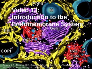
  • 1. Video 13: Introduction to the Endomembrane System Gergely, Z.R., Martinez, D.E., Donohoe, B.S. et al. J of Biol Res-Thessaloniki 25, 15 (2018). https://doi.org/10.1186/s40709-018-0086-2
  • 2. The Endomembrane System Derived from the cytoplasmic membrane and forming a coordinated unit Include the ER, Golgi, endosomes, lysosomes, and vacuoles (NOT the mitochondria and chloroplasts) Allow transportation of proteins, lipids, and complex polysaccharides
  • 3. Transport System Transport vesicles allow movement between cellular compartments Travel along the cytoskeleton mediated by motor proteins Sorting signals within amino acid sequences or oligosaccharides allow targeting to appropriate cellular compartment
  • 4. Transport Pathways All begin with the the biosynthetic pathway Proteins produced in RER ● Vesicles released from RER fuse with the cis Golgi complex, undergoing further modifications ● Transport vesicles released from the trans Golgi Karp’s Cell & Molecular Biology (2020) Fig 8.2
  • 5. Transport Pathways Secretory pathways result in exocytosis and may be constitutive (continuous) or regulated (require signal for release) Materials are also imported through endocytosis and digested within the lysosome Karp’s Cell & Molecular Biology (2020) Fig 8.2
  • 6. Pulse-Chase & Autoradiography Monitor movement of silver grains (in red) through secretory pathway Karp’s Cell & Molecular Biology (2020) Fig 8.3
  • 7. (Green) Fluorescent Proteins Karp’s Cell & Molecular Biology (2020) Fig 8.4 Temperature-sensitive mutation prevents movement of VSVG from ER at 40OC
  • 8. Other Experimental Approaches Subcellular fractionation (differential centrifugation) ● Idenification of fraction containing particular enzymes or functions ● Now extend cell-free systems to include synthetic liposomes Effects of genetic mutations ● Introduction of mutations in identify function of particular gene ● Newer methods include RNA interference and CRISPR
  • 10. The Endoplasmic Reticulum Likely from invagination of cytoplasmic membrane Differentiated into rough and smooth due to presence or absence of bound ribosomes on the cytoplasmic surface
  • 11. ER Similarities RER and SER are joined, having a continual luminal space which allows for free diffusion Number of proteins are common to both RER & SER Both synthesize some shared lipids and cholesterol Karp’s Cell & Molecular Biology Fig. 8.10
  • 12. Highly curved, tubular network with large numbers of reticulons (cause bending of the membranes) Karp’s Cell & Molecular Biology Fig. 8.10 Smooth Endoplasmic Reticulum Detoxification of drugs such as ethanol and barbituates in the liver by oxygenases Ca2+ sequestering in muscle cells (called sarcoplasmic reticulum) Synthesis of steroidal hormones in endocrine cells
  • 13. Continuous with the outer nuclear membrane and composed of flattened sacs (cisternae) connected by helical “ramps” Karp’s Cell & Molecular Biology Fig. 8.10 Rough Endoplasmic Reticulum Increased in secretory cells (acinar cells of pancreas & mucous cells in digestive tract) The site of synthesis of the majority of phospholipids for inclusion in cellular membranes
  • 14. Karp’s Cell & Molecular Biology Fig. 8.10 Rough Endoplasmic Reticulum Undergo co-translational translocation from bound ribosomes to ER lumen Synthesizes carbohydrate chains and initiates N- linked glycosylation of proteins Site of synthesis of approx. 1/3 of cellular proteins: those destined for secretion or inclusion in endomembrane organelles, integral membrane proteins
  • 15. Membrane-bound or Free Ribosome? Remainder of proteins are synthesized by free (cytoplasmic) ribosomes ● Cytosolic proteins ● Peripheral membrane proteins for cytosolic surface) ● Nuclear proteins (contain nuclear localization signals) ● Proteins for other organelles (lysosomes, mitochondria, chloroplasts) All translation is initiated on free ribosomes Proteins which should go to ER contain an N-terminal signal sequence at N-terminus: results in ribosome binding to ER surface and co-translational translocation
  • 16. Co-translational Translocation SRP binds signal sequence and guides ribosome to translocon channel protein by binding to the SRP receptor SRP = signal recognition particle Karp’s Cell & Molecular Biology Fig. 8.13 Polypeptide binding to translocon opens “plug” to allow polypeptide to enter ER Signal removal and folding
  • 17. Synthesis of Integral Proteins Hydrophobic “stop-transfer” sequence blocks translocation into ER lumen The next steps dependent upon location of positive and negative charges at ends of the transmembrane region Karp’s Cell & Molecular Biology Fig. 8.14 ● If N-terminus side is negative, the lateral gate of the translocon allows the transmembrane region to migrate into the membrane ● Results in C-terminus on cytosolic side
  • 18. Synthesis of Integral Proteins ● If N-terminus side is positive, the translocon first reorients the transmembrane region, which is then able to migrate through the lateral gate ● This results in the N-terminus being located on the cytosolic side ● Multi-pass polypeptides utilize translocon many times in an antiparallel manner ● Some proteins utilize alternative pathways Karp’s Cell & Molecular Biology Fig. 8.14
  • 20. Protein Processing Many secretory cells have a polarized or directional structure Reflects movement of polypeptides from synthesis in the RER, processing in the RER & Golgi, and secretion via transport vesicles or secretory granules Processing is a highly regulated multi-step process required for the production of proteins (biologically-active form) Karp’s Cell & Molecular Biology Fig 8.12
  • 21. Protein Processing Removal of signal peptide by signal peptidase Protein folding & molecular chaperones Formation and shuffling of disulfide bonds by disulfide isomerase Addition of carbohydrates by oligosaccharyltransferase
  • 22. Protein Folding Occurs spontaneously during translation Folding directed by amino acid sequence and the production of favorable interactions
  • 24. Membrane Asymmetry Cellular membranes “grow” rather than being produced through de novo synthesis Protein and lipid composition are altered as membranes are moved from one compartment to the next Through the means of membrane fusion, specific asymmetry of membrane is maintained Karp’s Cell & Molecular Biology Fig 8.15
  • 25. Altering Lipid Composition Karp’s Cell & Molecular Biology Fig 8.16 Enzymatic conversion Preferential inclusion of specific lipids within vesicles Movement from one compartment to another by lipid transfer proteins Newly synthesized lipids are inserted in cytosolic leaflet, but may be moved by flippases
  • 26. Glycosylation Majority of proteins produced in the RER become glycoproteins: starts in the RER & completed in the Golgi Addition of sugars to oligosaccaride catalyzed by specific glycosyltransferases ● Transfer a monosaccharide from a nucleotide sugar Oligosaccharide first synthesized on dolichol phosphate Transferred to protein by oligosaccharyltransferase enzyme
  • 28. Modification & Quality Control Following acquisition, two terminal glucose residues removed Bound by calnexin or calreticulin (chaperone) Final glucose removed Karp’s Cell & Molecular Biology Fig 8.18
  • 29. Modification & Quality Control Conformation-sensing enzyme (UGGT) determines whether it is misfolded based upon whether hydrophobic regions are exposed Karp’s Cell & Molecular Biology Fig 8.18
  • 30. Modification & Quality Control Eventually, a mannose is removed & it is transported to proteasome for destruction Karp’s Cell & Molecular Biology Fig 8.18 If it is misfolded, glucose residue again added and cycle repeated
  • 31. Transport from ER to Golgi Special exit sites on edges of ER lack ribosomes and are sites of vesicle budding These small vesicles fuse together to form larger transport vesicles and interconnected tubules Vesicular-tubule carriers (VTCs) move towards the Golgi complex and fuse with the cis-Golgi network ● VTC transport occurs via microtubule tracks The Golgi will allow for further modification of proteins and sorting for final transport
  • 32. Video 16: Golgi Complex & Transport https://www.cancer.gov/about-nci/organization/dcb/research-programs
  • 33. Golgi Complex Tubule networks on cis and trans faces are involved in sorting of proteins: ● cis will send back to ER or on to the cisternae ● trans will sort to plasma membrane or intracellular organelles
  • 34. Golgi Complex Golgi cisternae are involved in the sequential modification of the proteins synthesized by the ER ● Differential composition in each compartment Osmium tetroxide stains unsaturated fatty acids in the Cis Cisternae Mannosidase II enzyme in the Medial Cisternae Nucleoside diphosphatase Enzyme in the Trans Cisternae
  • 35. Golgi Complex Alters the N-linked oligosaccharides added within the ER as they progress through the cisternae Assembles O-linked oligosaccarides Majority of complex polysaccharide assembly Different enzymes found in each compartment
  • 36. Glycosylation Enzymes involved in glycosylation are integral proteins with active sites facing the interior of the Golgi Those involved in adding sugars are transferases Karp’s Cell and Molecular Biology (2020) Fig. 8.23
  • 37. Vesicular Transport Model Popular in the 1980s & 90s Vesicles move from one cisternae to the next, while the cisternae themselves are “static” In vitro evidence of budding and fusing vesicles Believed to occur in vivo in an anterograde direction Karp’s Cell and Molecular Biology (2020) Fig. 8.24
  • 38. Cisternal Maturation Model First & currently accepted Cisternae contents change over time and develop in an anterograde direction Vesicles travel in retrograde: are they returning enzymes? Could both potentially be occurring? Karp’s Cell and Molecular Biology (2020) Fig. 8.24
  • 39. Transport from ER to Golgi:Revisited ERGIC = ER Golgi intermediate compartment Comprised of the larger vesicles & tubules formed through the fusion of transport vesicles 3 kinds of coated transport vesicles: ● COPI = posterograde from Golgi ● COPII = anterograde from ER ● Clathrin-coated = from plasma membrane & trans Golgi network to other organelles Karp’s Cell and Molecular Biology (2020) Fig. 8.26
  • 40. Coated Vesicles Transport vesicles are coated in proteins which mediate ● Membrane curvature ● Selective uptake of cargo ● Selective binding to target compartment Karp’s Cell and Molecular Biology (2020) Fig. 8.25
  • 41. COPII Vesicles Karp’s Cell and Molecular Biology (2020) Fig. 8.25 Form outer scaffold = framework Remainder of proteins are inner layer of adaptors
  • 42. COPII Vesicles Guanine exchange factor catalyzes Sar1-GDP to Sar1-GTP: implants into cytoplasmic membrane of RER: recruits Sec23-Sec24 Karp’s Cell and Molecular Biology (2020) Fig. 8.26 Sec13- Sec31 form outer scaffold Sec24 recruits Golgi enzymes, membrane proteins for docking/fusion, and soluble cargo receptors
  • 43. Outer Vesicle Coats Karp’s Cell and Molecular Biology (2020) Fig. 8.28 Transport vesicle shapes and sizes are dependent upon the external coat proteins & their interactions Disassemble & released into cytoplasm to allow docking and fusion For COPII, catalyzed by hydrolysis of GTP to GDP
  • 44. COPI: Retrograde Transport Karp’s Cell and Molecular Biology (2020) Fig. 8.29 ● Majority of ER-resident proteins are retained and unlikely to be included in transport vesicles ● Soluble ER-resident proteins contain KDEL (lys-asp-glu- leu) amino acid retrieval sequence in C-terminus which bind specific KDEL receptors for return in COPI vesicle ● Similarly KKXX signal used for membrane proteins ● Unique signals likely for specific Golgi compartments
  • 45. Video 17: Trans-Golgi Transport & Protein Sorting http://www.nsf.gov/news/mmg/media/images/myosin_nucleus_h.jpg
  • 46. Sorting to the Membrane Both secretory proteins & cytoplasmic membrane proteins must be targeted to the cytoplasmic membrane. Both may be transported through secretory vesicles in the constitutive pathway Some secretory proteins also follow regulated pathways: aggregates in granules require hormone or nervous stimulus Karp’s Cell & Molecular Biology (2020) Fig. 8.2
  • 47. Targeting Vesicles 4 Steps: targeted transport, tethering, docking & fusion of membranes Karp’s Cell & Molecular Biology (2020) Fig. 8.32 Tethering may be through fibrous or multiprotein complexes ● Specificity mediated by G proteins called Rabs ● Rabs specific to particular compartments Rabs also recruit motor proteins for transport through cytoplasm
  • 48. Targeting Vesicles Docking due to interaction of v-SNARE & t-SNARE Karp’s Cell & Molecular Biology (2020) Fig. 8.32 v-SNARE t-SNARE
  • 49. Model of Synaptic Vesicle Exocytosis Karp’s Cell & Molecular Biology (2020) Fig. 8.33 Synaptobrevin (v-SNARE) & Syntaxin (t-SNARE) both contain transmembrane domains that anchor them to the vesicle & plasma membrane Including SNAP-25 (t-SNARE), 4 helix bundle forms through hydrophobic interactions
  • 50. Model of Synaptic Vesicle Exocytosis Karp’s Cell & Molecular Biology (2020) Fig. 8.33 ● For regulated release, may stay in interlocked formation until stimulus received ● Leads to formation of a water-filled pore as a transition state ● Transmembrane domains become embedded in single bilayer to form the fusion pore
  • 51. Synaptic Vesicle Exocytosis Karp’s Cell & Molecular Biology (8th ed) Fig. 4.57 ● Binding of Ca2+ by v-SNARE triggers fusion and release of neurotransmitters
  • 52. Exocytosis Karp’s Cell & Molecular Biology (2020) Fig. 8.33 Contacts between vesicle & membrane result in pore formation on outside of an alveolus Fusion Pore Fusion of vesicle and plasma membrane results in release of contents into the extracellular environment Luminal part of vesicle is now on the surface of the plasma membrane
  • 53. Plant Cell Central Vacuoles Karp’s Cell & Molecular Biology (2020) Fig. 8.38 Used for storage of many biomoleucles including sugars, polysaccharides, & amino acids Tonoplast (membrane) site of active transport of ions to ensure turgor pressure Site of cellular digestion, in lieu of lysosomes: contain hydrolases
  • 54. Posttranslational Uptake The proteins destined for organelles which are not a part of the endomembrane system are synthesized by free ribosomes Following translation, they are transported to the target organelle Relies on presence of amino acid localization signal Organelles with posttranslational uptake: ● Peroxisomes ● Nucleus ● Mitochondria ● Chloroplasts Interaction of specific chaperones or receptors allow directed transport from cytoplasm Pore proteins regulate entry into the organelle
  • 55. Mitochondrial Uptake Majority of polypeptides synthesized in cytoplasm and transported in as unfolded polypeptides by specific chaperones. Different amino acid sequences interact with different chaperones and protein complexes to determine appropriate localization Chaperones TOM Complex TIM Complex Karp’s Cell & Molecular Biology (2020) Fig. 8.49
  • 56. Mitochondrial Uptake Binding of chaperones involved in targeting to TOM complex on outer mitochondrial membrane TOM receptor recognizes mitochondrial protein and channel complex allows its translocation Karp’s Cell & Molecular Biology (2020) Fig. 8.49 Location of a positively charged localization signal determines compartment
  • 57. Mitochondrial Uptake Internal sequence results in transport through the TIM22 complex = inner mitochondrial membrane protein Karp’s Cell & Molecular Biology (2020) Fig. 8.49 A terminal sequence (presequence) results in transfer through TIM23 = matrix protein ● Bound by mitochondrial chaperones, folded and presequence removed
  • 58. Chloroplast Uptake Karp’s Cell & Molecular Biology (2020) Fig. 8.50 Sequential import through Toc and Tic complexes for entry into the stroma Stroma domain removed Also imported in unfolded state and interact with molecular chaperones Targeted through presence of the N-terminal transit peptide
  • 59. Chloroplast Uptake Karp’s Cell & Molecular Biology (2020) Fig. 8.50 Chaperones assist folding of stromal proteins Thylakoid transfer domain allows transfer into membrane or lumen Some thylakoid membrane proteins synthesized for direct insertion
  • 61. Lysosomes Are the digestive organelle of the cell, containing multiple acid hydrolases for the digestion of: ● Food particles ● Extracellular debris ● Pathogens ● Biomolecule degradation ● Autophagy Proton transport used to create acidic environment Karp’s Cell & Molecular Biology (2020) Fig. 8.35
  • 62. Autophagy Digestion of cellular organelles: ● Turnover of worn out/ defective components ● Source of biomolecules/ energy ● Protection from intracellular pathogens ● Prevents accumulation of specific cellular components: protective Karp’s Cell & Molecular Biology (2020) Fig. 8.37 Retained indefinitely
  • 63. Sorting of Lysosomal Proteins 1)Mannose residue phosphorylated in the cis-Golgi 2)Incorporation into clathrin-coated vesicles 3)Mannose 6- phosphate receptors interact with enzymes and adaptors Karp’s Cell & Molecular Biology (2020) Fig. 8.30
  • 64. Clathrin-coated Vesicles Mannose-6-P acts as targeting mechanism for lysosomal enzymes Interaction of receptor and/or lysosomal membrane proteins with adaptor Adaptor also required for assembly of clathrin coat Karp’s Cell & Molecular Biology (2020) Fig. 8.30
  • 65. Sorting of Lysosomal Proteins 4) Clathrin coat & receptors disassemble 5) Receptors return to trans-Golgi 6) Develop into endosomes, then lysosomes 7) Secreted lysosomal enzymes also returned by mannose- 6-P receptors Karp’s Cell & Molecular Biology (2020) Fig. 8.30
  • 66. Cellular Importation Endocytosis involves the importation of extracellular fluid, dissolved solutes, & suspended macromolecules ● May be bulk phase (general) or receptor- mediated (specific) Karp’s Cell & Molecular Biology (2020) Fig. 8.39 Phagocytosis is the uptake of particulate matter RECEPTOR-MEDIATED ENDOCYTOSIS
  • 67. Clathrin Coat Assembly Karp’s Cell & Molecular Biology (2020) Fig. 8.42 Assembly of clathrin coats for both endocytosis and lysosomal vesicles involves interaction of clathrin subunits and adaptor proteins
  • 68. Clathrin-coated Pits Phosphorylated phosphoinositol (phosophoinositides) recruit the AP2 adaptor proteins to the plasma membrane Binding allows m-subunit to interact with tails of receptors b-adaptin recruits clathrin Results in concentration of specific receptors within the pit Karp’s Cell & Molecular Biology (2020) Fig. 8.42
  • 69. Vesicle Formation Dynamin proteins polymerize to form ring around the “stalk” of the forming vesicle GTP hydrolysis leads to separation of the vesicle from the membrane Blocking of hydrolysis with analog shows helical association of dynamin Karp’s Cell & Molecular Biology (2020) Fig. 8.42
  • 70. Endocytic Pathway Karp’s Cell & Molecular Biology (2020) Fig. 8.42 Fate of receptors & ligands are dependent on their purpose Housekeeping receptors (red) Ligands include needed substances (eg. iron and cholesterol) ● Receptors are recycled to the plasma membrane ● Ligands to late endosomes
  • 71. Endocytic Pathway Karp’s Cell & Molecular Biology (2020) Fig. 8.42 Signaling receptors (green) Ligands include insulin and growth factors ● Both receptors & ligands to late endosomes Late endosomes fuse with additional vesicles containing lysosomal enzymes & fuse with lysosomes
  • 72. Phagocytosis Food acquisition (unicellular organisms), immune cell engulfment of pathogens Karp’s Cell & Molecular Biology (2020) Fig. 8.48 Yeast cell Leukocyte Some pathogens escape digestion: ● M. tuberculosis prevents fusion with lysosome ● L. monocytogenes phospholipases allow escape from lysosome into cytoplasm
  • 73. Entrapment of particle, followed by engulfment to form a phagosome Fuse with a lysosome for digestion: usable nutrients transferred to cytoplasm Residual body forms and contents released through exocytosis or retained permanently as lipofuscin pigment granule Phagocytosis Karp’s Cell & Molecular Biology (2020) Fig. 8.48
  • 74. Public Domain doi:10.7295/W9CIL240 Video 19: Introduction & Microtubule Structure
  • 75. The Cytoskeleton Divided into three classes: microfilaments (actin), intermediate filaments (IFs), and microtubules (MT) Noncovalently bonded protein polymers: highly dynamic Microfilaments Microtubules Merge Intermediate filaments
  • 76. Major Functions All three classes play various roles in providing structure and support Microtubules act as “tracks” for the movement of organelles and materials Also a framework for organization of organelles Karp’s Cell & Molecular Biology (2020) Fig 9.1
  • 77. Major Functions Microtubule network and peroxisome transport Peroxisomes Peroxisomes Peroxisomes Karp’s Cell & Molecular Biology (2020) Fig 9.2
  • 78. Major Functions Microfilaments involved in growth of axons Also form the force-generating apparatus for cell motility Karp’s Cell & Molecular Biology (2020) Fig 9.1
  • 79. Major Functions Movement of chromosomes Contractile ring for cytokinesis Formation of the mitotic spindle Karp’s Cell & Molecular Biology (2020) Fig 9.1
  • 80. Microtubules Components of cytoskeleton, mitotic spindle, cilia & flagella Hollow tubular structures of 25 nm outer diameter Karp’s Cell & Molecular Biology (2020) Fig 9.3 13 protofilaments shown in cross-section
  • 81. Microtubules Karp’s Cell & Molecular Biology (2020) Fig 9.3 Plus End Minus End Protofilaments formed of alternating a- and b-tubulin subunits Offset alignment results in helical structure Plus end (b-tubulin terminus) and minus end (a-tubulin terminus) results in microtubule structural and functional polarity
  • 82. Microtubule-Associated Proteins (MAPs) Karp’s Cell & Molecular Biology (2020) Fig 9.4 Multiple other proteins typically associated with microtubules Roles in increasing microtubule stability and promoting assembly of tubulin subunits Many regulated by phosphorylation Alterations in Tau function linked to various forms of dementia
  • 83. Tau & Dementia Debate: cause or effect of Alzheimer’s? Brain 2006 129(11):3035-3041 Hyperphosphorylated Tau protein incapable of binding to microtubules: found in neurofibrillary tagles/plaques
  • 84. Structural Support Organization of microtubules determines the overall shape of the cell: ● Radial arrangement seen in cultured animal cells results in round, flattened shape ● Basal to apical arrangement in columnar epithelial cells ● Lengthwise arrangement along axons Karp’s Cell & Molecular Biology (2020) Fig 9.5
  • 85. Structural Organization Association with integral membrane proteins influences organization Cellulose synthase association determines location/orientation of cellulose microfibrils which determines plant cell shape and manner of growth Organelle localization eg. Golgi apparatus Karp’s Cell & Molecular Biology (2020) Fig 9.6 Cellulose synthase Microtubules
  • 87. Intracellular Motility Microtubule networks act as “roads” through the cell, allowing specific transport of materials to a given target Karp’s Cell & Molecular Biology (2020) Fig 9.7 Microtubule tracks within for transport of organelles, transport vesicles and molecules such as mRNA & ribosomes Neurofilaments support structure of axons
  • 88. Motor Proteins Large variety of motor proteins in a given cell, each specialized for a specific type of cargo Three broad classes: kinesins, dyneins, & myosins ● Both kinesins & dyneins use microtubule pathways ● Myosins use microfilaments Movement based on binding and hydrolysis of ATP to cause conformational changes allowing stepwise movement from one subunit to the next
  • 89. Kinesins Heads are highly conserved motor region: binding and hydrolysis of ATP for movement along the microtubule Tail sequences very diverse bind the cargo specific for that particular type of kinesin Karp’s Cell & Molecular Biology (2020) Fig 9.11
  • 90. Kinesins Each “step” requires the binding and hydrolysis of one molecule of ATP Rate of movement is dependent upon ATP concentration Karp’s Cell & Molecular Biology (2020) Fig 9.11
  • 91. Kinesins The neck region of the molecule plays an important role in “transmitting” the conformational changes from one head subunit to the other Majority of kinesins move toward the plus end of the microtubule, but some do move toward the minus end ● Neck region also determines the direction of movement along the microtubule Small group of kinesins are involved in microtubule depolymerization instead: called microtubule depolymerases ● Critical in cell division processes
  • 92. Kinesins & Organelle Transport Normal Kinesin mutant Normal Microtubu les Mitochond ria Karp’s Cell & Molecular Biology (2020) Fig 9.12
  • 93. Cytoplasmic Dynein Dynein first discovered as the motor protein associated with cilia and flagella Karp’s Cell & Molecular Biology (2020) Fig 9.13 Composed of two identical heavy chains Involved in positioning of spindle & chromosome movement Transport towards minus end of the microtubule: positioning of centrosomes & Golgi, and in various transport
  • 94. Cellular Transport Model for use of kinesins and dyneins in cellular transport based upon microtubule polarity Vesicles may have both types bound at one time: inactive vs active forms, or a tug- of-war? Karp’s Cell & Molecular Biology (2020) Fig 9.13
  • 96. Microtubule-Organizing Centers (MTOCs) Specialized structure where the initial formation (or nucleation) of a small section of the microtubule occurs, followed by its rapid elongation MTOCs include: ● Centrosomes ● Basal body: associated with cilia/flagella & similar to centrosomes ● Plant MTOC: more dispersed on outer surface of nuclear envelope
  • 97. The Centrosome Karp’s Cell & Molecular Biology (2020) Fig 9.14 Contains two barrel-shaped centrioles surrounded by electron dense pericentriolar material (PCM) Major site of microtubule initiation in animal cells & is the centre of the cell’s microtubule network
  • 98. Nucleation Karp’s Cell & Molecular Biology (2020) Fig 9.15 Microtubules always assembled with the minus end associated with the centrosome Growth occurs through the addition of tubulin dimers to the plus end May remain associated or severed and anchored in other regions of the cell
  • 99. Nucleation Karp’s Cell & Molecular Biology (2020) Fig 9.16 Ring of 13 g-tubulin monomers in complex with additional proteins at site of nucleation g-tubulin, a/b-tubulin, DNA
  • 100. Microtubule Dynamics Karp’s Cell & Molecular Biology (2020) Fig 9.17 Noncovalent association of dimers allows rapid reorganization or polymerization/depolymerization as needed Interphase Preprophase band: future division plane Mitotic spindle formation Phragmoplast: cell wall formation between cells
  • 101. Role of GTP Karp’s Cell & Molecular Biology (2020) Fig 9.21 b-tubulin is a GTPase and must be bound to GTP for polymerization to occur GTP is hydrolyzed shortly after polymerization GDP-bound tubulin lacks stability on its own, & will rapidly depolymerize in the absence of MAPs Structural cap model describes effect
  • 102. Dynamic Instability Karp’s Cell & Molecular Biology (2020) Fig 9.24 Plus-end tracking proteins (+TIPs) bind to ends of microtubules to regulate rate of growth or shrinkage +TIPs mediate attachment to structures (eg. kinetochores, actin) Growing & shrinking microtubules found within the same region of the cell Rapid shift between growing & shrinking phases
  • 103. Video 22: Cilia & Flagella https://www.mdpi.com/2313-7673/3/2/5/htm
  • 104. Cilia & Flagella Hair-like cellular projections which are usually motile Both are constructed from microtubules in the same fashion Both may be used for cellular motility by unicellular organisms, and flagella present on animal gametes Differentiation based upon cell type and type of motion
  • 105. Cilia Single, non-motile cilium on almost all cells used as sensory “antennae” Used for cellular motility, but also found on surface of non-motile cells for the purpose of moving extracellular materials Use a coordinated oar-like motion Karp’s Cell & Molecular Biology (2020) Fig 9.25 & 9.26
  • 106. Flagella Karp’s Cell & Molecular Biology (2020) Fig 9.27 Found in singles or pairs for locomotion Variety of patterns of motions, including assymetric waveforms and symmetrical propellar-like movements
  • 107. Cilia/Flagella Structure Karp’s Cell & Molecular Biology (2020) Fig 9.30 Membrane is continuous with the plasma membrane Basal body is an MTOC identical to the centriole Primary cilium derived from centriole, new basal bodies in secondary cilia A and B tubules of basal body form doublets of the axoneme
  • 108. Karp’s Cell & Molecular Biology (2020) Fig 9.28 Axoneme is the core structure of longitudinal microtubules and associated proteins Axoneme Structure 9 peripheral doublets surrounding 2 central single microtubules (9 + 2 array) Central sheath connects to A tubules by radial spoke proteins
  • 109. Karp’s Cell & Molecular Biology (2020) Fig 9.28 & 9.29 Axoneme Structure Nexin-dynein regulatory complex (N-DRC) Movement of outer & inner axonemal dynein arms result in bending motion of flagella/cilia
  • 110. Karp’s Cell & Molecular Biology (2020) Fig 9.31 Intraflagellar Transport Growth occurs only at the (+) end Transport uses IFT particles (protein complexes) to carry tubulin or other cargo Kinesin transports outward, and cytoplasmic dynein inward
  • 111. Karp’s Cell & Molecular Biology (2020) Fig 9.32 & 9.33 Mechanism Dynein stem is anchored to the A tubule Movement due to binding & release of dynein heads 1) Heads bind to B tubule 2) Conformational change in dynein causes sliding of anchored doublet 3) Heads release B tubule allowing return to original position 4) Cycle repeats Nexin bridges results in limited movement and bending
  • 112. Karp’s Cell & Molecular Biology (2020) Fig 9.34 Sliding Mechanism Central position occurs when outer doublets are all at the same level Sliding of doublets in relation to the position of its neighbours will determine the direction of bending Inner tubules slide towards base of the cilium/flagellum *Different positional relationships can result in different stroke patterns
  • 114. Intermediate Filaments Provide mechanical strength to the cells, particularly those which undergo large amounts of stress (nerves, muscles, epithelia) Only in animals so far, but other insoluble fibres of diverse protein sequences found in other eukaryotes Chemically heterogeneous, with approx. 70 different genes in humans ● All of similar structure: solid & unbranched with 10-12 nm diameter ● 5 classes based on cell type of expression, and biochemical, genetic, and immuonologic criteria – Cytoplasmic filaments (Types I – IV) – Lamins (Type V): support nuclear membrane
  • 115. IF Structure All IFs share same basic structure: ● a-helical central fibrous domain: homologous sequences ● Terminal globular domains of variable size/sequence ● Different N- and C-terminal sequences and their association together results in polarity within the dimers ● Anti-parallel association of dimers into nonpolar tetramer = basic unit Karp’s Cell & Molecular Biology Fig 9.36
  • 116. IF Structure ● Subunits associate in groups of 8 to form a unit length ● End-to-end association to form the polymerized IF ● Growth through intercalation within existing IF ● Polymerization & depolymerization regulated through phosphorylation ● Only cytoskeletal fibres that lack polarity Karp’s Cell & Molecular Biology Fig 9.36 ~60 nm
  • 117. Cytoplasmic IFs Include proteins such as keratin (epithelia), desmin (muscle), and the neurofilaments (CNS and PNS nerves) Interconnected to other cytoskeletal filaments by plectin cross- bridges Karp’s Cell & Molecular Biology Fig. 9.35
  • 118. Epithelial Cells IFs (Types I and II) are composed of keratin Found radiating throughout the cell Often terminate on desmosomes (communication points between cells) Karp’s Cell & Molecular Biology Fig. 9.43 & 7.27
  • 119. At least three different proteins (Type IV Ifs) Unique in structure due to presence of sidearms : ensure appropriate spacing between parallel fibres Increasing amounts of IFs as neuron matures Karp’s Cell & Molecular Biology Fig. 9.43 & 7.27 Neurofilaments
  • 120. Lamins Form a strong, mesh-like grid on the nucleoplasmic side of the inner nuclear membrane called the nuclear lamina Disassembled during prophase of mitosis/meiosis
  • 122. Video 24: Microfilaments and Myosin By Howard Vindin - Own work, CC BY-SA 4.0, https://commons.wikimedia.org/w/index.php? curid=39436133
  • 123. Microfilaments = Actin filaments = F-actin Roles: ● Cellular motility ● Intracellular motile processes (vesicle transport, phagocytosis, cytokinesis) ● Muscle contraction Highly conserved, with different isoforms for specialized roles Karp’s Cell & Molecular Biology (2020) Fig 9.39
  • 124. Microfilaments Assembled from monomers into polar fibre with “barbed” and “pointed” ends Organized into: ● Ordered arrays ● Highly branched networks ● Tightly anchored bundles Karp’s Cell & Molecular Biology (2020) Fig 9.39 (+) “barbed” end (-) “pointed” end
  • 125. Assembly & Disassembly Regulated through presence of ATP/ADP with hydrolysis after incorporation Critical concentration for the addition of ATP-actin to either end is different: lower concentrations required for addition to (+) end At 0.3 mM (-) end removal = (+) end addition Karp’s Cell & Molecular Biology (2020) Fig 9.41
  • 126. Myosin Karp’s Cell & Molecular Biology (2020) Fig 9.43 Superfamily of motor proteins which associate with actin filaments Minimum of 17 classes identified, with about 40 different myosins in humans Type II (conventional) myosins first identified in muscle Actin subunit comprised of 6 polypeptide chains ATP-binding cleft & motor function associated with the head
  • 127. Myosin Type II Filaments Karp’s Cell & Molecular Biology (2020) Fig 9.45 Tails associate with heads on either end to form a bipolar filament Myosin moves towards (+) end of the microfilament Bipolar arrangement results in microfilaments being pulled towards one another
  • 128. Unconvensional Myosins Karp’s Cell & Molecular Biology (2020) Fig 9.61 Do not form filaments and generally operate independently Myosin I has a single head and acts as attachment point for microfilaments to the plasma membrane ● Potential involvement in the alteration of membrane shape
  • 129. Unconvensional Myosins Karp’s Cell & Molecular Biology (2020) Fig 9.46 Many involved in the transport of vesicles and organelles, or as organelle tethers Myosin V shows movement similar to kinesin, with one head bound to actin at all times
  • 130. Unconvensional Myosins Karp’s Cell & Molecular Biology (2020) Fig 9.47 Believed that long distance transport occurs on microtubules, with microfilaments used for local movement
  • 132. Muscle Organization Karp’s Cell & Molecular Biology (2020) Fig 9.49 Skeletal muscle cell (fiber) formed through fusion of mononucleated cells early in development Each myofibril is composed of repeating contractile units called sarcomeres
  • 133. Sarcomeres Visible striations due to the presence of thin and thick filaments and their regions of overlap Z-lines are borders between sarcomeres Karp’s Cell & Molecular Biology (2020) Fig 9.49
  • 134. Sarcomeres Composed of actin (thin) filaments and conventional myosin (thick filaments) Karp’s Cell & Molecular Biology (2020) Fig 9.50 Actin Myosin
  • 135. Sliding Filament Model Contraction results in decrease in width of the I and H bands as actin moves inward, shortening the sarcomere Karp’s Cell & Molecular Biology (2020) Fig 9.51
  • 136. Composition & Organization Thin filaments are the microfilaments and associated proteins ● Tropomyosin fits within the goove of the microfilament, 7 actin subunits in length ● Troponin globular protein which interacts with the end of tropomyosin and with actin Karp’s Cell & Molecular Biology (2020) Fig 9.52
  • 137. Composition & Organization Thick filaments are composed of several hundred myosin molecules with bipolar orientation Titin is an elastic protein stretching from M line to Z line ● Maintains overall structure, aids relaxation Karp’s Cell & Molecular Biology (2020) Fig 9.53
  • 138. Molecular Basis of Contraction Each myosin filament head undergoes conformational change to move actin filament 10 nm. Each filament interacts with approx 100 myosin molecules, resulting in continuous contraction of several hundred nm Karp’s Cell & Molecular Biology (2020) Fig 9.52
  • 139. Swinging Lever Mechanism 1) Myosin tightly bound in absence of ATP 2) ATP binding results in release 3) ATP hydrolysis results in conformational change 4) Rebinding to actin 5) ADP realeased and repeat http://www.esrf.fr/UsersAndScience/Publications/Highlights/2003/MX/MX03/ Lever arm swings
  • 140. Excitation-Contraction Coupling Transverse tubules transmit impulses from action potentials to interior of cell Sarcoplamic reticulum stores calcium ions needed to induce muscle contraction Karp’s Cell & Molecular Biology (2020) Fig 9.52 Muscle fibers organized into motor units stimulated by a single nerve
  • 141. Excitation-Contraction Coupling https://schoolbag.info/biology/mcat/53.html Action potential results in release of calcium ions from SR Binding of calcium to troponin causes change in conformation
  • 143. Cellular Motility Relies upon the presence of actin networks in the cell cortex (below the cell membrane) Processes include: ● Endocytosis & phagocytosis ● Extension of processes ● Cytokinesis Organization/function dependent on actin binding proteins Karp’s Cell & Molecular Biology (2020) Fig 9.39
  • 144. 1) Nucleating proteins ● Spire contains multiple actin-binding domains to form cluster for nucleation ● Formins used to create and lengthen unbranched filaments at the (+) end ● Arp 2/3 complex is template for addition of actin monomers to add branches Actin Binding Proteins Karp’s Cell & Molecular Biology (2020) Fig 9.66
  • 145. Actin Binding Proteins Karp’s Cell & Molecular Biology (2020) Fig 9.60 2) Monomer-sequestering proteins (thymosins) ● Bind actin-ATP monomers to prevent polymerization ● Regulates rate of polymerization/depolymerization 3) End-blocking (capping) proteins ● Prevent addition or loss of actin ● May be associated with the (+) or (-) end
  • 146. Actin Binding Proteins 4) Monomer polymerizing proteins ● Profilin binds to same site as thymosin ● Promotes removal of ADP, allowing ATP to bind and polymerization to occur 5) Depolymerizing proteins (cofilin family) ● Bind to pointed end to cause depolymerization 7) Filament-severing proteins ● Cause fragmentation (eg. cofilin and gelsolin) ● May create additional free barbed ends for growth, or may act as capping proteins
  • 147. Actin Binding Proteins Karp’s Cell & Molecular Biology (2020) Fig 9.60 6) Cross-linking and bundling ● Flexible rod-like proteins for forming mesh-like network for support (eg. filamin) ● Globular bundling proteins for parallel groupings as seen in microvilli (eg. villin & fimbrin) 8) Membrane-binding proteins ● Anchorage for changes in membrane shape
  • 148. Cell Locomotion: “Crawling” Karp’s Cell & Molecular Biology (2020) Fig 9.63 Protrusion formed on leading edge Lower surface of protrusion attaches to substratum Majority of cell is pulled forward Rear contacts broken
  • 149. Lamellipodia Karp’s Cell & Molecular Biology (2020) Fig 9.64 Dynamic fan-like or “ruffled” protrusion on leading edge of motile cells
  • 150. Directed Motility Karp’s Cell & Molecular Biology (2020) Fig 9.65 Stimulus results in activation of filament nucleating proteins (Arp 2/3 complex) by WASP/WAVE Polymerization of actin-ATP mediated by profilin
  • 151. Directed Motility Karp’s Cell & Molecular Biology (2020) Fig 9.65 Capping of older filaments occurs, while allowing new branches to continue growing, resulting in the outgrowth of the lamellipodium
  • 152. Directed Motility Karp’s Cell & Molecular Biology (2020) Fig 9.65 Older filaments are comprised of actin- ADP Depolymerization of the (-) ends of capped microfilaments mediated by cofilin Profilin allows release of ADP to allow formation of actin-ATP for further use
  • 153. Traction Forces Karp’s Cell & Molecular Biology (2020) Fig 9.65 Focal adhesions are formed between the leading edge of the lamellipodium and the substratum These focal adhesions allow for the bulk of the cell to be moved forward by myosin and actin Vinculin staining at focal adhesions
  • 154. Axonal Outgrowth Karp’s Cell & Molecular Biology (2020) Fig 9.69 Growth cone at tip resembles a highly motile fibroblast ● Broad lamellipodium ● Short microspikes towards edge of lamellipodia ● Elongated filipodia Actin Tubulin
  • 155. Axonal Outgrowth Karp’s Cell & Molecular Biology (2020) Fig 9.70 Direction of growth is determined by physical and chemical stimuli detected by receptors in the growth cone Tubulin Ephrin f ● Response to chemoattractants (eg. netrin) which diffuse through extracellular environment ● Interacting with membrane-bound molecules (eg. ephrin) to encourage growth in that particular direction.
  • 156. Video 27: Video 27: Introduction to Introduction to Cell Signalling Cell Signalling https://www.kurzweilai.net/using-cells-chemical-signaling-to-control-cancer-or-detect-toxins
  • 157. Cell Signalling & Communication Cells receive signals from the environment, which results in a particular response: ● Alterations in cell behavior (eg. Chemotaxis) ● Alterations in gene expression (eg. Gene expression) Multicellular organisms need to coordinate their behavior ● Embryonic development ● Cell division
  • 158. Vocabulary Signal molecules: extracellular molecules which act as messengers between cells Signalling cell: the cell which produces the signal molecule Target cell: the cell receiving the signal Receptor protein: a protein in the target cell which recognizes and responds specifically to that signal Signal transduction: means by which the message is translated into a response
  • 159. Receptors may be found on the cell surface or within the cell Hydrophilic signals bind to surface receptors Hydrophobic signals bind to intracellular receptors Receptor Proteins
  • 160. Autocrine Signalling Receptors on the surface of the cell allow it to respond to a signalling molecule it produces itself Karp’s Cell & Molecular Biology (2020) Fig 15.1
  • 161. Paracrine Signalling Karp’s Cell & Molecular Biology (2020) Fig 15.1 Messenger produced by signalling cell acts on nearby target cells: signal travels short distance through extracellular space Limited due to their stability, digestion by enzymes, or through interactions with the extracellular matrix
  • 162. Endocrine Signalling Karp’s Cell & Molecular Biology (2020) Fig 15.1 Messenger produced for long distance signalling: signal travels through bloodstream Many hormones are produced in organs distant from the site of the target cell
  • 163. GAP JUNCTIONS ● Hydrophilic channels allow communication between cells that form during development CNX OpenStax / Wikimedia Commons Other Forms of Cell Signalling https://projects.ncsu.edu/project/bio183de/ Lab/cells_celldivision/cells_celldivision1.html SYNAPTIC
  • 164. Signalling Overview Karp’s Cell & Molecular Biology (2020) Fig 15.2 Diverse signalling molecules ranging from small soluble compounds and proteins to large cell surface bound glycoproteins Specific receptors on the target cell will bind to their ligand (signalling molecule)
  • 165. Diversity of Extracellular Messengers Amino acids & their derivatives (eg. Epinephrine & dopamine) act as neurotransmitters and hormones Gases (NO, CO) Cholesterol-derived steroid hormones Eicosanoids (derived from fatty acids) act in regulating various systemic responses (inflammation, blood pressure) Polypeptides/proteins on membrane of signalling cell or secreted into extracellular environment
  • 166. Cell Signalling Receptors G protein-coupled receptors (GPCR): binding of ligand results in activation of GTP-binding protein Receptor protein-tyrosine kinases (RTK): binding of ligand results in activation of kinase activity and phosphorylation of target proteins Ligand-gated channels: alterations in ion concentrations lead to altered cellular activities (neurotransmission, muscle contraction, calcium as a second messenger) Steroid hormone receptors: intracellular, act as transcription factors
  • 167. Signalling Overview Ligand binding to receptor results in conformational change: transmits signal across membrane Effector enzyme generates small molecule second messenger Karp’s Cell & Molecular Biology (2020) Fig 15.2 Recruitment of signalling proteins OR
  • 168. Signalling Overview Sequential interaction of proteins, resulting in conformational changes which affect their activity Final target protein is activated leading to alterations in cellular responses Karp’s Cell & Molecular Biology (2020) Fig 15.2 RESPONSE
  • 169. The addition (kinases) or removal (phosphatases) of phosphate groups alter protein conformation and therefore activity Termination of signalling requires the destruction or inactivation of receptors, second messengers, and transduction proteins Karp’s Cell & Molecular Biology (2020) Fig 15.2 Signal Transduction Pathways Phosphorylation occurs on serine, threonine or tyrosine Proteins are cytoplamic or membrane-bound
  • 171. Superfamily of receptors that mediate signalling through interaction with G proteins G Protein-Coupled Receptors (GPCR) Karp’s Cell & Molecular Biology (2020) Fig. 15.5 G proteins are trimeric proteins that bind to GDP/GTP Following activation, G proteins interact with specific effectors to initiate signalling
  • 172. Largest group of proteins in animals, with highly varied roles in the cell G Protein-Coupled Receptors Karp’s Cell & Molecular Biology (2020) Table 15.1
  • 173. G Protein-Coupled Receptor Action Karp’s Cell & Molecular Biology (2020) Fig. 15.5 Consist of 7 a-helical transmembrane domains (also called 7TM receptors) Extracellular loops form binding site for ligand Ligand binding will results in conformational change in transmembrane domains N-term C-term
  • 174. G Protein-Coupled Receptor Action Karp’s Cell & Molecular Biology (2020) Fig. 15.5 Conformational change results in strong affinity for the G protein Heterotrimeric G proteins consist of three subunits: ● a (contains GDP/GTP binding site), b, and γ ● Binding to receptor results in exchange of GDP for GTP N-term C-term a GDP/GTP binding site
  • 175. GPCR Action Karp’s Cell & Molecular Biology (2020) Fig. 15.6 GTP binding results in dissociation of a from b/γ and its interaction with the effector molecule to initiate the signalling cascade Hydrolysis of GTP to GDP results in a dissociation and the effector being “turned off”
  • 176. GPCR Action GDP-bound a loses affinity for effector molecule and associates with b/γ again 4 different types of G proteins with different a subunits which interact with different effectors: ● Gs activate adenyl cyclase ● Gi inhibit adenyl cyclase ● Gq with phospholipase C b (PLCb) ● G12/13 not characterized Karp’s Cell & Molecular Biology (2020) Fig. 15.6 b/γ also involved in signalling and interact with adenyl cyclase PLCb, and K+ and Ca2+ channels
  • 177. Regulation of G Protein Activity G proteins with GTP bound interact with the effector molecule to activate them G proteins are slow-acting GTPases, resulting in their eventual hydrolysis of bound GTP to GDP + Pi Acceleration of this process occurs through the action of regulators of G protein signaling (RGSs)
  • 178. Termination of Response ● G protein-coupled receptor kinase (GRK) are serine-threonine kinases which phosphorylate activated GPCR ● Phosporylated residues are bound by arrestin, which compete with G proteins for binding to GPCR Karp’s Cell & Molecular Biology (2020) Fig. 15.6 Effectors are no longer active after dissociation of the a subunit, but the receptors need to undergo desensitization in order to deactivate them:
  • 179. Arrestin & Internalization ● Inclusion in signalling complexes & activation of transcription factors (3) ● Digestion (4) ● Recycled to cell surface (5/6) Karp’s Cell & Molecular Biology (2020) Fig. 15.6 Arrestins associate with AP2 in clathrin-coated pits, which results in GPCR accumulation within the pits Endocytosis occurs, resulting in 3 potential roles:
  • 180. GPCRs and Bacterial Toxins Some bacterial toxins target the functioning of GPCRs through targeting the function of the a subunits ● Cholera toxin modifies a subunits by inhibiting GTPase activity, leading to continual activation of adenylate cyclase. – This is the cause of diarrhea due to altered water retention by intestinal cells. ● Pertussis toxin inhibits function of a subunits, interfering with signalling required for immune defense
  • 182. Interact with multiple targets, resulting in a coordinated rapid response to external stimuli Most are soluble (except phosphoinositides & DAG) and will rapidly diffuse through the cell Include: ● cyclic AMP/GMP (cAMP/cGMP) ● phosphoinositides, diacylglycerol (DAG), inositol triphosphate (IP3) ● Ca2+ ● NO Second Messengers
  • 183. Cyclic AMP Karp’s Cell & Molecular Biology (2020) Fig. 15.13 Production catalyzed by adenylyl cyclase ● Interacts with GTP-bound G protein a subunits ● Recall that G proteins may either activate adenylyl cyclase (as) or inhibit its function (ai) Each activated adenylyl cyclase allows a rapid amplification of the signal initiated through ligand binding
  • 184. Karp’s Cell & Molecular Biology (2020) Fig. 15.12 Reg ulati on of Bloo d Gluc ose In response to glucagon (from pancreatic a-cells) or epinephrine ● Through G protein- coupled receptors ● Also cause inhibition of glycogen synthase Activated by insulin from pancreatic b-cells ● Mediated by a receptor protein-tyrosine kinase
  • 185. Glucose Mobilization Karp’s Cell & Molecular Biology (2020) Fig. 15.14 2) Adenylyl cyclase in cell membrane is activated & cAMP is produced ● Downregulation may occur through action of phosphodiesterase 1) Ligand binding results in G protein activation & mobilization of the as subunit 3) cAMP then binds to & activates protein kinase A (PKA)
  • 186. PKA https://www.creative-enzymes.com/resource/protein-kinase-a_15.html Inactive form is tetramer of 2 catalytic and 2 regulatory subunits Following binding of cAMP, regulatory subunits dissociate, releasing 2 active PKA PKA phosphorylates targets within the cell, regulating their activity
  • 187. Glucose Mobilization Karp’s Cell & Molecular Biology (2020) Fig. 15.14 4) Inactivates glycogen synthase 5) Activates phosphorylase kinase 6) Phosphorylase kinase activates glycogen phosphorylase 7) Glycogen phosphorylase depolymerizes glycogen Phosphatases involved in downregulation PKA
  • 188. Glucose Mobilization Karp’s Cell & Molecular Biology (2020) Fig. 15.14 9) PKA which enters the nucleus phosphorylates the cAMP response element-binding protein (CREB) 10) CREB binds response elements within the promoter of genes to upregulate their transcription
  • 189. Regulation of PKA Targeting PKA is known to phosphoylate over 100 different substrates Which responses occur in a given cell type due to: ● Differential expression of these targets ● Presence of PKA- anchoring proteins (AKAPs) Karp’s Cell & Molecular Biology (2020) Fig. 15.15
  • 190. Regulation of PKA Targeting Different AKAPs found within specific membranes in different cell types Karp’s Cell & Molecular Biology (2020) Fig. 15.16 Sequester PKAs to certain cellular regions to coordinate their interactions with targets
  • 191. Phosphatidylinositol (PI) Numerous lipid derivatives function as second messengers Enzymatically altered by phospholipases, phospholipid kinases, and phospholipid phosphatases Those derived from PI called phosphoinositides Hbf878, CC0, via Wikimedia Commons
  • 192. Phosphoinositides Number of derivatives are created through the actions of kinases which remain associated with the membrane PI(4)P, PI(4,5)P2, and PIP3 (not shown) all act though interaction of the phosphorylated inositol ring with lipid- binding domain containing proteins Karp’s Cell & Molecular Biology (2020) Fig. 15.10 Domain acts to recruit these proteins to the membrane for protein- protein interactions
  • 193. Phospholipase C (PLC) Karp’s Cell & Molecular Biology (2020) Fig. 15.10 GPCR binding may also lead to activation of phosphatidylinositol- specific phospholipase C-b, which cleaves PI(4,5)P2 Diacylglycerol Inositol 1,4,5- triphosphate
  • 194. Diacylglycerol (DAG) https://www.mdpi.com/2072-6694/6/2/860 DAG remains associated with the membrane and binds to proteins containing the DAG-binding domain Protein kinase C (PKC) associates with DAG and phosphorylates various targets: ● Extracellular signal molecule release ● Cell growth & differentiation ● Metabolism ● Apoptosis ● Immunity
  • 195. Inositol 1,4,5-triphosphate Karp’s Cell & Molecular Biology (2020) Fig. 15.10 IP3 is a soluble second messenger which will rapidly diffuse through the cell IP3 receptor on SER is a calcium channel, which opens upon binding Ca2+ also a second messenger, interacting with various targets: ● Muscle contraction ● Exocytosis ● Cell shape
  • 196. Video 30: Receptor Protein-Tyrosine Kinases By Emw - Own work, CC BY-SA 3.0, https://commons.wikimedia.org/w/index.php?curid=8762948
  • 197. Receptor Protein-Tyrosine Kinases Large number of kinases expressed by different cell types Approximately 2/3 of tyrosine kinases are membrane- bound receptors which are activated through ligand binding and dimerization ● Ligands inclued growth factors & hormones Regulate diverse processes: ● Cell growth & proliferation ● Differentiation ● Cellular motility
  • 198. Receptor Protein-Tyrosine Kinases Karp’s Cell & Molecular Biology (2020) Fig. 15.17 Monomeric receptors undergo dimerization due to ligand binding
  • 199. Receptor Protein-Tyrosine Kinases Karp’s Cell & Molecular Biology (2020) Fig. 15.17 In the absence of phosphorylation, the ATP-binding active site of the kinase domain is blocked by the activation loop Phosphorylation results in activation loop being in stable conformation away from the active site, allowing trans- autophosphorylation of adjacent cytoplasmic domains
  • 200. Receptor Protein-Tyrosine Kinases Karp’s Cell & Molecular Biology (2020) Fig. 15.17 The phosphorylated tyrosines and their surrounding amino acid sequences determine the specificity of interactions with signalling proteins Most characterized domains: ● Src-homology 2 (SH2) ● Phosphotyrosine- binding (PTB)
  • 201. RTK Signal Transduction Karp’s Cell & Molecular Biology (2020) Fig. 15.19 May be bound by adaptor or scaffolding proteins which allow the recruitment of other proteins Docking proteins may themselves be phosphorylated by the receptor to allow interaction with signalling molecules
  • 202. RTK Signal Transduction Karp’s Cell & Molecular Biology (2020) Fig. 15.19 Many transcription factors are able to bind to activated RTKs Binding results in phosphorylation, leading to dimerization Following dimerization, the transcription factor is transported into the nucleus, where it binds specific DNA sequences to allow transcription
  • 203. RTK Signal Transduction Karp’s Cell & Molecular Biology (2020) Fig. 15.19 Activation of enzymes through: ● Recruitment to membrane & co-localization with substrate ● Conformational change due to binding to receptor ● Phosphorylation Regulation of enzymatic activity by RTKs is also common ● Kinases, phosphatases, lipases
  • 204. Turning RTK Signals Off Karp’s Cell & Molecular Biology (2020) Fig. 8.42 Ubiquitin ligases contain SH2 domains which allow them to associate with RTKs and label them for internalization Ubiquitination results in interaction with the AP2 adaptor which localizes the receptors into clathrin- coated pits for endocytosis ● Destruction in the lysosome ● Recycling back to cell surface ● Use in endosomes for intracellular signalling
  • 205. Signalling Cascades Karp’s Cell & Molecular Biology (2020) Fig. 15.22 Most well known RTK pathway is the Ras-MAP kinase pathway: ● Activation of Ras (a small G protein) through GTP binding ● Sequential activation of protein kinases ● Phosphorylation of targets including transcription factors, other kinases, cytoskeletal components, and more
  • 206. https://erc.bioscientifica.com/view/journals/erc/26/6/ERC-19-0098.xml Small G Proteins Monomeric G proteins, similar in function to the heterotrimeric G proteins (see GPCRs) ● Interaction with adaptor (Grb2/Sos) makes nucleotide binding site of Ras available ● Ras active when GTP is bound ● GTPase function, resulting in hydrolysis to GDP and inactivity Ras functions by recruiting Raf to the membrane, leading to its phosphoryation and activation
  • 207. Karp’s Cell & Molecular Biology (2020) Fig. 15.21 Regulation of Small G Proteins 3 types of accessory proteins are involved in the regulation of small G proteins 1) Guanine nucleotide dissociation inhibitors (GDIs) ● Prevent the release of GDP ● Results in maintenance of inactive G proteins
  • 208. Karp’s Cell & Molecular Biology (2020) Fig. 15.21 Regulation of Small G Proteins 2) Guanine nucleotide-exchange factors (GEFs) ● Binding allows the exchange of GDP for GTP, resulting in activation of the G protein ● Binding of the G protein to its target results in GEF release, activation of the target & signal transduction
  • 209. Karp’s Cell & Molecular Biology (2020) Fig. 15.21 Regulation of Small G Proteins 3) GTPase-activating proteins (GAPs) ● Binding to G protein/target complex results in acceleration of GTPase activity ● Once GTP hydrolysis occurs, complex disassociates
  • 210. Regulation of MAP Kinase Pathways Scaffolding proteins allow specific assembly of components for mediating particular processes Kinases only phosphorylate specific substrates Inactivation of MAP kinases by phosphatases (MKP) https://erc.bioscientifica.com/view/journals/erc/26/6/ERC- 19-0098.xml
  • 211. Karp’s Cell & Molecular Biology (2020) Fig. 15.12 Reg ulati on of Bloo d Gluc ose In response to glucagon (from pancreatic a-cells) or epinephrine ● Through G protein- coupled receptors ● Also cause inhibition of glycogen synthase Activated by insulin from pancreatic b-cells ● Mediated by a receptor protein-tyrosine kinase
  • 212. a and b chains produced from single polypeptide a chain contains the insulin binding domain b chain is transmembrane protein with tyrosine kinase domain Exists as stable dimer even in the absence of insulin Insulin binding causes confromational change in kinase domains Karp’s Cell & Molecular Biology (2020) Fig. 15.24 Insulin Receptor
  • 213. Autophosphorylaed insulin receptor binds to PTB domain of insulin receptor sustrates (IRS-1 & IRS-2) IRSs also have PH domain (interact with phospholipids on membrane) Phosphorylated tyrosines interact with signal molecules with SH domains Karp’s Cell & Molecular Biology (2020) Fig. 15.24 Insulin Receptor
  • 214. Following activation by binding to IRS, PI3K produces phosphoinositide PIP3 which leads to the recruitment of PKB & PDK1 Karp’s Cell & Molecular Biology (2020) Fig. 15.24 Role of PI 3-kinase PDK1 & mTOR activate PKB for signal transduction PKB mediates glucose uptake by causing fusion of GLUT4 receptor vesicles with membranes
  • 216. Karp’s Cell and Molecular Biology (2020) Fig 4.56 Calcium plays important roles in cell functions ● Synaptic transmission ● Muscle contraction https://schoolbag.info/biology/mcat/53.html Calcium as an Intracellular Messenger
  • 217. Inositol 1,4,5-triphosphate & Calcium Karp’s Cell & Molecular Biology (2020) Fig. 15.10 IP3 receptor on SER is a calcium channel, which opens upon binding GPCR binding may also lead to activation of Phospholipase C- b, which cleaves PIP2 to produce DAG and IP3 Cytoplasmic calcium concentration normally low (10-7 M)
  • 218. IP3-mediated signalling due to oscillation in cytoplasmic calcium levels IP3 and Calcium Oscillation Karp’s Cell & Molecular Biology (2020) Fig. 15.11 & Table 15.2
  • 219. Opening of voltage-gated Ca2+ channels (1) leads to entry of some Ca2+ ions Binds to ryanodine receptors (RyR) (2) on the surface of the SER Triggers opening of these channels and release of stored Ca2+ from ER (3) Karp’s Cell & Molecular Biology (2020) Fig. 15.28 Calcium-induced Calcium Release
  • 220. Cytoplasmic concentration restored through action of: ● Ca2+ pumps in ER (4) ● Na+/Ca2+ exchanger in plasma membrane (5) Response may be localized in small region or may result in increasing, spreading wave throughout cell Karp’s Cell & Molecular Biology (2020) Fig. 15.28 Calcium-induced Calcium Release
  • 221. Sperm contain a phospholipase, which cleave PIP2 contained within the egg to produce PI3 Karp’s Cell & Molecular Biology (2020) Fig. 15.29 Calcium wave in Fertilization PI3 binds receptors on the SER, resulting in calcium wave Image produced here every 10 seconds
  • 222. Karp’s Cell & Molecular Biology (2020) Fig. 15.30 Store-Operated Calcium Entry STIM1 are calcium-sensing proteins which are normally dispersed throughout the ER membrane
  • 223. Karp’s Cell & Molecular Biology (2020) Fig. 15.30 Store-Operated Calcium Entry STIM1 become clustered near plasma membrane & recruit Orai1 Cellular stores may become depleted through repeated responses Orai1 Ca2+ ion channels allow import of Ca2+ & storage in ER
  • 224. Cell-type specificity due to ability to: ● Activate/inhibit enzymes ● Alter transport ● Change membrane permeability ● Cause membrane fusion ● Impact structure and function of the cytoskeleton Cellular Effects of Ca2+
  • 225. Mediate effects of calcium through interacting with it and target proteins Calmodulin most widely studied and found in all eukaryotes ● Low affinity for calcium, so only binds when calcium increased in cytoplasm ● Alters conformation and binds to cell- specific targets ● Targets include the Na+/Ca2+ exchanger, thereby acting to bring a halt to further signalling as well. Calcium-binding Proteins Karp’s Cell & Molecular Biology (2020) Fig. 15.31
  • 226. Karp’s Cell and Molecular Biology (2020) Fig 15.33 Nitric Oxide Acts as both an extracellular and a second messenger to regulate various processes Acetylcholine signalling (1) results in the influx of Ca2+ (2), which activates nitric oxide synthase (NOS) NOS produces NO from arginine (3), & NO diffuses from the endothelial cell into neighbouring smooth muscle cells (4)
  • 227. Karp’s Cell and Molecular Biology (2020) Fig 15.33 Nitric Oxide In the muscle cell, NO binds and activates guanylyl cyclase, which produces cGMP (5) cGMP is a second messenger, which mediates muscle relaxation through the activation of a cGMP- dependent protein kinase, which results in blood vessel dilation Nitroglycerine is treatment for angina: catabolized to NO Converted to GTP by cGMP phosphodiesterase
  • 228. Multiple different ligands can result in the activation of a single effector = convergence Convergence, Divergence & Crosstalk eg. Both GPCR & TKR activate PLCs Karp’s Cell and Molecular Biology (2020) Fig 15.33
  • 229. Signals from the same ligand can result in the activation of more than one effector = divergence Convergence, Divergence & Crosstalk eg. PLC, PI3K, & GAP all activated by TKR Karp’s Cell and Molecular Biology (2020) Fig 15.33
  • 230. Different pathways may regulate each other = crosstalk Convergence, Divergence & Crosstalk eg. Calcium can inhibit PKC activity Karp’s Cell and Molecular Biology (2020) Fig 15.33
  • 231. Video 32: Apoptosis https://www.thoughtco.com/apoptosis-372446 Credit: Steve Gschmeissner/Science Photo Library/Getty Images
  • 232. Karp’s Cell and Molecular Biology (2020) Fig 15.37 Apoptosis (Programmed Cell Death) Ordered destruction of the cell which is essential in the process of development and maintenance of health in multi-cellular organisms
  • 233. https://www.novusbio.com/research-areas/apoptosis Apoptosis (Programmed Cell Death) External or internal signals initiate apoptosis Apoptotic bodies are removed by phagocytes
  • 234. Apoptosis vs Necroptosis Apoptosis results in safe clearance of cellular components Karp’s Cell and Molecular Biology (2020) Fig 15.37 Rupturing of cellular membranes during necroptosis results in release of cellular components, resulting in inflammation https://www.creative-diagnostics.com/ necroptosis-signaling-pathway.htm
  • 235. Karp’s Cell and Molecular Biology (2020) Fig 15.38 Apoptosis in Development Cells which are located between the eventual digits undergo apoptosis, with macrophages (blue fluorescence) ingesting the apoptotic bodies.
  • 236. Apoptosis in Maintaining Health ● Prevention of autoimmunity: eliminate T cells and B cells with interact with “self” antigens. ● Down-regulation of immunity following clearance of pathogens. ● Removal of cells with irreparable DNA damage – Loss of appropriate triggering of apoptosis is a central feature of cancerous cells ● Inappropriate triggering of apoptosis leads to disease (eg. neurodegeneration)
  • 237. Intrinsic Pathway Regulated by members of the Bcl-2 family of mitochondrial proteins upon receiving internal stimuli ● Both pro-apoptotic and anti-apoptotic members with several BH domains are normally expressed and associate with one another in the membrane ● Proapoptotic BH3-only members act as initiators of apoptosis by inhibiting the anti-apoptotic members and activating the proapoptotic members ● Following activation, pro-apoptotic Bax/Bak form pores in the mitochondrial outer membrane, allowing the release of cytochrome c (& other mediators) from the intermembrane space
  • 238. Karp’s Cell and Molecular Biology (2020) Fig 15.40 Intrinsic Pathway Cytochrome c then interacts with various cytoplamic proteins and procaspase-9 This is the activated initiator or apoptosome Caspase-9 cleaves numerous other “executioner” procaspases, leading to their activation
  • 239. Caspases Family of cysteine proteases which are central in the activation of cell death pathways. Targets are varied cellular components including: ● Proteins regulating cell adhesion: isolates cell from its neighbours ● Proteins involved in regulating anti-apoptotic signal pathways ● Cytoskeletal proteins, including the lamins ● Inhibitors of DNA digestion: initiates the destruction of the genome
  • 240. Karp’s Cell and Molecular Biology (2020) Fig 15.39 Extrinsic Pathways Tumor-necrosis factor (TNF) is a cytokine produced in response to infection by intracellular pathogens and other environmental stresses. Following activation, the TNF receptor death domains bind to an adaptor protein (TRADD) for the recruitment of effector proteins (RIP1K). Signalling results in apoptosis, necroptosis, or cell survival?
  • 241. Karp’s Cell and Molecular Biology (2020) Fig 15.39 Extrinsic Pathways: Apoptosis RIP1K can associate with FADD to recruit 2 molecules of procaspase-8 Procaspases cleave each other to produce activated caspase-8 Caspase-8 acts of various “executioner” procaspases to activate them and initiate the apoptotic pathway
  • 242. Karp’s Cell and Molecular Biology (2020) Fig 15.39 Extrinsic Pathways: Necroptosis Much less understood then apoptotic pathway Association of RIP1K with RIP3K leads to their phosporylation and formation of the necrosome Phosphorylation of MLKL and its insertion in the membrane interferes with phospholipid structure and membrane stability
  • 243. Extrinsic Pathways: Cell Survival? TNF binding to the TNFR also results in activation of the NF-kB transcription factor ● First identified in B cells, but utilized in all animal cells as a positive regulator for cell survival and proliferation ● Normally sequestered in cytoplasm through association with IkBs (inhibitor proteins), which become phosphorylated following TNF binding and inactivated ● Following release, NF-kB relocates to the nucleus to initiate transcription. ● Inappropriate expression linked to the development of cancer