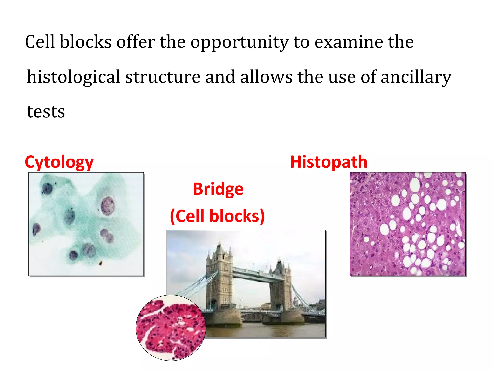
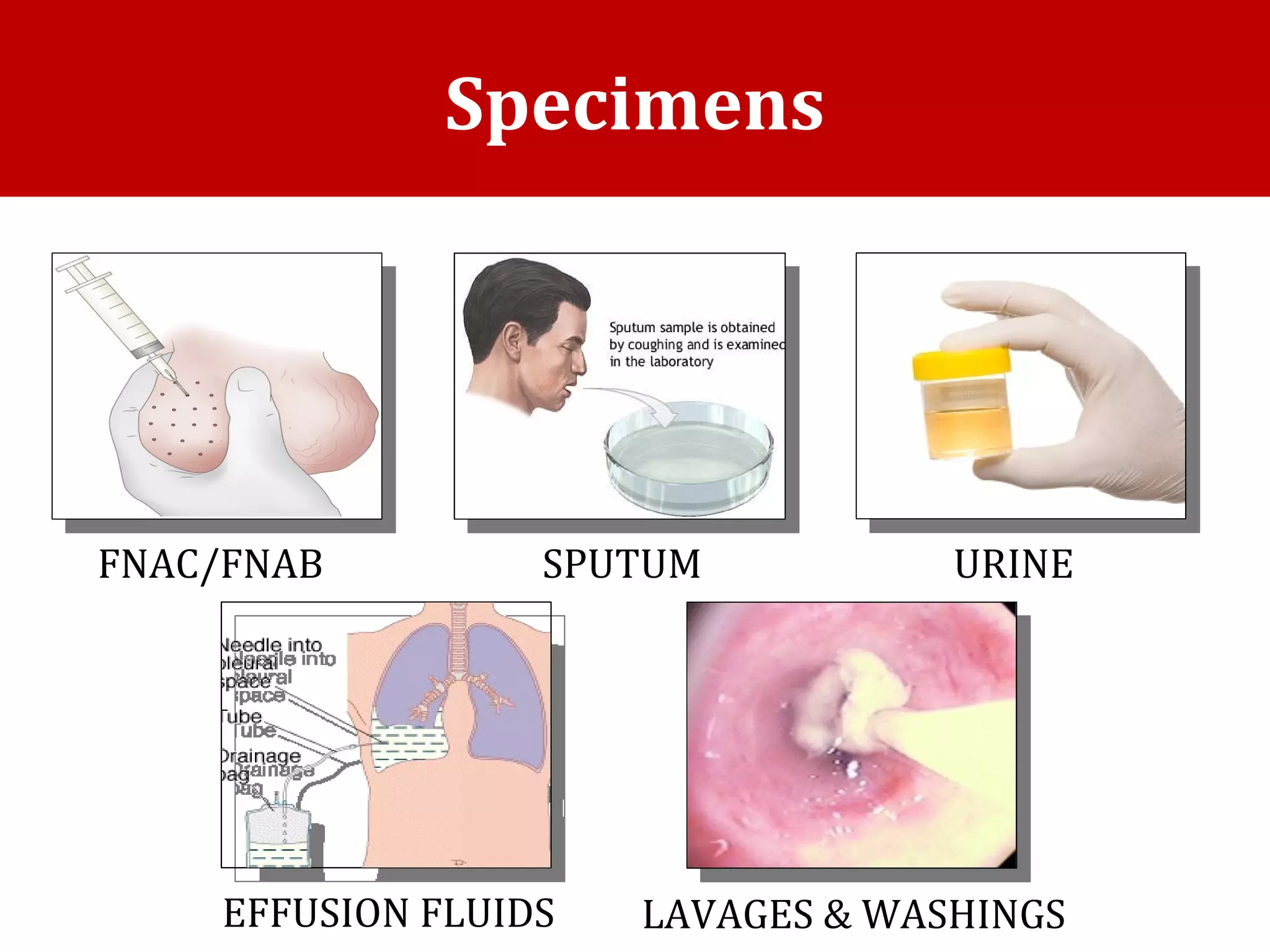
CELL BLOCK
AND
ITS UTILITY IN
CYTOPATHOLOGY
CELL BLOCK
AND
ITS UTILITY IN
CYTOPATHOLOGY
Dr. Anam
Moderator: Dr. Bawana Raina
Dr. Anam
Moderator: Dr. Bawana Raina
• A cell block is a method of preparing cytologic material so that it can be
processed, sectioned, stained, and viewed as a histology section.
• It can provide diagnostic information in addition to that obtained from cytology
slides.
• It is easier to do special stains, and immunohistochemistry on cell block slides
rather than on regular cytology slides because the slides often require
adaptations of the staining protocols and different controls.
• It should be understood that the term 'block' in this context is not a reference to
placing the cells in a paraffin block, but rather it refers to the technique of
'blocking' something to hold it together and maintain its shape.
What is a cell block ?What is a cell block ?
Cell blocks offer the opportunity to examine the
histological structure and allows the use of ancillary
tests
Cytology Histopath
Bridge
(Cell blocks)
• Acquisition of tissue for cell block can increase both
diagnostic sensitivity and specificity (through both cellular
morphology and ancillary testing)
• It requires minimal effort and is extremely cost efficient.
• Moreover, tissue preserved in cell block can be readily
shared for second opinions and research without fear of
losing the original diagnostic smear specimen
Need for Cell Block
Specimens
FNAC/FNAB SPUTUM
EFFUSION FLUIDS
URINE
LAVAGES & WASHINGS
1. Direct processing of tissue fragments present in fluids.
2. Plasma thrombin method
3. Fixed sediment method
4. Bacterial agar method
5. Technique using alcohol, acetone and paraffin
6. Compact technique
7. Cell blocks from Millipore
8. Histogel method
9. Gelatin embedding
10. Celloidin bag method
11. Scraping of cytology smears
12. Automated preparation
13.Albumin method.
Methods of cell block preparation
Fixed Sediment MethodFixed Sediment Method
Residual clot /tissue in the hub of the needle or spontaneous clot in a
fluid container is carefully removed & rinsed in 50% ethanol/buffered
formalin
Centrifuged at 4000rpm for 6min to form pellets
Supernatant is discarded
Sediment is removed and put into filter paper, placed in cassette.
& processed as regular paraffin specimen and then
stained routinely by H&E
Normal Saline Needle Rinse MethodNormal Saline Needle Rinse Method
Commonly used method ( FNA)
Rinse the aspirate needle with 20-30 ml saline
centrifugation
discard supernatant
collect the sediment and process routinely
Plasma Thrombin methodPlasma Thrombin method
Principle : to enmesh the cellular material in a
clot.
1. Centrifugation of the cell suspension. Discard
the supernatant.
2. For approximately 1 ml of sediment, add 4
drops of plasma.
3. Add 4 drops of Thrombin.
4. Mix with a vortexer or by tapping gently on
the counter .
5. Allow the mixture to sit for a few minutes
until a soft ball forms.
6. Place this mass into an appropriately
labeled cassette.
7. Place the cassette in fixative and submit
for processing as tissue.
• centrifuge the cell suspension.
discard supernatant.
• Melt 4% agar by placing the tube of
agar in a small beaker of water and
heating it in a microwave for a brief
period of time.
• Add approximately 4 ml of
completely melted agar to the
sediment in a centrifuge tube. Mix
gently.
• Refrigerate to get a cell button.
• place in a cassette and process
routinely.
Principle: the concentrated sedimented is
supported by a cell adjuvant. (agar) . Agar
solidifies below 50 degree C. Cell pellet
can be otained. This is a more time-
consuming method; but it creates an
excellent block, with the various cells
distributed in layers for optimal
visualization.
The Agar MethodThe Agar Method
Other cell adjuvantsOther cell adjuvants
• Collodion bag ( scanty cellularity)- nitrocellulose material,
to make blocks of friable tissue e.g.. brain.
• HistoGel
• Gelatin foam
• Albumin
• pre-gelatinised starch
• The CellientTM
automated cell block system concentrates,
processes, and embeds loose cells into a paraffin block
ready for sectioning. User variability is minimized and tech
time is reduced.
• Cells are concentrated into a well in the cassette by vacuum,
and subsequent washes with alcohol and xylene fix and clear
the specimen.
• The cell button is then infiltrated with paraffin in the same
cassette. The entire process takes approximately 45 minutes.
• The instrument can accommodate only one specimen block
at a time, so high volume labs may require multiple
instruments.
• The standard process uses alcohol fixation. However a
formalin fixed protocol is available, giving the user options in
case immunohistochemistry may be required.
The Cellient Automated Cell Block SystemThe Cellient Automated Cell Block System
Cell Block Fixation and ProcessingCell Block Fixation and Processing
• Formalin fixation -Formalin has been used to fix CBs from FNA rinses and has
been claimed as ‘the poor man’s CB’ as it does not require any special equipment
or reagents.
• Alcohol fixation - The Cellient system uses methanol as a fixative. Although
immunostains and molecular analysis may be performed on samples fixed in
alcohol, the laboratory needs to validate tests appropriately to avoid false negative
or false positive results in IHC.
• Nathan alcohol formalin substitute (NAFS) fixation- A CB technique using an
ethanol–formalin fixative (nine parts of 100% ethanol and one part of 40%
formaldehyde), also known as NAFS, followed by paraffin processing, has been
used to obtain good cytological details with less toxicity.
• Microwave fixation - Microwave fixation can expedite the overall process of CB
preparation and can reduce the turnaround time significantly.
Advantages of cell block
✓Slides are more readily interpretable by histopathologists.
✓Availability of a block facilitates more sections.
✓Imp residual material is salvaged which is generally not
available in cytology smears .Loose cells, cell aggregates
and microscopic tissue fragments are easily recoverable.
✓Concentrated in a small area of the slide so examination
less time consuming
✓Special stains mucicarmine, congo stain, melanin etc
✓Stains for immunocytochemistry
✓For microorganisms esp fungi and bacteria
✓Pattern and architectural recognition of tumor possible
✓CB is simple ,reproducible and readily available in
routine laboratory
✓No necessity of biopsy
✓Storage of cell blocks is easier than unstained slides
Disadvantages
✓Compared to routine smears takes longer
time
✓Sparse cellularity
✓Distortion artifacts
• Cytologic studies of body fluids are done to
ascertain the etiology of effusion.
• Cytologic evaluation is the best method to detect
malignancy in body cavity fluids.
• Diagnostic accuracy of routine smears for
malignancy ranges from 50-80 % with a false
negative rate of about 25%. To improve diagnostic
accuracy, cell block preparation can be done.
Cell blocks in body fluids
Few advantages:
✓Spontaneous formed clot in the body fluid may enmesh
virtually all cells.
✓Induced clot prepared from the sediment obtained by
centrifugation of the fluid specimen would also contain
many cells .
✓Identification of primary site of malignancy can be
enhanced with the use of cell block technique.
✓Histologic patterns of cancer (acinar ,papillary ,duct like)
can be readily identified on a cell block.
✓ Psammoma bodies ,granulation tissue , cholesterol clefts , microorganisms,
fragments of collagenous stroma, hyperpalstic mesothelium, etc can be
identified.
✓ Fluid samples or tissue fragments can be preserved in a refrigerator for 72
hrs before processing in cell blocks.
✓ IHC on cell blocks are comparable to that of surgical specimens.
( differentiation of reactive mesothelial cell clusters from metastatic
adenocarcinoma and mesothelioma)
✓ Microarray technique, Molecular tests like FISH ,In situ PCR.
✓ Electron microscopy.
Role of cell block in non
neoplastic effusion
✓Reactive mesothelial cells
✓Differentiation between polymorphous lymphocytes of
non neoplastic effusion & monomorphous lymphoid cells
of lymphoproliferative diseases
✓Rheumatic effusion (elongated & giant cell
multinucleated histiocytes , granular background ,
cholesterol crystals)
✓L E cells, granulomas in TB, number of parasitic, fungal
and viral diseases can be picked up.
Cell blocks in neoplastic effusion
✓Adenocarcinoma
✓Signet ring cell carcinoma
✓Small cell anaplastic carcinoma
✓Mesotheliomas
CB In IHC and Molecular analysis
• CBs are the preferred choice for ICC as they are comparable with surgical
biopsies, can use the same control slides, provide the best milieu for
morphological interpretation and are without background staining.
• avoids subjecting patients to unnecessary sampling.
• Biopsy tissue is not available
• CB is preferable to a biopsy for molecular testing as the ratio of tumour to
background cells is usually high ( fnac).
✓
Gynaecological cytology -
• With the advent of liquid-based collections, the opportunity for CB preparation is now available because a
residual specimen is almost always present.
• The occasional presence of stromal invasion and tumour necrosis in CB sections is useful for differentiating HSIL
from squamous cell carcinoma in cervical cytology.
• ICC of p16INK4a, a surrogate marker of human papilloma virus (HPV) infection, and ISH studies for HPV can be
performed on CBs.
Thyroid-
• minimal role of CBs because of low cellularity
• Direct smears in combination with LBC ( liquid based cytology) are preferred.
Breast-
• CB is considered to be suitable, with some reservations, for the assessment of hormone receptors and Her-2/neu
analysis.
• Ethanol fixation may result in spurious Her-2/neu IHC staining.
• .Alcohol fixation may also alter the antigenicity of progesterone receptors.
Lung -
•The role of CB has also been highlighted in the sub typing of lung carcinoma by
morphologyand IHC for squamous and adenocarcinoma markers.
•CBs can be prepared from samples from endobronchial ultrasound-guided
transbronchial needle aspirates (EBUS- TBNAs), and subjected to a panel of
antibodies.
•Studies indicate that combining conventional smears, LBC and CB improves the
sensitivity of EBUS-TBNA for the staging of lung carcinoma.
•The clinical utility of cytological specimens, including CB, for molecular testing in
lung cancer has been validated recently for the personalized treatment of non-
small-cell lung carcinoma. CBs permit the analysis of molecular alterations in lung
cancer using different cytogenetic and molecular technologies- epidermal
growth factor receptor (EGFR) mutational analysis .
Deep Seated Abdominal organs-
•CB with on-site cytopathological evaluation of direct FNA smears can
increase diagnostic yield, improve tumour sub classification and further
molecular test- ing in solid organ tumours.
•Cytology microarray using CBs
Similar to tissue microarrays, CBs of FNA material or effusion fluids have
been increasingly used for array construction for immunocytochemical
marker validation. More recently, cultured cells have been used to make
CBs for array construction. Although technically complex and expensive,
their immense role in clinical research make CB microarrays a potentially
useful tool in cytology.
Figure 1: Metastatic lung adenocarcinoma in a cell block of pleural fluid.
a H&E stain showed relatively monotonous tumor cells arranged in tubules. ×400.
b TTF-1/napsin A double stain showed positive brown nuclear TTF-1 and red cytoplasmic napsin A
staining in the tumor cells. ×400.
c TTF-1/napsin A double stain. ×600.
a) Pap smear interpreted HSIL,
b) H and E cell block section containing “microbiopsies”,
c) p16-stained cell block section showing true nuclear positivity,
d) biopsy showing invasive squamous cell carcinoma.
Thank you

Cell block in cytology

  • 1.
    CELL BLOCK AND ITS UTILITYIN CYTOPATHOLOGY CELL BLOCK AND ITS UTILITY IN CYTOPATHOLOGY Dr. Anam Moderator: Dr. Bawana Raina Dr. Anam Moderator: Dr. Bawana Raina
  • 2.
    • A cellblock is a method of preparing cytologic material so that it can be processed, sectioned, stained, and viewed as a histology section. • It can provide diagnostic information in addition to that obtained from cytology slides. • It is easier to do special stains, and immunohistochemistry on cell block slides rather than on regular cytology slides because the slides often require adaptations of the staining protocols and different controls. • It should be understood that the term 'block' in this context is not a reference to placing the cells in a paraffin block, but rather it refers to the technique of 'blocking' something to hold it together and maintain its shape. What is a cell block ?What is a cell block ?
  • 3.
    Cell blocks offerthe opportunity to examine the histological structure and allows the use of ancillary tests Cytology Histopath Bridge (Cell blocks)
  • 4.
    • Acquisition oftissue for cell block can increase both diagnostic sensitivity and specificity (through both cellular morphology and ancillary testing) • It requires minimal effort and is extremely cost efficient. • Moreover, tissue preserved in cell block can be readily shared for second opinions and research without fear of losing the original diagnostic smear specimen Need for Cell Block
  • 5.
  • 6.
    1. Direct processingof tissue fragments present in fluids. 2. Plasma thrombin method 3. Fixed sediment method 4. Bacterial agar method 5. Technique using alcohol, acetone and paraffin 6. Compact technique 7. Cell blocks from Millipore 8. Histogel method 9. Gelatin embedding 10. Celloidin bag method 11. Scraping of cytology smears 12. Automated preparation 13.Albumin method. Methods of cell block preparation
  • 7.
  • 8.
    Residual clot /tissuein the hub of the needle or spontaneous clot in a fluid container is carefully removed & rinsed in 50% ethanol/buffered formalin Centrifuged at 4000rpm for 6min to form pellets Supernatant is discarded Sediment is removed and put into filter paper, placed in cassette. & processed as regular paraffin specimen and then stained routinely by H&E
  • 9.
    Normal Saline NeedleRinse MethodNormal Saline Needle Rinse Method Commonly used method ( FNA) Rinse the aspirate needle with 20-30 ml saline centrifugation discard supernatant collect the sediment and process routinely
  • 10.
    Plasma Thrombin methodPlasmaThrombin method Principle : to enmesh the cellular material in a clot. 1. Centrifugation of the cell suspension. Discard the supernatant. 2. For approximately 1 ml of sediment, add 4 drops of plasma. 3. Add 4 drops of Thrombin. 4. Mix with a vortexer or by tapping gently on the counter . 5. Allow the mixture to sit for a few minutes until a soft ball forms. 6. Place this mass into an appropriately labeled cassette. 7. Place the cassette in fixative and submit for processing as tissue.
  • 11.
    • centrifuge thecell suspension. discard supernatant. • Melt 4% agar by placing the tube of agar in a small beaker of water and heating it in a microwave for a brief period of time. • Add approximately 4 ml of completely melted agar to the sediment in a centrifuge tube. Mix gently. • Refrigerate to get a cell button. • place in a cassette and process routinely. Principle: the concentrated sedimented is supported by a cell adjuvant. (agar) . Agar solidifies below 50 degree C. Cell pellet can be otained. This is a more time- consuming method; but it creates an excellent block, with the various cells distributed in layers for optimal visualization. The Agar MethodThe Agar Method
  • 12.
    Other cell adjuvantsOthercell adjuvants • Collodion bag ( scanty cellularity)- nitrocellulose material, to make blocks of friable tissue e.g.. brain. • HistoGel • Gelatin foam • Albumin • pre-gelatinised starch
  • 13.
    • The CellientTM automatedcell block system concentrates, processes, and embeds loose cells into a paraffin block ready for sectioning. User variability is minimized and tech time is reduced. • Cells are concentrated into a well in the cassette by vacuum, and subsequent washes with alcohol and xylene fix and clear the specimen. • The cell button is then infiltrated with paraffin in the same cassette. The entire process takes approximately 45 minutes. • The instrument can accommodate only one specimen block at a time, so high volume labs may require multiple instruments. • The standard process uses alcohol fixation. However a formalin fixed protocol is available, giving the user options in case immunohistochemistry may be required. The Cellient Automated Cell Block SystemThe Cellient Automated Cell Block System
  • 14.
    Cell Block Fixationand ProcessingCell Block Fixation and Processing • Formalin fixation -Formalin has been used to fix CBs from FNA rinses and has been claimed as ‘the poor man’s CB’ as it does not require any special equipment or reagents. • Alcohol fixation - The Cellient system uses methanol as a fixative. Although immunostains and molecular analysis may be performed on samples fixed in alcohol, the laboratory needs to validate tests appropriately to avoid false negative or false positive results in IHC. • Nathan alcohol formalin substitute (NAFS) fixation- A CB technique using an ethanol–formalin fixative (nine parts of 100% ethanol and one part of 40% formaldehyde), also known as NAFS, followed by paraffin processing, has been used to obtain good cytological details with less toxicity. • Microwave fixation - Microwave fixation can expedite the overall process of CB preparation and can reduce the turnaround time significantly.
  • 15.
    Advantages of cellblock ✓Slides are more readily interpretable by histopathologists. ✓Availability of a block facilitates more sections. ✓Imp residual material is salvaged which is generally not available in cytology smears .Loose cells, cell aggregates and microscopic tissue fragments are easily recoverable. ✓Concentrated in a small area of the slide so examination less time consuming ✓Special stains mucicarmine, congo stain, melanin etc
  • 16.
    ✓Stains for immunocytochemistry ✓Formicroorganisms esp fungi and bacteria ✓Pattern and architectural recognition of tumor possible ✓CB is simple ,reproducible and readily available in routine laboratory ✓No necessity of biopsy ✓Storage of cell blocks is easier than unstained slides
  • 17.
    Disadvantages ✓Compared to routinesmears takes longer time ✓Sparse cellularity ✓Distortion artifacts
  • 18.
    • Cytologic studiesof body fluids are done to ascertain the etiology of effusion. • Cytologic evaluation is the best method to detect malignancy in body cavity fluids. • Diagnostic accuracy of routine smears for malignancy ranges from 50-80 % with a false negative rate of about 25%. To improve diagnostic accuracy, cell block preparation can be done. Cell blocks in body fluids
  • 19.
    Few advantages: ✓Spontaneous formedclot in the body fluid may enmesh virtually all cells. ✓Induced clot prepared from the sediment obtained by centrifugation of the fluid specimen would also contain many cells . ✓Identification of primary site of malignancy can be enhanced with the use of cell block technique. ✓Histologic patterns of cancer (acinar ,papillary ,duct like) can be readily identified on a cell block.
  • 20.
    ✓ Psammoma bodies,granulation tissue , cholesterol clefts , microorganisms, fragments of collagenous stroma, hyperpalstic mesothelium, etc can be identified. ✓ Fluid samples or tissue fragments can be preserved in a refrigerator for 72 hrs before processing in cell blocks. ✓ IHC on cell blocks are comparable to that of surgical specimens. ( differentiation of reactive mesothelial cell clusters from metastatic adenocarcinoma and mesothelioma) ✓ Microarray technique, Molecular tests like FISH ,In situ PCR. ✓ Electron microscopy.
  • 21.
    Role of cellblock in non neoplastic effusion ✓Reactive mesothelial cells ✓Differentiation between polymorphous lymphocytes of non neoplastic effusion & monomorphous lymphoid cells of lymphoproliferative diseases ✓Rheumatic effusion (elongated & giant cell multinucleated histiocytes , granular background , cholesterol crystals) ✓L E cells, granulomas in TB, number of parasitic, fungal and viral diseases can be picked up.
  • 22.
    Cell blocks inneoplastic effusion ✓Adenocarcinoma ✓Signet ring cell carcinoma ✓Small cell anaplastic carcinoma ✓Mesotheliomas
  • 23.
    CB In IHCand Molecular analysis • CBs are the preferred choice for ICC as they are comparable with surgical biopsies, can use the same control slides, provide the best milieu for morphological interpretation and are without background staining. • avoids subjecting patients to unnecessary sampling. • Biopsy tissue is not available • CB is preferable to a biopsy for molecular testing as the ratio of tumour to background cells is usually high ( fnac).
  • 24.
    ✓ Gynaecological cytology - •With the advent of liquid-based collections, the opportunity for CB preparation is now available because a residual specimen is almost always present. • The occasional presence of stromal invasion and tumour necrosis in CB sections is useful for differentiating HSIL from squamous cell carcinoma in cervical cytology. • ICC of p16INK4a, a surrogate marker of human papilloma virus (HPV) infection, and ISH studies for HPV can be performed on CBs. Thyroid- • minimal role of CBs because of low cellularity • Direct smears in combination with LBC ( liquid based cytology) are preferred. Breast- • CB is considered to be suitable, with some reservations, for the assessment of hormone receptors and Her-2/neu analysis. • Ethanol fixation may result in spurious Her-2/neu IHC staining. • .Alcohol fixation may also alter the antigenicity of progesterone receptors.
  • 25.
    Lung - •The roleof CB has also been highlighted in the sub typing of lung carcinoma by morphologyand IHC for squamous and adenocarcinoma markers. •CBs can be prepared from samples from endobronchial ultrasound-guided transbronchial needle aspirates (EBUS- TBNAs), and subjected to a panel of antibodies. •Studies indicate that combining conventional smears, LBC and CB improves the sensitivity of EBUS-TBNA for the staging of lung carcinoma. •The clinical utility of cytological specimens, including CB, for molecular testing in lung cancer has been validated recently for the personalized treatment of non- small-cell lung carcinoma. CBs permit the analysis of molecular alterations in lung cancer using different cytogenetic and molecular technologies- epidermal growth factor receptor (EGFR) mutational analysis .
  • 26.
    Deep Seated Abdominalorgans- •CB with on-site cytopathological evaluation of direct FNA smears can increase diagnostic yield, improve tumour sub classification and further molecular test- ing in solid organ tumours. •Cytology microarray using CBs Similar to tissue microarrays, CBs of FNA material or effusion fluids have been increasingly used for array construction for immunocytochemical marker validation. More recently, cultured cells have been used to make CBs for array construction. Although technically complex and expensive, their immense role in clinical research make CB microarrays a potentially useful tool in cytology.
  • 28.
    Figure 1: Metastaticlung adenocarcinoma in a cell block of pleural fluid. a H&E stain showed relatively monotonous tumor cells arranged in tubules. ×400. b TTF-1/napsin A double stain showed positive brown nuclear TTF-1 and red cytoplasmic napsin A staining in the tumor cells. ×400. c TTF-1/napsin A double stain. ×600.
  • 29.
    a) Pap smearinterpreted HSIL, b) H and E cell block section containing “microbiopsies”, c) p16-stained cell block section showing true nuclear positivity, d) biopsy showing invasive squamous cell carcinoma.
  • 35.