Anatomy of cornea by alamin
•Download as PPTX, PDF•
1 like•148 views

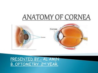
A view of the anatomy of Cornea.

Recommended
More Related Content
What's hot
What's hot (20)
Similar to Anatomy of cornea by alamin
Similar to Anatomy of cornea by alamin (20)
Recently uploaded
Recently uploaded (20)
Anatomy of cornea by alamin
- 1. PRESENTED BY - AL AMiN B. OPTOMETRY 2nd YEAR.
- 2. Word cornea has come from ‘Kerato’. Kerato in Greek mean ‘Horn’ or ‘Shield like’. Ancient Greek used to believe that cornea is derived from thin sliced horn of animal.
- 3. ¤ The cornea is a transparent avascular tissue with a smooth , convex outer surface and concave inner surface with uniform curvature , which resembles a small watch glass. ¤ The cornea is composed of proteins and cells. ¤ It does not contain blood vessels, unlike most of the tissues in the human body. Blood vessels may cloud the cornea, which may prevent it from refracting light properly and may adversely affect vision. ¤ Since there are no nutrient-supplying blood vessels in the cornea, tears and the aqueous humor (a watery fluid) in the anterior chamber provide the cornea with nutrients. ¤ Light enters the eye through the major refractive structure of the eye, focusing light onto the retina.
- 4. ¤ Anterior Surface : - Elliptical , about 11.7mm horizontally and 10.6mm vertically. ¤ Posterior Surface : - Circular, about 11.7mm in diameter. ¤ Thickness : - Centrally about 0.52mm. - Peripherally about 0.67mm. ¤ Optical zone : - Cornea is almost a sphere , the central 1/3rd is called optical zone about 5.4mm. ¤ Radius of curvature : - Anterior surface is about 7.8mm. - Post. Surface is about 6.5mm.
- 6. ¤ Resistance provide protective layer. ¤ Resist ocular pressure due to collagenous components of stroma. ¤ The cornea acts as the eye’s outermost lens. It functions like a window that controls and focuses the entry of light into the eye.
- 7. ¤ There are mainly 5 layers of cornea – 1. A nterior Epithelium. 2. B owman’s membrane. 3. C entral Stroma. 4. D ecemet’s membrane. 5. E ndothelium. There is one more layer which comes between central stroma and decement’s membrane – 6. Dua’s layer.
- 9. 50-90µ thick. Stratified squamous type. Tight junction in basal cells account for epithelium’s transparency. It acts as a barrier to protect the cornea , resisting the free flow of fluids from the tears, and prevent bacteria from entering the epithelium and corneal stroma. The corneal epithelium consists of several layers of cells. The cells of the deepest layer are columnar, known as basal cells which are attached by multiprotein complexes known as hemidesmosomes to an underlying basement membrane. Then follow two or three layers of polyhedral cells, commonly known as wing cells. Lastly, there are three or four layers of squamous cells, with flattened nuclei. The layers of the epithelium are constantly undergoing mitosis. Basal and wing cells migrate to the anterior of the cornea, while squamous cells age and slough off into the tear film. Corneal epithelium sheds at regular interval. It takes normally about 7days for replacement of entire corneal epithelium.
- 11. The Bowman's membrane a smooth, acellular, nonregenerating layer, located between the superficial epithelium and the stroma in the cornea of the eye. It is composed of strong, uniformly oriented collagen fibrils in which the smooth anterior surface faces the epithelial basement membrane and the posterior surface merges with the collagen lamellae of the corneal stroma proper. In adult humans, Bowman's membrane is 8-12 μm thick. With ageing, this layer becomes thinner.
- 12. The corneal stroma is a mesenchymal connective tissue making up 90% of the corneal thickness, with physical properties that provide the cornea its essential character. The stroma is formed during late embryogenesis by a population of neural crest cells migrating from the periocular mesenchyme. They are each about 1.5-2.5 μm in thickness. At its centre, human corneal stroma is composed of about 200 flattened lamellæ (layers of collagen fibrils), superimposed one on another.
- 13. Dua’s layer is a layer of the cornea that had not been detected previously. The fourth caudal layer, and located between the corneal stroma and Descemet's membrane. Despite its thinness, the layer is very strong and impervious to air. Dua’s layer is well-defined, acellular and strong, consisting of five to eight lamellae of type-1 collagen bundles totalling about six to 15 microns thickness. The bundles are coarse and arranged in transverse, longitudinal and oblique directions. Bundle spacing is similar to that in stromal tissue, but Dua’s layer is entirely free of keratocytes in the zone that forms the posterior wall of the bubble.
- 14. Descemet's membrane is the basement membrane that lies between the corneal proper substance, also called stroma, and the endothelial layer of the cornea. It is composed of different kinds of collagen (Type IV and VIII) than the stroma. The endothelial layer is located at the posterior of the cornea. Descemet's membrane, as the basement membrane for the endothelial layer, is secreted by the single layer of squamous epithelial cells that compose the endothelial layer of the cornea. Its thickness ranges from 3 μm at birth to 8–10 μm in adults.
- 15. The corneal endothelium is a single layer of endothelial cells on the inner surface of the cornea. It faces the chamber formed between the cornea and the iris. The corneal endothelium are specialized, flattened, mitochondria-rich cells that line the posterior surface of the cornea and face the anterior chamber of the eye. The corneal endothelium governs fluid and solute transport across the posterior surface of the cornea and maintains the cornea in the slightly dehydrated state that is required for optical transparency. The number of endothelial cells in the fully developed cornea decreases with age up until early adulthood, stabilizing around 50 years of age. The corneal endothelium is attached to the rest of the cornea through Descemet's membrane, which is an acellular layer composed mostly of collagen IV.
- 16. ¤ Cornea is an avascular structure. ¤ Small loops derived from the anterior ciliary vessels invade its periphery for about 1mm and provide nourishment. ¤ These loops are not in cornea but in the subconjunctival tissue which overlaps the cornea .
- 17. ¤ The cornea is one of the most sensitive tissues of the body and has one of the richest sensory nerve supplies in the body. ¤ It is supplied by long and short cilliry nerves from the ophthalmic division of the trigeminal nerve.
- 18. At limbus, ¤ Corneal epithelium becomes bulbar conjunctival epithelium. ¤ Bowman’s membrane becomes continuous with the lamina propria of the conjunctiva and tenon’s capsule. ¤ Stroma becomes sclera. ¤ Decements membrane becomes schwalbe’s line. ¤ Endothelium lines the trabecular meshwork and becomes continuous with the anterior surface of the epithelium.
- 19. ¤ Tight junctions of the epithelial cells . ¤ Endothelial pump mechanism. ¤ Absence of blood vessels. ¤ Absence of pigments. ¤ Scarcity of cell nuclei in stroma. ¤ Uniform structure of stroma.
- 20. ¤ The cornea, with the anterior chamber and lens, refracts light.The cornea accounting for approximately two-thirds of the eye's total optical power. In humans, the refractive power of the cornea is approximately +43 dioptres. ... While the cornea contributes most of the eye's focusing power, its focus is fixed.