
Recommended
More Related Content
What's hot
What's hot (20)
Similar to Acute cholecystitis causes symptoms diagnosis management
Similar to Acute cholecystitis causes symptoms diagnosis management (20)
Recently uploaded
Recently uploaded (20)
Acute cholecystitis causes symptoms diagnosis management
- 1. Prepared by: Almas khan
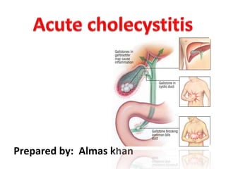
- 3. Cholecystitis is an inflammation of the gallbladder. gallbladder is a small, pear-shaped organ on the right side of your abdomen, beneath your liver. The gallbladder holds a digestive fluid that's released into your small intestine (bile)
- 4. 1 .. Calculus: it’s obstructive type & 90% occur due to gall stones. 2 .. Acalculus: its non obstructive types occur in critically ill patients like burn , severe sepsis , shock,DM,etc 3... Acute emphysematous cholecystitis is an uncommon condition caused by gas- forming organisms and characterized by the presence of gas in the wall and lumen of the gall bladder.
- 5. •Gall stones : 90% •Tumor •Bile duct blockage •Infection : AIDS and certain viral infections can trigger gall bladder inflammation. •Blood vessel problems: A very severe illness can damage blood vessels and decrease blood flow to the gallbladder, leading to cholecystitis.
- 7. 1…Pain •Onset : it may be sudden in onset or superimposed on the pain of chronic cholecyctitis usually participated by large or fatty meal •Site : epigastrium or right hypochondria •Radiation :it radiates through the trunk to the tip of the right scapula •Aggressive factors: movement and breathing •Relieving factors: no except analgesic
- 8. 2.. Nausea 3.. Vomiting 4.. Anorexia 5.. Sweating 6.. Chills 7.. Bloating
- 9. On examination •Patient anxious ,lying quietly with shallow breathing •Tachycardia •Tenderness •In severe case there may be palpable inflammatory mass around gall bladder •Jaundice in 25 % cases
- 10. •Boas’s sign : the area below the scapula at 9,10,11 rib become hypersensitive •Murphy sign: Murphy's sign is elicited in patients with acute cholecystitis by asking the patient to take in and hold a deep breath while palpating the right sub costal area. If pain occurs on inspiration, when the inflamed gallbladder comes into contact with the examiner's hand, Murphy's sign is positive
- 11. Blood CP: TLC raised (12000-15000) LFTs : Raised serum ALT ,AST and Alkaline phosphates Ultrasound : show Gall stones HIDA scan: cystic duct obstruction CT Scan
- 12. Perforated peptic ulcer Acute pancreatitis Colon CA Liver Abscess Hepatitis Basal pneumonia with pleurisy
- 13. oChronic cholecystitis oCholangitis oEmpyema (pus in the gallbladder) oGangrene oPerforation of GB oPancreatitis oPeritonitis
- 14. Conservative management consist of 3A 1)Aspiration: nasogastric aspiration and iv fluids 2)Analgesics: pentazocine, Meperidine 3) Antibiotics : Inj cefuroxime 1.5g iv 8 hourly + Inj Metronidazole 500mg 8 hourly
- 15. Cholecystectomy: removel of GB •Open cholecystectomy or • laparoscopic cholecystectomy
Editor's Notes
- i