
Recommended
More Related Content
What's hot
What's hot (20)
Viewers also liked
Similar to Mitochondrial dna
Similar to Mitochondrial dna (20)
Recently uploaded
Recently uploaded (20)
Mitochondrial dna
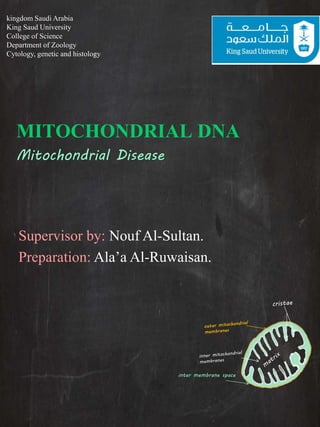
- 1. MITOCHONDRIAL DNA Mitochondrial Disease Supervisor by: Nouf Al-Sultan. Preparation: Ala’a Al-Ruwaisan. inter membrane space kingdom Saudi Arabia King Saud University College of Science Department of Zoology Cytology, genetic and histology
- 2. Objectives: 1. Who Discovered the Mitochondria? 2. What are Mitochondria? 3. Structure of Mitochondria. 4. What is mitochondrial DNA? 5. Characteristics of mitochondria dna. 6. NUCLEAR DNA VS MITOCHONDRIAL DNA. 7. Schematic representation of mammalian mtDNA. 8. The RITOLS model of mitochondrial DNA replication. 9. What is Mitochondrial Disease 1.Replicative Segregation 2.Homoplasmy and Heteroplasmy 3.Maternal Inheritance 1o- Why is mitochondrial mutation so high? 11- Type Disease of Mitochondria 12- New gene therapy for mitochondrial diseases a step closer thanks to ONPRC inter membrane space
- 3. Discovered the Mitochondria: • There isn't one single person credited with discovering the mitochondria, as over the years a number of scientists have made important contributions to the study of the discovery of this important cellular structure: • 1800s In 1857, Albert von Kölliker described what he called “granules” in the cells of muscles. - Other scientists of the era also noticed these “granules” in other cell types. • 1886 , when Richard Altman, a cytologist, identified the organelles using a dye technique, and dubbed them “bioblasts.” He postulated that the structures were the basic units of cellular activity. • 1898, Carl Benda coined the term mitochondria. He derived the term from the Greek language for the words thread, mitos, and granule, chondros. -Though mitochondria are an integral part of the cell, evidence shows that they evolved from primitive bacteria. inter membrane space
- 4. What are Mitochondria? • Mitochondria are specialized structures unique to the cells of animals, plants and fungi. • They serve as batteries, powering various functions of the cell and the organism as a whole.. • In nearly every cell in the body, Mitochondria are responsible for producing energy (called ATP) that the cell needs to function. • They are like power stations in our bodies, supplying the energy every cell needs to function. inter membrane space
- 5. Mitochondria Structure: • Mitochondria are rod shaped structure found in both animal and plant cells. • It is a double membrane bound organelle. It has the outer membrane and the inner membrane. The membranes are made up of phospholipids and proteins.
- 6. Mitochondria Structure: • The components of mitochondria are as follows: • Outer membrane: It is smooth and is composed of equal amounts of phospholipids and proteins. • It has a large number of special proteins known as the porins. • The porins are integral membrane proteins and they allow the movement of molecules that are of 5000 daltons or less in weight to pass through it. • The outer membrane is freely permeable to nutrient molecules , ions, energy molecules like the ATP and ADP molecules.
- 7. • Inner membrane: The inner membrane of mitochondria is more complex in structure. • It is folded into a number of folds many times and is known as the cristae. • This folding help to increase the surface ares inside the organelle. • The cristae and the proteins of the inner membrane aids in the production of ATP molecules. • Various chemical reactions takes place in the inner membrane of the mitochondria. • Unlike the outer membrane, the inner membrane is strictly permeable, it is permeable only to oxygen, ATP and it also helps in regulating transfer of metabolites across the membrane. Mitochondria Structure:
- 8. • Intermembrane space: It is the space between the outer and inner membrane of the mitochondria, it has the same composition as that of the cell's cytoplasm. • There is a difference in the protein content in the intermembrane space. • Matrix: The matrix of the mitochondria is a complex mixture of proteins and enzymes. These enzymes are important for the synthesis of ATP molecules, mitochondrial ribosomes, tRNAs and mitochondrial DNA. Mitochondria Structure:
- 9. What is mitochondrial DNA • Mitochondrial DNA or mtDNA or mDNA is the DNA in the mitochondria, rest of the DNA present in the eukaryotic cells is in the nucleus, in plants DNA is also found in chloroplasts. • The mitochondria have a small amount of DNA of their own. • Human mitochondrial DNA spans about 16,500 DNA base pairs, it represents a small fraction of the total DNA in cells. The mtDNA contains 37 genes. All these genes are essential for normal function of the mitochondria. inter membrane space
- 10. Characteristics of mitochondria DNA: • Is inherited exclusively from the mother! mtDNA is a circular shape single chromosome • It is only 16 kb in length - contains 16,600 bp. • Codes for 37 genes. • Contains 22 tRNA and 2 rRNA coding genes. • Encodes 13 proteins that are subunits of oxidative phosphorilation. • Contains only exons, no introns. • Has no reparation system – high mutation rate especially in D-loop! • No crossing over. • Replicative segregation, homoplasmy & heteroplasmy inter membrane space
- 11. • Nuclear DNA – found in nucleus of the cell – 2 sets of 23 chromosomes – maternal and paternal – can "discriminate between individuals of the same maternal lineage“ – double helix – bounded by a nuclear envelope – DNA packed into chromatin • Mitochondrial DNA – found in mitochondria of the cell – each mitochondria may have several copies of the single mtDNA molecule – maternal only – cannot "discriminate between individuals of the same maternal lineage“ – Circular – free of a nuclear envelope – DNA is not packed into chromatin Nuclear DNA vs. Mitochondrial DNA
- 12. Schematic representation of mammalian mtDNA. Chan Bae Park, and Nils-Göran Larsson J Cell Biol 2011;193:809-818 © 2011 Park and Larsson 1. the double-stranded circular mammalian mtDNA 2. molecule of ∼16.5 kb contains 3. Asingle longer noncoding region, the displacement loop (D loop) region. 4. harboring the promoters for transcription of both mtDNA strands (HSP and LSP) and the origin of leading strand replication (OH). 5. The origin of lagging strand replication (OL) is embedded in a cluster of tRNA genes. 6. The genes for the two rRNAs (12S and 16S rRNA),. 7. 13 mRNAs (ND1–6, ND4L, Cyt b, COI–III, ATP6, and ATP8),. 8. 22 tRNAs (F, V, L1, I, M, W, D, K, G, R, H, S1, L2, T, P, E, S2, Y, C, N, A, and Q) are indicated by boxes. Illustration by Annika Röhl.
- 13. The RITOLS model of mitochondrial DNA replication. Ian J. Holt, and Aurelio Reyes Cold Spring Harb Perspect Biol 2012;4:a012971 ©2012 by Cold Spring Harbor Laboratory Press The RITOLS model of mitochondrial DNA replication. Replication initiates at one of two sites (OH or Ori-b), here conflated to OR, for origin of replication. Leading strand DNA synthesis progresses with concurrent incorporation of RNA on the lagging strand. At some point (frequently OL), lagging strand DNA synthesis initiates and the lagging strand RNA is replaced by, or converted to, DNA.
- 15. Disease of Mitochondria • More than 100 different rearrangements and 100 different point mutations have been identified in mtDNA that can cause human disease, often involving the CNS and musculoskeletal system • The diseases that result from these mutations show distinctive pattern of inheritance because of 3 unusual features of mitochondria. 1.Replicative Segregation 2.Homoplasmy and Heteroplasmy 3.Maternal Inheritance
- 16. 1.Replicative Segregation • At cell division, multiple copies of mtDNA in each of the mitochondria in a cell replicate and sort randomly among newly synthesized mitochondria. • The mitochondria in turn are distributed randomly between the 2 daughter cells. This is called as replicative segregation. • The first unique feature of mitochondria is the absence of tightly controlled segregation seen during mitosis and meiosis of the 46 nuclear chromosomes. inter membrane space
- 18. 2.Homoplasmy and Heteroplasmy • One daughter cell may by chance receive mitochondria that contain only a pure population of normal mtDNA or a pure population of mutant mtDNA (Homoplasmy) • The daughter cell may receive a mixture of mitochondria some with and some without mutation (Heteroplasmy)
- 20. 3.Maternal Inheritance of mtDNA • Sperm mitochondria are generally eliminated from the embryo so that mtDNA is inherited from the mother. • All children of a female who is homoplasmic for a mtDNA mutation will inherit the mutation • None of the offspring of a male carrying the same mutation will inherit the defective DNA • Maternal inheritance of a homoplasmic mtDNA mutation causing Leber Hereditary optic neuropathy is known. inter membrane space
- 22. Why is mitochondrial mutation so high? • The mitochondrial genome has a very high mutation rate, 10- to 17-fold higher than that observed in nuclear DNA. • Although mtDNA repair systems do exist and , they are not sufficient to counteract the oxidative damage sustained by the mitochondrial genome. • Protective histones are also lacking. • Oxygenation process is high percentage. • The mtDNA mutation rate can be increased by environmental agents or by mutation of nuclear genes involved in mtDNA maintenance inter membrane space
- 23. • It has been identified in mtDNA: (1) missense mutations in the coding regions of genes that alter the activity of an oxidative phosphorylation protein; (2) point mutations in tRNA or rRNA genes that impair mitochondrial protein synthesis; (3) Deletions or duplications of the mtDNA molecule. They are generally somatic in origin, although a small proportion is inherited, in some diseases. THREE TYPES OF MUTATIONS inter membrane space
- 24. Type Disease of Mitochondria 1. Alpers Disease 2. Barth Syndrome / LIC (Lethal Infantile Cardiomyopathy) 3. Beta-oxidation Defects 4. Carnitine-Acyl-Carnitine Deficiency 5. Carnitine Deficiency 6. Creatine Deficiency Syndromes 7. Co-Enzyme Q10 Deficiency 8. Complex I Deficiency 9. Complex II Deficiency 10.Complex III Deficiency 11. Complex IV Deficiency / COX Deficiency 12.Complex V Deficiency 13.CPEO 14.CPT I Deficiency 15.CPT II Deficiency 16.KSS 17.Lactic Acidosis 18.LBSL - Leukodystrohpy 19.LCAD 20.LCHAD inter membrane space
- 25. Type Disease of Mitochondria 21.Leigh Disease or Syndrome 22.Luft Disease 23.MAD / Glutaric Aciduria Type II 24.MCAD 25.MELAS 26.MERRF 27.MIRAS 28.Mitochondrial Cytopathy 29.Mitochondrial DNA Depletion 30.Mitochondrial Encephalopathy 31.Mitochondrial Myopathy 32.MNGIE 33.NARP 34.Pearson Syndrome 35.Pyruvate Carboxylase Deficiency 36.Pyruvate Dehydrogenase Deficiency 37.POLG Mutations 38.Respiratory Chain 39.SCAD 40.SCHAD 41.VLCAD inter membrane space
- 27. 1.LHON • Disease Description: • Leber’s Hereditary Optic Neuropathy, also called LHON or Leber’s (LAY-bers), is a rare condition which can cause sudden, profound, painless loss of central vision. • While symptoms can begin at any age and in men or women, it is most common among men around age 20. • LHON is an extremely rare genetic disorder that is passed through the egg cell of the mother. • These mutations can lead to the reduction in cellular energy production, which in turn results in cell damage and death of certain optic nerve cells. • average 50% of men and 15% of women with a LHON mutation will lose vision in their lifetime.
- 28. 2.MERRF (Myoclonic Epilepsy with Ragged Red Fibres) • Long Name: Myoclonic Epilepsy and Ragged-Red Fiber Disease. • Symptoms: Myoclonus, epilepsy, progressive ataxia, muscle weakness and degeneration, deafness, and dementia. • Cause: Mitochondrial DNA point mutations: A8344G, T8356C • MERRF is a progressive multi-system syndrome usually beginning in childhood, but onset may occur in adulthood. • The rate of progression varies widely. • Onset and extent of symptoms can differ among affected siblings. inter membrane space
- 30. 3.CPEO • Long Name: Chronic Progressive External Ophthalmoplegia Syndrome. • Symptoms: visual myopathy, retinitis pigmentosa, dysfunction of the central nervous system. • Cause: Single mitochondrial DNA deletions. Mitochondrial DNA point mutations: A3243G (most common) inter membrane space
- 32. 4. MELAS • Long Name: Mitochondrial Encephalomyopathy Lactic Acidosis and Strokelike Episodes. • Cause: Mitochondrial DNA point mutations: A3243G (most common) • MELAS is a progressive neurodegenerative disorder with typical onset between the ages of 2 and 15, although it may occur in infancy or as late as adulthood. Initial symptoms may include stroke-like episodes, seizures, migraine headaches, and recurrent vomiting. • Typically, the age of death is between 10 to 35 years, although some patients may live longer. • Death may come as a result of general body wasting due to progressive dementia and muscle weakness, or complications from other affected organs such as heart or kidneys. inter membrane space
- 33. • Mitochondrial DNA schematically depicted as circular double-strand DNA with some mutation syndromes. • MIDDM = maternally inherited deafness and diabetes mellitus; MELAS = mitochondrial encephalomyopathy, lactic azidosis and stroke-like episodes; DIDMOAD = diabetes insipidus, diabetes mellitus, optic atrophy, and deafness; KSS = Kearns-Sayre syndrome; LHON = Leber‘s hereditary optic neuropathy; MERRF = myoclonic epilepsy, ragged-red fibers; NARP = neuropathy, ataxia, and retinitis pigmentosa.
- 34. Figure 2 The mitochondrial morbidity map DiMauro, S. et al. (2013) The clinical maze of mitochondrial neurology Nat. Rev. Neurol. doi:10.1038/nrneurol.2013.126
- 35. Other disorders • A buildup of somatic mutations in mitochondrial DNA has been associated with an increased risk of certain age- related disorders such as heart disease, Alzheimer disease, and Parkinson disease. Additionally, research suggests that the progressive accumulation of these mutations over a person's lifetime may play a role in the normal aging process. inter membrane space
- 36. New gene therapy for mitochondrial diseases a step closer thanks to ONPRC
- 37. Any question
- 38. References • Human Mitochondrial DNA Replication , Ian J. Holt and Aurelio Reyes • The mitochondrial genome: structure, transcription, translation and replication, Taanman JW1, 1999 Feb. http://www.sciencedirect.com/science/article/pii/S00 05272898001613 • Mitochondrial DNA mutations in disease and aging Chan Bae Park1 and Nils-Göran Larsson2 • Mitochondrial DNA mutations in human disease Robert W. Taylor & Doug M. Turnbull • http://www.childneurologyfoundation.org/disorders/mi tochondrial-diseases/ • http://www.smw.ch/content/smw-2012-13690/ • Novel techniques for the prevention of mitochondrial DNA disorders an ethical review
- 39. Good luck
Editor's Notes
- Schematic representation of mammalian mtDNA. The double-stranded circular mammalian mtDNA molecule of ∼16.5 kb contains a single longer noncoding region, the displacement loop (D loop) region, harboring the promoters for transcription of both mtDNA strands (HSP and LSP) and the origin of leading strand replication (OH). The origin of lagging strand replication (OL) is embedded in a cluster of tRNA genes. The genes for the two rRNAs (12S and 16S rRNA), 13 mRNAs (ND1–6, ND4L, Cyt b, COI–III, ATP6, and ATP8), and 22 tRNAs (F, V, L1, I, M, W, D, K, G, R, H, S1, L2, T, P, E, S2, Y, C, N, A, and Q) are indicated by boxes. Illustration by Annika Röhl.
- The RITOLS model of mitochondrial DNA replication. Replication initiates at one of two sites (OH or Ori-b), here conflated to OR, for origin of replication. Leading strand DNA synthesis progresses with concurrent incorporation of RNA on the lagging strand. At some point (frequently OL), lagging strand DNA synthesis initiates and the lagging strand RNA is replaced by, or converted to, DNA.