1. Breast disease
How to make breast examination :-
-Tripple assessment
.Physical examination
.Radiological examination (U.S , for patient < 35 years , > 35 Mamography)
.Pathological assessment : (Needle aspiration and Exsicion biopsy)
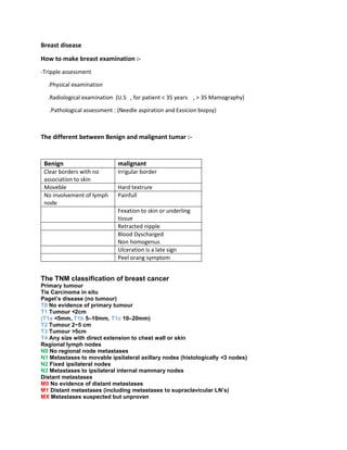
The different between Benign and malignant tumar :-
Benign malignant
Clear borders with no
association to skin
Irrigular border
Moveble Hard textrure
No involvement of lymph
node
Painfull
Fexation to skin or underling
tissue
Retracted nipple
Blood Dyscharged
Non homogenus
Ulceration is a late sign
Peel orang symptom
The TNM classification of breast cancer
Primary tumour
Tis Carcinoma in situ
Paget’s disease (no tumour)
T0 No evidence of primary tumour
T1 Tumour <2cm
(T1a <5mm, T1b 5–10mm, T1c 10–20mm)
T2 Tumour 2−5 cm
T3 Tumour >5cm
T4 Any size with direct extension to chest wall or skin
Regional lymph nodes
N0 No regional node metastases
N1 Metastases to movable ipsilateral axillary nodes (histologically <3 nodes)
N2 Fixed ipsilateral nodes
N3 Metastases to ipsilateral internal mammary nodes
Distant metastases
M0 No evidence of distant metastases
M1 Distant metastases (including metastases to supraclavicular LN’s)
MX Metastases suspected but unproven
2. Acute Mostitis :-
Acute bacterial infection of the breast is related to lactation in most cases
Types of acute mastitis:-
-Suppurative (phlegmon , purulent , gangrenous )
-Serous
-Lacteting mostits (treatment: flucloxacillin ,Breast drainage of affected segment)
-Non lactating mastitis (treatment : Amoxiclane)
Classification of tumer :-
- Benign fibroadinoma
- Benign papilloma (ductel , lopular)
- Malignant (Ductel , lopular)