
Recommended
More Related Content
What's hot
What's hot (20)
Viewers also liked
Viewers also liked (20)
Similar to Genetics of hemophilia A
Similar to Genetics of hemophilia A (20)
More from Ahmad Qudah
More from Ahmad Qudah (10)
Recently uploaded
Recently uploaded (20)
Genetics of hemophilia A
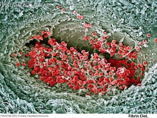
- 1. Fibrin Clot.
- 2. The Molecular Genetics of Haemophilia A Ahmad A. Al-Qudah Supervisor : Dr. Laila F. Nimri
- 3. Genetics of Haemophilia A - Hematology & Coagulation Cascade . - Coagulopathies . - Haemophilia A : - Genetics . - Diagnosis . - Treatment ( Management ) . - Acquired Haemophilia - Abstract . - ‘’ The Royal Disease ‘’ . - Bibliography .
- 4. Hematology & Coagulation Cascade Hematology : is the study of blood and it’s components .
- 5. Hematology & Coagulation Cascade Coagulation Cascade : is the process by which blood forms clots , prevention of blood loss from a damaged vessel . I – fibrinogen II – Prothrombin III – Tissue thromboplastin/tissue factor IV – Calcium V – Proacelerin VII – Proconvertin VIII – Hemophilia A (antihemophilic factor) IX – Hemophilia B (Christmas factor) X – Stuart-Prower factor XI – Plasma thromboplastin antecedent (PTA) XII – Hageman factor XIII – Fibrin stabilizing factor Prekallikrein/Fletcher factor High molecular weight kininogen (HMWK)/Fitzgerald factor
- 6. Hematology & Coagulation Cascade
- 7. Coagulopathies is a condition in which the blood’s ability to clot is impaired. This condition can cause prolonged or excessive bleeding, which may occur spontaneously or following an injury or medical and dental procedures. Genetic disorder or Acquired , Resulting in : 1- insufficient amount of Plt or Coagulation factor . 2- nonfunctional Plt or Coagulation factor . 3- vessel wall damage .
- 8. Coagulopathies Also we can classify Coagulopathy into : Hyper-Coagulobility : ( thrombophilia ) is an abnormality of blood coagulation that increases the risk of thrombosis . - DIC , Antithrombin III deficiency , Thrombocytosis . Hypo-Coagulobility : is an unusual susceptibility to bleeding due to an abnormality in coagulation. - Thrombocytopenia , Haemophilia , vWD .
- 9. Haemophilia is a group of hereditary genetic disorders that impair the body's ability to control blood clotting or coagulation . Haemophilia A (clotting factor VIII deficiency) Haemophilia B (clotting factor IX deficiency) Haemophilia C (clotting factor XI deficiency)
- 10. Haemophilia A clotting factor VIII deficiency - Most common type ( 85 % ) - Frequency of 1 per 10,000 males - In 1/3 of cases there is no family history - Inherited as an X-linked recessive disorder - Commonly an inversion of intron 1 and 22
- 11. Haemophilia A Molecular Genetics The factor VIII gene is situated near the tip of the long arm of the X chromosome (Xq28 region). It is extremely large and consists of 26 exons. Commonly an inversion of intron 1 and 22.
- 12. Haemophilia A Signs and symptoms Characteristic symptoms vary with severity. In general symptoms are internal or external bleeding episodes. Patients with more severe hemophilia suffer more severe and more frequent bleeds, while patients with mild hemophilia typically suffer more minor symptoms except after surgery or serious trauma.
- 13. Haemophilia A Bleeding may occur anywhere in the body. Superficial bleeding, or shallow. Most serious sites of bleeding are : - Joints - Muscles - Digestive tract - Brain The muscle and joint hemorrhages are indicative of hemophilia.
- 15. Haemophilia A
- 16. Haemophilia A
- 17. Haemophilia A
- 18. Haemophilia A
- 19. Haemophilia A Diagnosis Clinical Diagnosis A specific diagnosis of hemophilia A cannot be made on clinical findings , but The muscle and joint hemorrhages are indicative . Testing Coagulation screening tests. Evaluation of an individual with a suspected bleeding disorder includes: platelet count and platelet function analysis (PFA closure times) or bleeding time; activated partial thromboplastin time (APTT); and prothrombin time (PT), In Haemophilia , PTT and APTT is prolonged . This Prolongation is vary according to the severity of the case .
- 20. Haemophilia A Management - There is no cure for haemophilia - Controlled with regular infusions of the deficient clotting factor. - Factor replacement can be either isolated from human blood serum, recombinant, or a combination of the two.
- 21. Haemophilia A - Some haemophiliacs develop antibodies (inhibitors) against the replacement factors given to them, so the amount of the factor has to be increased or non-human replacement products must be given, such as porcine factor VIII.
- 22. Acquired Haemophilia A - Due to antibody to factor VIII (most common autoimmune factor deficiency) - Most patients elderly - Often presents with severe soft tissue or mucosal bleeding (different bleeding pattern than inherited hemophilia)
- 23. Abstract Hemophilia A is an X-linked bleeding disorder resulting from a defect in coagulation factor VIII. Clinical severity, the level of factor VIII activity and coagulant antigen vary widely. However the phenotypic expression directly reflects the genetic defect. In about 5%, hemophilia A results from partial deletion of the factor VIII gene and is clinically severe. In another 5% single base mutations have been found, which destroy the binding sites for the restriction enzyme TaqI. They can be "nonsense" mutations resulting in stop codons and clinically severe hemophilia, or "missense" mutations resulting in amino acid changes and milder forms of hemophilia. By the use of the polymerase chain reaction and denaturing gradient gel electrophoresis we have detected several point mutations. The formation of anti-factor VIII antibodies appears to be more frequent in patients with defects resulting in absence of factor VIII protein. Carrier detection and prenatal diagnosis can be made with 100% certainty in families with identified mutations. In the absence of any informative polymorphism the conventional methods for carrier detection are still helpful
- 24. Haemophilia “ The Royal Disease “ - Queen Victoria , through two of her five daughters - passed the mutation to various royal houses across the continent, including the royal families of Spain, Germany and Russia - Her son Leopold suffered from frequent hemorrhages before dying from a brain hemorrhage at 31
- 25. Genetics of Haemophilia A Bibliography • THROMBIN GENERATION AND BLEEDING IN HEMOPHILIA A http://www.ncbi.nlm.nih.gov/pmc/articles/PMC3395070/ • A homozygous female hemophilia A http://www.ncbi.nlm.nih.gov/pmc/articles/PMC3385172/ • Molecular genetics of hemophilia A http://www.ncbi.nlm.nih.gov/pubmed/2508218 • Porcine model of hemophilia A http://www.ncbi.nlm.nih.gov/pubmed/23209578 • A Strategy for the Molecular Diagnosis in Hemophilia A in Chinese Population http://www.ncbi.nlm.nih.gov/pubmed/23111982 • The gene therapy journey for hemophilia: are we there yet? http://www.ncbi.nlm.nih.gov/pubmed/22829631 • Acquired Haemophilia A http://www.ncbi.nlm.nih.gov/pubmed/23067181