More Related Content
Similar to AN INTERESTING CASE OF EXTRAMEDULLARY HEMATOPOIESIS IN A PATIENT WITH NEWLY DIAGNOSED HEMOGLOBIN H DISEASE
Similar to AN INTERESTING CASE OF EXTRAMEDULLARY HEMATOPOIESIS IN A PATIENT WITH NEWLY DIAGNOSED HEMOGLOBIN H DISEASE (20)
AN INTERESTING CASE OF EXTRAMEDULLARY HEMATOPOIESIS IN A PATIENT WITH NEWLY DIAGNOSED HEMOGLOBIN H DISEASE
- 1. RESEARCH POSTER PRESENTATION DESIGN © 2015
www.PosterPresentations.com
Introduction: Extramedullary hematopoiesis (EMH) is a rare entity, which consists of the formation of
hematopoietic tissue in various parts of the human body due to the existence of a hematological disorder. A–
thalassemia is a great example of a disease that can result in extramedullary hematopoiesis.
Hemoglobin H disease is a type of a-thalassemia consisting of the loss of 3 genes (--/-a) and is often
manifested as a mild anemia with thalassemic morphological changes and the existence of erythrocyte
inclusions. Hemoglobin H is a tetramer composed of four beta globin chains and a great decrease in alpha
chain availability. In this hemolytic disorder, splenomegaly may occur, as well as bone marrow hyperplasia
and pathological fractures.
Case description: The present study reports an unusual case of a 64-yr-old male, former blood donor, who
was admitted to our clinic for investigation recent occurrence of mild anemia with splenomegaly, the
appearance of a paraspinal mass and monoclonal gammopathy. Eventually, thalassemia-associated tests
confirmed the diagnosis of hemoglobin H disease. Further examinations with bone marrow aspiration and
biopsy, as well as an MRI scan revealed that the patient had extramedullary hematopoiesis due to
hemoglobin H disease, with monoclonal gammopathy of undetermined significance (MGUS)
Discussion: The question remains what is to be done in this kind of situations. Surely these patients should
remain under observation. Whether or not they should receive a blood transfusion relays upon the better
judgement of the physician in charge, who is after all responsible for weighing the advantages and
disadvantages of multiple blood transfusions, especially since many of these patients will later on require
chelation therapy in order to treat the iron overload.
ABSTRACT
CASE REPORT DISCUSSION - CONCLUSIONS
REFERENCES
1.An J, Weng Y, He J, Li Y, Huang S, Cai S, Zhang J: Intrathoracic extramedullary hematopoiesis presenting
as tumor-simulating lesions of the mediastinum in α-thalassemia: A case report. Oncology Letters 10: 1993-
1996,2015
2.Chu KA, Lai RS, Lee CH,Lu JY, Chang HC, Chiang HT:Intrathoracic extramedullary haematopoiesis
complicated by massive haemothorax in alpha-thalassaemia. Thorax 1999, 54:466-468
3.Kalchiem- Dekel O, Greenbaum U:Extramedullary Hematopoiesis in β-Thalassemia. Mayo Clin Proc.
November 2015;90(11):1591-1592
4.McDonald K,Kermalli H, Majumder S, Naut E:An uncommon cause of abdominal pain in a patient with
thalassemia intermedia. The American Journal of Medicine,2014.03.005
5.Ueda T, Migita M, Yamanishi M, Maeda M, Harano K, Fukunaga Y:A 6-year-old girl with hemoglobin H
disease. J Nippon Med Sch 2011; 78(2)
6.Molina-Urra R, Martinez D, Sagasta A, Carrio A, Setoain X, Nomdedeu B, Campo E: Paraspinal
extramedullary hematopoiesis in hereditary spherocytosis with a concurrent follicular lymphoma: case
report and review of the literature. Diagnostic Pathology (2015) 10:158
7.Bobylev D, Zhang R, Haverich A, Krueger M :Extramedullary haematopoiesis presented as intrathoracic
tumour in a patient with alpha-thalassaemia. Journal of Cardiothoracic Surgery 2013, 8:120
8.Winichakoon P, Tantiworawit A, Rattanathammethee T, Hantrakool S, Chai-Adisaksopha C, Rattarittamrong e,
Norasetthada L, Charoenkwan P : Prevalence and risk factors for complications in patients with Non-
transfusion dependent alpha- and beta- thalassemia. Anemia. 2015; 2015:793025
Introduction: Extramedullary hematopoiesis functions as a compensatory phenomenon in cases of
insufficient bone marrow function and refers to the hematopoiesis that takes place outside the medulla of the
bone. It is usually associated with various hematologic disorders, including thalassemia.1–4
Hemoglobin H disease is a type of a-thalassemia (inherited, autosomal disorder) consisting of the loss of 3
genes (--/-a) and is often manifested as a mild microcytic, hypochromic anemia with the existence of
erythrocyte inclusions. Hemoglobin H is a tetramer composed of four beta globin chains and a great
decrease in alpha chain availability.5 It is frequently observed in Mediterranean countries, South-East Asia
and Africa. In this hemolytic disorder, splenomegaly may occur, as well as and bone marrow hyperplasia
and pathological fractures. As a compensatory response to inefficient hematopoiesis due to thalassaemia ,
extramedullary hematopoiesis may develop.
The present study describes a case of extramedullary hematopoiesis presenting as a paraspinal lesion with
monoclonal gammopathy in a 64-year –old patient undergoing medical examination after an accident.
Case Report : A 64-year-old patient presented with intense back pain to our hospital. He was a former blood
donor, smoker, heavy alcohol drinker without other health problems. Physical examination revealed blood
pressure 150/90 mmHg, respiratory rate 15/min and pulse rate 97/min but no other remarkable findings. His
laboratory studies revealed anemia (Hb 9,4 g/dl), normal WBC and PLTs (WBC 8100/μl and PLT 261.000
/μl respectively), elevated RBC (5.070.000 / μl), normal ESR (4mm/hr) and as well as a marked monoclonal
gammopathy (1,90 g/dl). Serum glucose, electrolytes, renal and hepatic function were normal. In addition to
the above, there was an elevated ferritin level (954 ng/ml). Subsequently, a CT scan revealed hepatomegaly,
splenomegaly and soft-tissue mass of 4.5 cm extending longitudinally in the left thoracic paravertebral space
at the T9-T10 vertebral bodies.
The existence of mild anemia and of a paraspinal mass in combination with the monoclonal gammapathy
lead to additional diagnostic tests. Quantitative immunoglobulin level measurement revealed IgG 1230
mg/dl, IgA 200 mg/dl and IgM 42 mg/dl and immunoblotting showed IgGλ. Bone marrow aspiration and
biopsy confirmed the findings of inefficient hematopoiesis with erythroid hyperplasia without invasion of
plasma cells or other malignant cells. The bone scan (Tc 99m nanocoloid) showed signs of an
overfunctioning bone marrow (Image 1) Hemoglobin electrophoretic studies showed HbA2 (1,3%), as well
as positive hemoglobin H and hemoglobin F (HbF<2,0%) Furthermore, thalassemia-associated molecular
tests with genotypic analysis were performed and confirmed the diagnosis of a-thalassemia. The magnetic
resonance imaging confirmed the possibility of the paraspinal mass being extramedullary hematopoiesis
(Image 2)
The hemoglobin H disease was actually diagnosed by the presence of erythrocyte inclusions (Image 3).
Eventually, the patient was diagnosed with extramedullary hematopoiesis due to hemoglobin H disease, with
concurrent monoclonal gammopathy of undetermined significance (MGUS).
Tsifi A.¹Mantzourani M.²Tsifis I.²Theodoridis D.³Vieru A-M.²Triantafyllou M.² Lontou S-P²Solomos Z.´Miltiadou K.µDaikos G.²
AN INTERESTING CASE OF EXTRAMEDULLARY HEMATOPOIESIS IN A PATIENT WITH
NEWLY DIAGNOSED HEMOGLOBIN H DISEASE
¹National and Kapodistrian University of Athens, 1st Department of Internal Medicine, ″Laikon″ General Hospital , Athens, Greece.
atsifi@hotmail.com ² National and Kapodistrian University of Athens, 1st Department of Internal Medicine, ″Laikon″ General Hospital,
Athens, Greece ³Biopathology Department, Pedi ″Ilion″, Athens, Greece ´1st Department of Internal Medicine, ″Pammakaristos″
General Hospital, Athens, Greece µDepartment of Medical Oncology, ″Metaxa″ Special Cancer Hospital, Piraeus, Greece
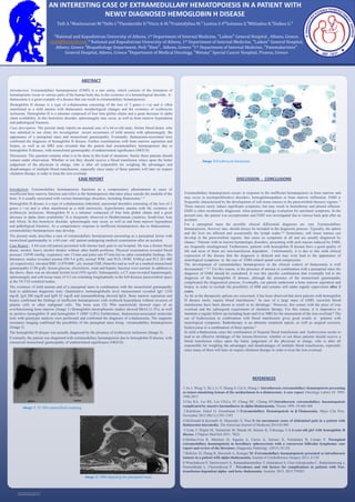
Image 2: MRI depicting the paraspinal mass
Image 1 :Tc 99m nanocolloid scanning
Image 3:Erythrocyte Inclusions
Extramedullary hematopoiesis occurs in response to the inefficient hematopoiesis in bone marrow and
may occur in myeloproliferative disorders, hemoglobinopathies or bone marrow infiltration. EMH is
frequently characterized by the development of soft tissue masses in the paravertebral thoracic regions. 6
These masses rarely induce significant symptoms, but may result in hemothorax and pleural effusion.
EMH is often incidentally diagnosed, when patients undergo evaluation for unrelated symptoms. In the
present case, the patient was asymptomatic and EMH was investigated due to intense back pain after an
accident.
For a paraspinal mass the possible clinical differential diagnoses are vast. Extramedullary
hematopoiesis, however rare, should always be included in the diagnostic process. Typically, the spleen
and the liver are affected and occasionally the lymph nodes.4,6 Sometimes, soft tissue masses can
develop in the paravertebral regions, that cause no symptoms at all and are usually diagnosed by
chance.1 Patients with no known hematologic disorders, presenting with such masses induced by EMH,
are frequently misdiagnosed. Furthermore, patients with hemoglobin H disease have a good quality of
life and most of them are non-transfusion dependent. Unfortunately, it is due to this mild clinical
expression of the disease that the diagnosis is delayed and may even lead to the appearance of
neurological symptoms in the case of EMH-related spinal cord compression.
The development of extramedullary hematopoiesis in the clinical context of thalassemia is well
documented.1–3,6,7 For this reason, in the presence of anemia in combination with a paraspinal mass the
diagnosis of EMH should be considered. It was this specific combination that eventually led to the
diagnosis of the hemoglobin H disease, although the presence of the monoclonal gammopathy
complicated the diagnostical process. Eventually, our patient underwent a bone marrow aspiration and
biopsy in order to exclude the possibility of MM and remains still under regular supervision after 3
years.
As far as the therapeutic options are concerned, it has been observed that most patients with hemoglobin
H disease rarely require blood transfusions.5 In case of a large mass of EMH, recurrent blood
transfusions have been shown to result in its shrinkage.1 However, this comes with the price of iron
overload and the subsequent requirement of chelation therapy. For this reason, it is imperative to
maintain a regular follow-up including heart and liver MRI for the measument of the iron overload.8 The
use of hydroxyurea in combination with blood transfusions gives good results in patients with
neurological symptoms. Radiotherapy is an alternate treatment option, as well as surgical excision,
hydroxyurea or a combination of these options.2
In mild a-thalassemia cases the combination of frequent blood transfusions and hydroxyurea seems to
lead to an effective shrinkage of the lesions.However, whether or not these patients should receive a
blood transfusion relays upon the better judgement of the physician in charge, who is after all
responsible for weighing the advantages and disadvantages of multiple blood transfusions, especially
since many of them will later on require chelation therapy in order to treat the iron overload.