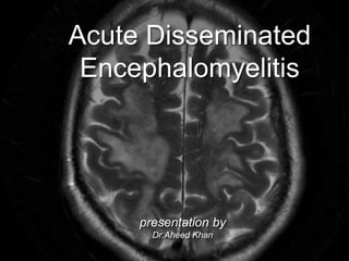
Acute Disseminated Encephalomyelitis
•Download as PPTX, PDF•
3 likes•1,666 views

This document discusses Acute Disseminated Encephalomyelitis (ADEM), a demyelinating disease of the central nervous system. It is an immune-mediated condition that mostly affects children and can be triggered by viral infections or vaccinations. Patients typically experience an acute neurological deficit with symptoms like encephalopathy, weakness, or vision loss. MRI scans show multifocal white matter lesions that are often large with ill-defined borders. Treatment involves high-dose steroids, and most patients fully recover, though some cases can be multiphasic with recurrence of symptoms. It is important to follow patients over time to distinguish ADEM from other conditions like multiple sclerosis.

Recommended
More Related Content
What's hot
What's hot (20)
Similar to Acute Disseminated Encephalomyelitis
Similar to Acute Disseminated Encephalomyelitis (20)
More from Aheed Khan
More from Aheed Khan (10)
Recently uploaded
Recently uploaded (20)
Acute Disseminated Encephalomyelitis
- 2. Demyelinating Disorders • inherent or acquired loss of myelin sheath in both PNS and CNS • Causes-genetic, metabolic, infectious or autoimmune mechanisms. • Demyelination can result progressively in ionic disequilibria, energy crisis, conduction block and eventually neurodegeneration.
- 4. types • Inflammatory/Immune-MS, ADEM, Optic Neuritis,SLE • Infectious- HIV,PML,Lyme’s disease • Granulomatous-Sarcoidosis, Wegeners • Myelin-Metachromatic Leukodystrophy, Adrenoleukodystrophy, Canavan disease,Alexander disease • Toxic/Metabolic-B12 deficiency,Cerebral pontine- myelinolysis,radiation, PRES
- 5. Acute Disseminated Encephalomyelitis • immune mediated self-limiting, inflammatory and demyelinating disease of CNS • mostly affects white matter of brain and spinal cord • acute-onset encephalopathy with neurological deficits, typically monophasic, • preceded by viral/bacterial infection or post-vaccination
- 6. . Epidemiology • pre-pubertal children • winter months • mean age 5 to 8 years, more fulminant in <2 years • slightly male predominance • 0.07-0.4 per 100’00 per year in paediatric population • mortality rates- <2%, commonly in <2 years • full recovery- 57-92% patients
- 7. History • preceding infection/vaccination- absent in 1/4rth patients • documentation of at least 1 fever-free day from prodromal illness s/o ADEM. • 1-2 days to several weeks hiatus following illness • neurological signs-mental status change/seizure/hemiparesis/weakness in extremities or bladder/bowel dysfunction/visual loss/craniopathies • posterior column abnormalities absent • age younger than 11-12 years
- 8. post-vaccination history • less than 5% follow vaccination • rabies, hepatitis B, influenza, Japanese B encephalitis, diphtheria/ pertussis/tetanus, measles, mumps, rubella, pneumococcus, polio, smallpox, and varicella. • Currently, measles, mumps, and rubella vaccination are most often associated with postvaccination ADEM
- 9. Clinical Features • fever, headache, vomiting, meningismus • encephalopathy-characteristic feature with neurological deficits [alteration in consciousness or behavioural change unexplained by fever, systemic illness or postictal symptoms] • level of consciousness-range from subtle lethargy to frank coma • rapid onset encephalopathy- a/w multifocal deficits or seizures[35% cases]
- 10. Neurological Symptoms • Encephalopathy-hallmark of ADEM • Long tract pyramidal signs like acute hemiparesis • cerebellar ataxia • cranial neuropathies, including optic neuritis • spinal cord dysfunction (transverse myelitis)
- 11. Pathophysiology • HPE-perivenular round cell inflammation • patchy demyelination with preservation of axon cylinders and the prominence of microglial cells in the inflammatory exudate • Possible Mechanism-T-helper cell mediated Molecular mimicry to auto-Ag • Serum IgG to Myelin Oligodendrocyte- glycoprotein[MGO]- 40%- a/w favourable outcome* perivenular round cell inflammation *Van Haren, K., Tomooka, B. H., Kidd, B. A., Banwell, B., Bar-Or, A., Chitnis, T., … Robinson, W. H. (2013). Serum autoantibodies to myelin peptides distinguish a
- 12. Examination • Irritability & Lethargy f/b headache or meningeal signs • Motor Defects- Weakness[75%]> Sensory Defects. Combination~localisation • Cranial Nerve Palsies-e.g visual loss[23-89%] • Ataxia-appendicular>axial ot gait [28-65%]
- 13. Diagnosis • Careful history and examination • Neuroimaging • Lab studies
- 14. Neuroimaging • MRI Brain>>CT Scan • T2/Flair Images for diagnosing demyelinati ng diseases Potential location for lesions in acquired demyelination
- 15. Lab findings • no specific marker for ADEM, • Basic Workup-CBC- thrombocytosis and Raised ESR • CSF Examination-non-specific: • pleocytosis-lymphocytic/monocytic • mild elevation of CSF WBC/RBC counts • elevated CSF protein • Antibodies to myelin oligodendrocyte-glycoprotein, anti-N-methyl-d-aspartate receptor • oligoclonal bands-10% ADEM; consider alternate diagnosis • EEG- generalised slowing~encephalopathy; polyregional demyelination-focal slowing
- 16. MRI Features • small punctate to tumefactive lesions-multifocal areas of hyperintensity on MRI • supratentorial or infratentorial white matter • bilateral, asymmetrical 7 year old ADEM MRI
- 17. ADEM Features • centrifugal at the junction of the deep cortical gray and subcortical white matter ADEM
- 18. ADEM Features • additional lesions-deeper white matter-basal ganglia,cerebellum, spinal cord ADEM FLAIR Image of ADEM showing lesions Type to enter a caption.
- 19. International Paediatric Multiple Sclerosis Study Group in 2012 Definitions: • CIS=Clinically Isolated syndrome: A first monofocal or multifocal CNS Demyelinating event • Monophasic ADEM: • first polyfocal CNS event • Encephalopathy-not explained by fever • MRI- large,poor margins, >1-2 lesions involving Cerebral white matter • no new symptoms or signs after 3 months • Multiphasic ADEM: new episode after 3 months or more from 1st episode with new or re-emergence or prior findings
- 20. .
- 21. Treatment • Pediatric MS- methylprednisolone[20-30mg/kg/day-max 1g/day] for 3-5 days for relapses, with or without taper; and for decreasing relapse frequency- Interferon alpha/beta or injectable DMA-natalizumab • NMO- Initial episodes and relapses -methylprednisolone for 3-5 days f/b tapering. Rituximab is effective in relapses • ADEM-high dose i/v steroids for 5 days, with oral prednisone taper of 1 month for relapses. Follow up at 3 months for checking signs of demyelination.
- 22. Follow-up • VERY IMPORTANT • first episode-encephalopathy-ADEM-needs follow up after 3 months-complete resolution=>Monophasic ADEM. if recurrence occurs- Multiphasic ADEM • first episode-no encephalopathy or signs of ataxia or focal sensory deficits, u/L loss of vision-> +/- MRI signs of MS=> label it as CIS; keep on follow up for recurrence. • if recurrence occurs with MRI findings consistent with MS with CSF findings=> label it as MS
- 23. THANK YOU