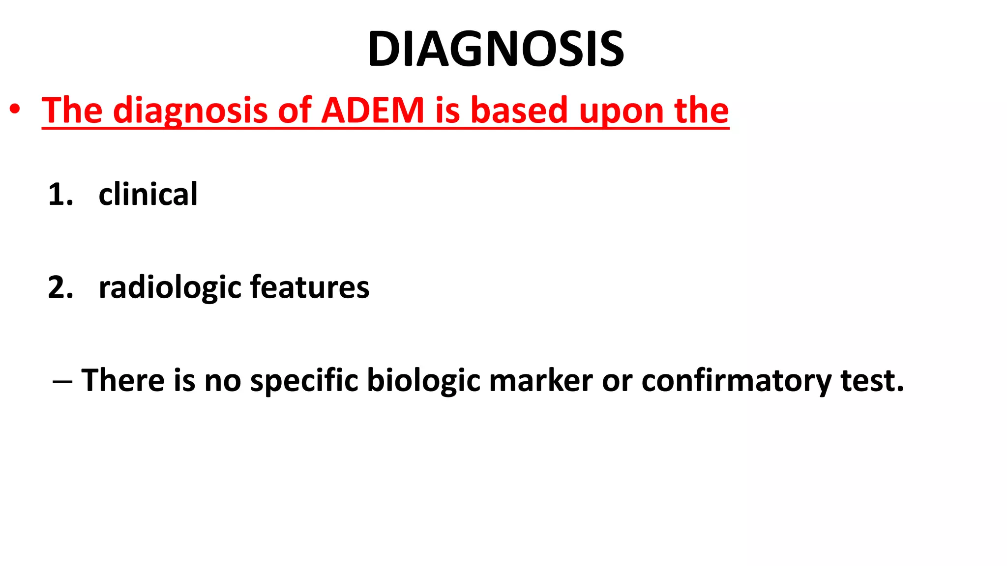
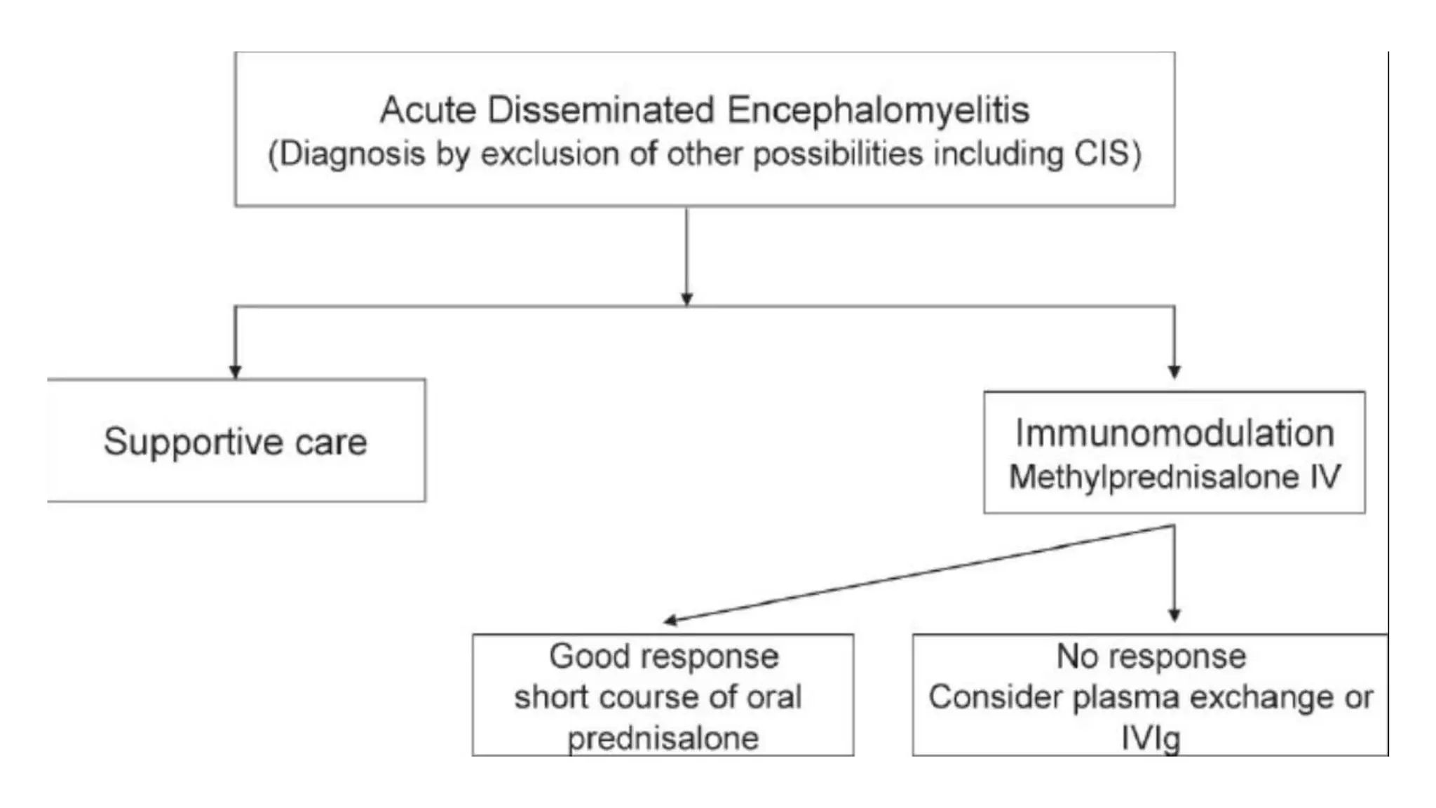
This document provides information about acute disseminated encephalomyelitis (ADEM). It defines ADEM as a demyelinating disease of the central nervous system that typically presents as a monophasic disorder with encephalopathy and multifocal neurological symptoms. The document discusses the pathogenesis, clinical features, diagnosis, differential diagnosis and treatment of ADEM. It states that ADEM is usually treated initially with high-dose intravenous corticosteroids over 3-5 days.
3. Demyelinating Diseases
A. Central nervous syetem:
1. Acute disseminated encephalomyelitis (ADEM)
2. Multiple sclerosis(MS)
3. Neuromyelitis optica(NMO)
B. Peripheral nervous syetem:
1. Acute inflammatory demyelinating polyneuropathy (AIDP)
4. Definition
is a demyelinating disease of the central nervous system
that typically presents as
1. monophasic disorder
2. encephalopathy
3. multifocal neurologic symptoms
5. INTRODUCTION
• Acute Disseminated Encephalomyelitis (ADEM)
– Usually monophasic
– Demyelinating disorder
– Characterized by diffuse neurologic signs and symptoms
(polysymptomatic)
– Nuroimaging ; multifocal lesions of demyelination
6. EPIDEMIOLOGY
ADEM is an uncommon illness
Mean age between 5 and 8 yr
estimated incidence is 0.4/100,000 population per year
Approximately three to six cases are seen each year
There is no specific ethnic distribution
indicate a slight male predominance
A seasonal distribution has been observed showing that most
ADEM cases occur in the winter and spring
7. PATHOGENESIS
Most of these conditions are thought to be
– caused by immune system dysregulation
– triggered by an infectious or other environmental agent
– genetically susceptible host.
9. PATHOGENESIS
The immunopathological events leading to ADEM
can be divided into two major phases :-
1. Initial T cell priming and activation
2. Subsequent recruitment and effector phase
10. PATHOGENESIS
typically following a recent (1-2 weeks prior) viral
infection or vaccination
• Sites of demyelination ;
1. white matter
2. Gray matter, especially that of the basal ganglia, is also often
involved
3. lesser extent, as is the spinal cord.
11. PATHOGENESIS
• Large number of viruses associated with these infections ;
– approximately 50-75 percent of ADEM cases
– including :
• Measles , Mumps, Rubella
• Varicella zoster, Epstein-Barr, Cytomegalovirus
• Herpes simplex, Hepatitis A, Influenza
• Enterovirus infections.
12. PATHOGENESIS
• Less than 5 percent of ADEM cases follow immunization.
• Associated with immunization for:
• Rabies, Hepatitis B, Influenza
• Japanese B encephalitis
• Diphtheria /Pertussis / Tetanus
• Measles, Mumps , Rubella,
• Pneumococcus, Polio, Smallpox, and Varicella.
15. CLINICAL FEATURES
• febrile illness occurs in 50 to 75 percent of children
• Neurologic symptoms typically appear 4 to 13 days after the
infection or vaccination
16. CLINICAL FEATURES
• The Neurological Signs :
1. Encephalopathy ,
2. multifocal neurologic deficits
18. CLINICAL FEATURES
• Encephalopathy results in symptoms, such as:
1. Altered level of consciousness
I. lethargy →coma
II. Confusion
III. Excessive irritability
2. Acute cognitive dysfunction
3. Behavioral changes
4. Seizures
• In about ⅓ of those diagnosed
19. CLINICAL FEATURES
• Other common neurologic signs of ADEM include:
1. Long tract pyramidal signs
2. Acute hemiparesis
3. Cerebellar ataxia
4. Cranial neuropathies
5. cranial neuropathies including optic neuritis
6. spinal cord dysfunction (transverse myelitis)
20. CLINICAL FEATURES
• less commonneurologic signs of ADEM include:
1. Aphasia
2. movement disorders
3. sensory deficits
21. CLINICAL FEATURES
• New clinical symptoms may develop during
hospitalization.
• Progression of neurologic signs to maximum deficits
usually occurs over four to seven days
22. CLINICAL FEATURES ; optic neuritis
• Symptoms of optic neuritis include
1. vision loss
2. pain with eye movement
3. afferent pupillary defect.
23. CLINICAL FEATURES
Transverse Myelitis
• Symptoms of transverse myelitis include
1. flaccid paralysis of the legs
2. sensory level on examination.
• The arms can be involved if the demyelinating lesion is in the cervical cord.
• Respiratory failure may occur with high cervical lesions that extend into the brainstem.
– Bowel and bladder involvement secondary to spinal cord disease results in
constipation and urinary retention
24. Clinical course
• The severe phase of ADEM typically lasts from two to four weeks.
• Children may deteriorate after hospital admission
– many develop new neurologic signs.
• Patients usually recover completely from the acute illness
– although some have neurologic sequelae.
25. hyperacute variants of ADEM
Inflammatory hemorrhagic demyelination of central nervous system white
matter is seen in rare conditions that are considered to be hyperacute
variants of ADEM
26. hyperacute variants of ADEM
o These include:
1.Acute hemorrhagic leukoencephalitis (AHL)
2.Acute hemorrhagic encephalomyelitis (AHEM)
3.Acute necrotizing hemorrhagic leukoencephalitis (ANHLE) of
Weston Hurst
27. Acute hemorrhagic leukoencephalitis
• These hemorrhagic variants are more rapidly progressive and more severe
than typical ADEM.
• symptomatology is similar to ADEM, with meningismus, headache, seizures,
multifocal neurologic signs, asymmetrical neurologic deficits, and coma.
• They typically follow an upper respiratory infection. (similar to ADEM)
28. Brain imaging with MRI
Acute hemorrhagic leukoencephalitis
o diffuse white matter lesions, often large
o associated with cerebral edema
29. Brain imaging with MRI
Acute hemorrhagic leukoencephalitis
o hemorrhage itself is not necessarily seen with conventional
T2-weighted and fluid-attenuated inversion recovery
(FLAIR) sequences.
o spin-echo MRI sequences are more readily able to identify
the acute hemorrhage associated with this form of ADEM.
30. Acute hemorrhagic leukoencephalitis
• Some patients recover with treatment.
• However, the prognosis for survival and recovery of
neurologic function is worse for AHL than ADEM.
31. Laboratory studies
• nonspecific findings of inflammation.
– Leukocytosis is common, occurring in up to two-thirds of patients
• predominantly ; lymphocytosis
– Platelet counts are elevated in a number of children with ADEM.
– C-reactive protein concentration may be increased.
– Sedimentation rates are elevated in a third of patients
32. Investigation
• Investigation for infectious agents usually includes
– viral cultures of the throat and nasopharynx, stool, and CSF
– serologic testing for a variety of agents, including influenza, Epstein-
Barr virus, herpes, varicella, mycoplasma, cytomegalovirus, and
rubella.
– These studies are rarely positive
33. The cerebrospinal fluid
• The cerebrospinal fluid typically shows both ;
– Cerebrospinal fluid (CSF) is abnormal in about two-thirds of patients
– CSF can also be normal
• Evidence of inflammation
1. white and red blood cells
2. increased protein concentration
3. increase in myelin basic protein.
• CSF myelin basic protein concentration level, reflecting demyelination, is frequently
elevated in ADEM.
34. Lumbar puncture
• Some patients with ADEM have oligoclonal bands in CSF.
• Oligoclonal bands
– are a nonspecific finding more often associated with multiple sclerosis;
– they may also occur in
1. chronic central nervous system infections
2. viral syndromes
3. neuropathies.
35. The cerebrospinal fluid
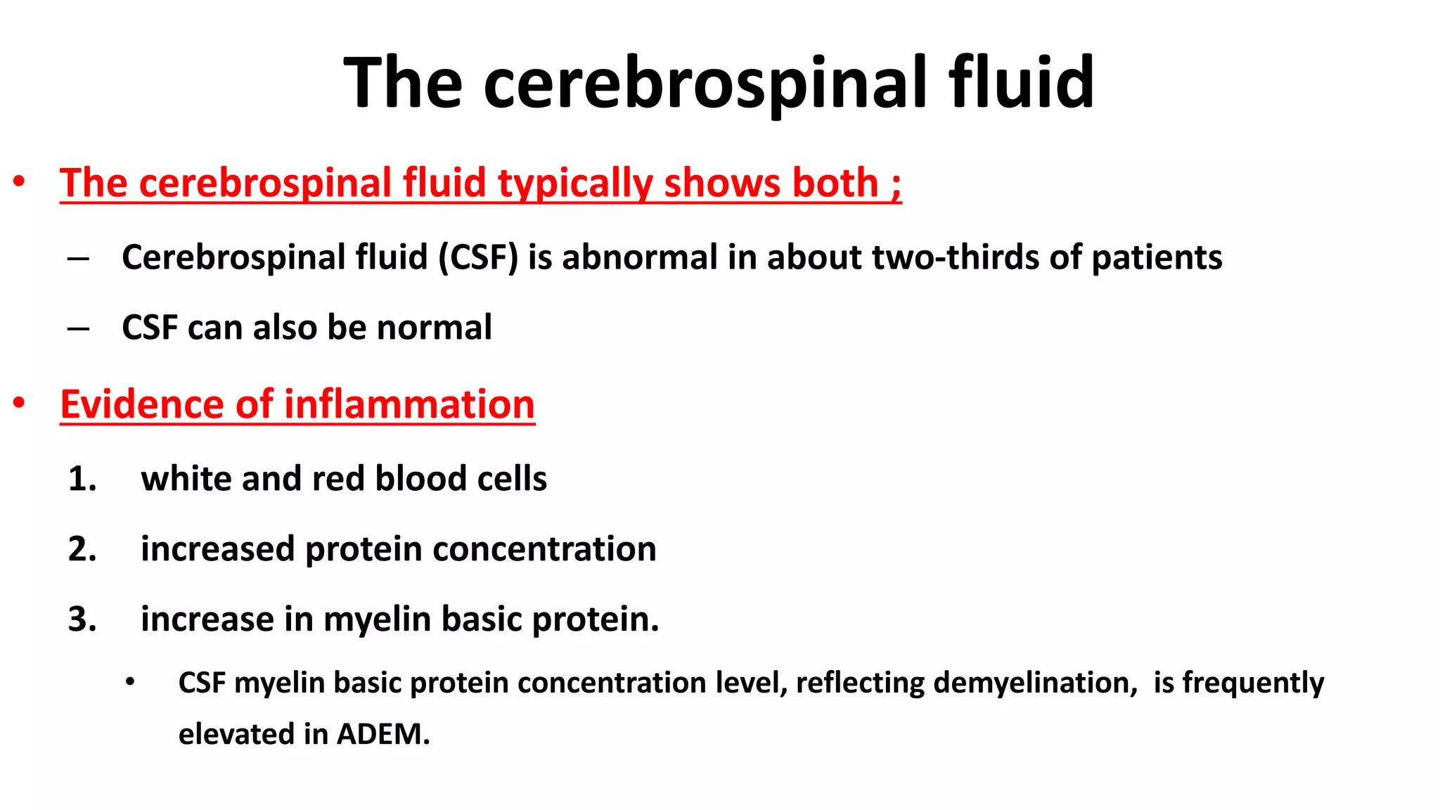
• Results of CSF immune profile testing (eg, CSF:serum
immunoglobulin G [IgG] index, CNS IgG synthetic rate,
oligoclonality) employing age-appropriate normative data
are positive in fewer than 10% of prepubertal children
with ADEM
36. The MRI abnormalities
• The MRI abnormalities are best defined by
1. T2-weighted images
2. fluid-attenuated inversion recovery (FLAIR) sequences
3. proton-density, or echo-planar trace diffusion MRI techniques
– characteristic high-signal lesions in virtually all cases of
ADEM
37. The MRI abnormalities
• The MRI abnormalities
– Contrast enhancement is seen at times in acute lesions.
• Findings may progress over a relatively short period of time,
consistent with evolution of the disease process.
• show high-signal changes consistent with vasogenic edema.
38. MRI abnormalities
• Abnormalities on MRI vary in location.
• Lesions associated with ADEM are
1. typically bilateral
2. asymmetric
3. poorly marginated.
• Almost all patients have multiple lesions in the deep and subcortical
white matter, characteristic of demyelination.
• The periventricular white matter is often spared.
39. MRI abnormalities
• Typical lesions of ADEM include
– centrifugal at the junction of the deep cortical gray and
subcortical white matter.
– Such lesions are found in more than 90% of children with
ADEM.
40. MRI abnormalities
• Additional lesions may be found in
– deeper white matter, basal ganglia (30-40%),
– thalamus (30-40%),
– brainstem (45-55%),
– cerebellum (30-40%),
– spinal cord (16-28%).
42. MRI abnormalities
• Diagnosis of ADEM
– should always rest on clinical grounds in children as in adults.
– Radiographic studies and other laboratory tests are especially
valuable in ruling in or out alternative diagnoses.
43. An Axial MRI of the brain and a saggital MRI of the spine of a patient with
acute disseminated encephalomyelitis (ADEM)
44. MRI abnormalities
• Brainstem and spinal cord abnormalities on MRI are
common in ADEM
• spinal cord, large confluent intramedullary lesions that
extend over multiple segments are typical
– Enhancement is variable.
46. MRI
• Magnetization transfer
– may help distinguish ADEM from MS, in that normal appearing
brain (on T2 weighted images) has normal magnetization transfer
ratio (MTR) and normal diffusivity,
– whereas in MS both measurements are significantly decreased
47. CT
• The CT scan
– low-density abnormalities
– found in more than half of childhood or adolescent ADEM cases
– but this technique is far less sensitive than MRI for the
disclosure of extent and number of lesions.
48. electroencephalogram (EEG)
• The electroencephalogram (EEG) is not diagnostic
– It may show background slow wave activity that is typical of an
encephalopathy
– Seizure activity is seen.
50. DIAGNOSIS
• The diagnosis of ADEM is based upon the
1. clinical
2. radiologic features
– There is no specific biologic marker or confirmatory test.
51. DIAGNOSIS
• An ADEM diagnosis :is considered when
– individuals develop multifocal neurologic abnormalities with:
1. Confusion
2. Excessive irritability
3. Altered level of consciousness (encephalopathy)
• Especially if the onset of symptoms occurs within 1 to 2 weeks
after a viral/bacterial infection or a vaccination
52. DIAGNOSIS
• Encephalopathy is a required feature for the diagnosis of ADEM,
– but is not a typical feature of multiple sclerosis.
• cerebrospinal fluid pleocytosis ≥50 white blood cells/mm can be
observed in ADEM,
– whereas this finding is highly atypical for multiple sclerosis.
53. DIAGNOSIS
• Bacterial and viral meningitis or encephalitis must be
considered and ruled out.
• Empiric treatment with broad-spectrum antibiotics and
acyclovir should be considered until an infectious etiology is
excluded
56. Multiphasic ADEM
• Individuals who have experienced typical ADEM are at
risk for recurrence.
– As many as 10% of children with an initial diagnosis of ADEM
experience another ADEM attack
– typically within the first 2-8 years after the initial attack.
57. Monophasic ADEM
1. A single clinical episode of ADEM may evolve over as
long as three months
2. Any new and fluctuating symptoms occurring within
three months of the initial event
3. symptoms that appear during glucocorticoid taper or
4. within one month of completing a glucocorticoid
taper
58. Recurrent And Multiphasic ADEM
• Recurrent ADEM and multiphasic ADEM are the
two relapsing forms of the disease.
1. more than three months after the initial event
and
2. more than one month after completion of
glucocorticoids.
– By definition for ADEM, both must include a clinical
presentation with encephalopathy.
59. Recurrent ADEM
1. three or more months after the first ADEM event
2. same symptoms that occurred at the time of the
initial presentation.
3. The MRI findings are also similar to the initial event
and are without new lesions,
– although there may be enlargement of the original lesions.
60. Multiphasic ADEM
1. recurrent disease that meets criteria for ADEM but involves new
anatomic areas of the central nervous system.
2. Symptoms and neuroimaging findings are different from the
initial event, with the exception that symptoms and signs of
encephalopathy may not differ from the initial episode.
3. The MRI must show new lesions compared with the first attack
and demonstrate complete or partial resolution of the lesions
associated with the first ADEM episode.
61. Differential Diagnosis
• other inflammatory demyelinating disorders should be
considered. These include:
1. Multiple sclerosis
2. Optic neuritis
3. Transverse myelitis
4. Neuromyelitis optica (Devic's disease)
5. Other rare conditions
66. TREATMENT
• It is important to first consider a treatment with
antibiotics and/or acyclovir until an infectious cause is
ruled out
• A high dose of intravenous corticosteroids, for 3-5 days
is the primary and most common first treatment of
ADEM
68. ADEM: Treatment
• High Dose Steroids
– High dose IV Methyl Prednisolone 30 mg/kg/day for 3-5
days
• IVIG (<1 yr, No improvement in 48-72 hrs , AHLE, Recurrent)
– 1 gm/kg/day iv for 2 days
• Plasmapheresis
• Symptomatic Rx
69. TREATMENT
• For children with ADEM, we recommend
immunosuppressive treatment
• We suggest initial therapy with high-dose glucocorticoids
• IV methylprednisolone (30 mg/kg per day, up to a
maximum dose of 1000 mg per day) for five days without
a taper.
70. TREATMENT
• For children with ADEM who have an insufficient response to IV
glucocorticoid treatment, we suggest intravenous immune
globulin treatment
• For children with ADEM who do not respond to glucocorticoids and
IVIG, we suggest treatment with plasma exchange
71. PROGNOSIS
• Prognosis for most children with ADEM is
good
• Recovery is usually a slow process lasting from four to
six weeks
• The majority of children with ADEM make a full
recovery
72. PROGNOSIS
• Between 60 to 90 percent are left with no neurological deficits
• Those children who do have residual symptoms are reported to have symptoms
from:
• Transverse myelitis
• Recurrent headaches
• Behavioral problems
73. PROGNOSIS
• Long-term clinical follow-up and sequential imaging by
MRI are normally required to confirm a diagnosis of
ADEM.
74. PROGNOSIS
• Development of a relapse with new lesions, it is not
compatible with a diagnosis of monophasic ADEM
75. PROGNOSIS
• Depending on the clinical and imaging features, it likely
suggests the correct diagnosis being either multiphasic
ADEM or MS.
76. PROGNOSIS
• Depending on the clinical and imaging features, it likely
suggests the correct diagnosis being either multiphasic
ADEM or MS.
77. PROGNOSIS
• The prognosis for survival and recovery of
neurologic function is worse for the hyperacute
hemorrhage variants of ADEM, such as acute
hemorrhagic leukoencephalitis, than for typical
ADEM
78. PROGNOSIS
• Complete recovery in 10 (77%) of the survivors
• Relapsing disease in 2 (15%)
• Mortality less than 2%
– fulminant cervical transverse myelitis or brain
swelling. Children younger than 2 years
79. • Acute disseminated encephalomyelitis
• Presents with
– Altered mental status/encephalopathy
(irritability to obtunded)
– Acute/subacute onset of focal symptoms
based on lesions
(max neuro symptoms over 4-7 days)
– Typically still during febrile illness
(typically URI)
– 1/3 with seizures
• Workup
– Labs: none are diagnostic
– CSF: pleocytosis, elevated protein, elevated IgG index; rule out infection!
– Imaging: MRIbrain with contrast
• Treatment: IV steroids (ok to start while r/o infection and prophylactic antibiotics
are on board) > IVIg > PLEX; PT
• Outcome
– Recovery over 4-6 weeks- 60-90% with no residual defecits
– Repeat MRI 6-12 months later to assess for lesion resolution
Editor's Notes
Symptoms of optic neuritis include
vision loss
pain with eye movement
afferent pupillary defect.
Inflammation of the optic disc may be seen on direct funduscopic examination if there is extensive involvement of the optic nerve.
Patients with retrobulbar optic neuritis typically have a normal funduscopic examination.
Direct imaging of the optic nerve with a gadolinium-enhanced MRI of the brain and orbits is a more sensitive means to diagnose optic neuritis in these patients
diffuse white matter lesions, often large and associated with cerebral edema
White matter lesions can be detected on MRI within 72 hours of the first symptoms, but hemorrhage itself is not necessarily seen with conventional T2-weighted and fluid-attenuated inversion recovery (FLAIR) sequences.
Specialized spin-echo MRI sequences are more readily able to identify the acute hemorrhage associated with this form of ADEM.
Positivity of studies for CSF oligoclonal bands and immunoglobulin elevation favors the diagnosis of MS in individuals younger than 20 years with first or recurrent bouts of acute CNS demyelinating illness
Abnormalities on MRI vary in location.
Lesions associated with ADEM are typically bilateral but may be asymmetric and tend to be poorly marginated.
Almost all patients have multiple lesions in the deep and subcortical white matter, characteristic of demyelination.
The periventricular white matter is often spared.
A) An Axial MRI of the brain (FLAIR image) in a child with acute disseminated encephalopmyelitis (ADEM) demonstrates multifocal areas of hyperintensity in both cerebral hemispheres involving cortical gray matter, centrum semiovale, and deep gray nuclei. B) A saggital MRI of the spine (T2 image) in the same child demonstrates high signal intrinsic to the spinal cord, consistent with longitudinally extensive transverse myelitis.
Abnormalities on MRI vary in location.
Lesions associated with ADEM are typically bilateral but may be asymmetric and tend to be poorly marginated.
Almost all patients have multiple lesions in the deep and subcortical white matter, characteristic of demyelination.
The periventricular white matter is often spared.
Axial T2-weighted MRI brain images show relatively symmetric high signal intensity in the deep gray matter, including bilateral thalami (panel A). There is also high T2 signal within the left aspect of the pons (panel B). Sagittal T2-weighted MRI of the cervical spine demonstrates expansion/swelling and high signal intensity within the spinal cord (panel C), and corresponding axial T2-weighted MRI reveal that the high signal is mostly posterior (panel D). One month later, T2-weighted axial images demonstrate resolution of the findings in the deep gray matter (panel E) and pons (panel F).
Is far more sensitive,
T2 - demonstrates regions of high signal, with surrounding oedema typically situated in subcortical locations ; the thalami and brainstem can also be involved.
T1 C+ (Gd) - punctate, ring or arc enhancement (open ring sign) is often demonstrated along the leading edge of inflammation; absence of enhancement does not exclude the diagnosis.
DWI - there can be peripheral resctricted diffusion; the center of the lesion, although high on T2 and low on T1 does not have increased restriction on DWI (c.f.cerebral abscess), nor however does it demonstrate absent signal on DWI as one would expect from a cyst. This is due to increase in extra cellular water in the region of demyelination.
Magnetization transfer may help distinguish ADEM from MS, in that normal appearing brain (on T2 weighted images) has normal magnetization transfer ratio (MTR) and normaldiffusivity, whereas in MS both measurements are significantly decreased
A single clinical episode of ADEM may evolve over as long as three months
Any new and fluctuating symptoms occurring within three months of the initial event are considered to be part of the same inciting event.
symptoms that appear during glucocorticoid taper or within one month of completing a glucocorticoid taper are considered to be part of the same inciting event.
ADEM: acute disseminated encephalomyelitis; HIV: human immunodeficiency virus; MDEM: multiphasic disseminated encephalomyelitis; SLE: systemic lupus erythematosus; CNS: central nervous system