Bradshaw - Human Performance and Biosystems - Spring Review 2013
Adwait Suratkar poster presentation new
1. CARBON NANOTUBES AS A FUNCTIONAL PLATFORM
FOR CELL CULTURE
Adwait P. Suratkar, Carol Lynam, Edwina Stack, Richard O’Kennedy
It is known that electrical stimulation may influence the function of electrically excitable cells such as nerve cells and muscle cells.
Many biomedical devices are dependent on efficient electrical communication with living cells. Advancement of these devices
depends upon effectively bridging the tissue/electrode interface. This project aims to:
Achieve greater biocompatibility of carbon nanotubes (CNTs) by immobilising proteins of interest
Investigate these CNT platforms as substrates for cell culture
PEM / CNT( -)
PEM / CNT(+)
CNT+/PSS-
CNT-/PAH+
CNT+/CNT-
CNT-/CNT+
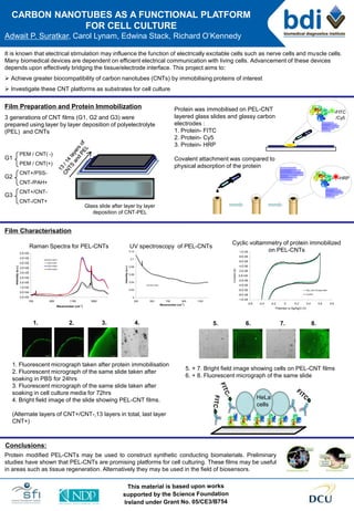
Film Preparation and Protein Immobilization
3 generations of CNT films (G1, G2 and G3) were
prepared using layer by layer deposition of polyelectrolyte
(PEL) and CNTs
Film Characterisation
Raman Spectra for PEL-CNTs
Cyclic voltammetry of protein immobilized
on PEL-CNTs
UV spectroscopy of PEL-CNTs
1. Fluorescent micrograph taken after protein immobilisation
2. Fluorescent micrograph of the same slide taken after
soaking in PBS for 24hrs
3. Fluorescent micrograph of the same slide taken after
soaking in cell culture media for 72hrs
4. Bright field image of the slide showing PEL-CNT films.
(Alternate layers of CNT+/CNT-,13 layers in total, last layer
CNT+)
5. 6. 7. 8.
Conclusions:
Protein modified PEL-CNTs may be used to construct synthetic conducting biomaterials. Preliminary
studies have shown that PEL-CNTs are promising platforms for cell culturing. These films may be useful
in areas such as tissue regeneration. Alternatively they may be used in the field of biosensors.
1. 2. 3. 4.
Protein was immobilised on PEL-CNT
layered glass slides and glassy carbon
electrodes :
1. Protein- FITC
2. Protein- Cy5
3. Protein- HRP
Covalent attachment was compared to
physical adsorption of the protein
G1
G2
G3
HeLa
cells
5. + 7. Bright field image showing cells on PEL-CNT films
6. + 8. Fluorescent micrograph of the same slide
- -FITC
/Cy5
Glass slide after layer by layer
deposition of CNT-PEL
- -HRP
0.E+00
5.E+04
1.E+05
2.E+05
2.E+05
3.E+05
3.E+05
4.E+05
4.E+05
5.E+05
150 650 1150 1650
Wavenumber (cm-1
)
Intensity(a.u.)
CNT-CNT+
CNT+CNT-
CNT+PSS-
CNT-PAH+
0
0.02
0.04
0.06
0.08
0.1
0.12
300 500 700 900 1100
Wavenumber (cm-1
)
Intensity(a.u.)
CNT+PSS-
-1.E-05
-8.E-06
-6.E-06
-4.E-06
-2.E-06
0.E+00
2.E-06
4.E-06
6.E-06
8.E-06
1.E-05
-0.6 -0.4 -0.2 0 0.2 0.4 0.6 0.8
Potential vs Ag/AgCl (V)
Current(A)
PEL-CNT-Protein-HRP
Control