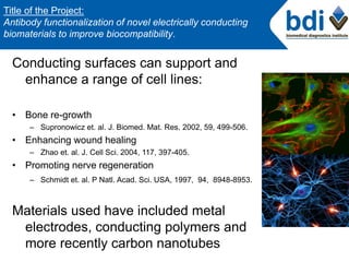
1. Conducting surfaces can support and
enhance a range of cell lines:
• Bone re-growth
– Supronowicz et. al. J. Biomed. Mat. Res. 2002, 59, 499-506.
• Enhancing wound healing
– Zhao et. al. J. Cell Sci. 2004, 117, 397-405.
• Promoting nerve regeneration
– Schmidt et. al. P Natl. Acad. Sci. USA, 1997, 94, 8948-8953.
Materials used have included metal
electrodes, conducting polymers and
more recently carbon nanotubes
Title of the Project:
Antibody functionalization of novel electrically conducting
biomaterials to improve biocompatibility.
2. • Good conductors of heat and electricity
• Remarkable mechanical properties
• Chemically inert and thermally stable
• Modification with biomolecules can lead
to biomedical applications
• The exploration of CNTs in biomedical
applications is just underway, but has
significant potential. CNTs have been
shown to be compatible with
physiological cells and tissues
Carbon Nanotubes
3. Project aims
• Functional modification of CNTs to achieve
biocompatibility
– Functionalise with antibodies to cell adhesion molecules
– Compare covalent attachment vs. physical adsorption
• Investigate whether these antibody-modified CNT
materials are good platforms for cell culture
4. Project outline
CNT film preparation and functionalisation:
Layer by layer deposition of CNTs
Antibody functionalisation
Film characterization :
Raman and UV spectroscopy
Fluorescent microscopy and scanning
Electrochemical analysis
Cell culture studies with CNT-films:
Antibody staining and fluorescent microscopy
5. Layer by layer deposition of CNTs
13 / 14
PEL
layers
Layer by layer deposition is a technique used to attach oppositely
charged polyelectrolytes (PEL) to a surface.
It forms functional and stable films
Polyelectrolytes used include: Sodium polystyrene sulfonate (PSS-)
Poly (allylamine hydrochloride) (PAH+)
Hyaluronic acid (HA-) + CNTs
Chitosan (Chit +) + CNTs
7. Layer by layer deposition of CNTs
PAH+/PSS-/…..CNT+ Generation1 – only CNT on top layer
PSS-/PAH+/…..CNT-
PSS-/CNT+/….. Generation 2 – CNT every second layer
PAH+/CNT-/…..
CNT+/CNT-/….. Generation3 – CNT every layer
CNT-/CNT+/…..
13 / 14
PEL
layers
8. Raman Spectroscopy of CNT films
• The laser used
for the Raman
analysis was a
488nm Ar+ laser
Raman Spectroscometer
9. Raman Spectroscopy of CNT films
Wavenumber (cm-1)
RamanIntensity(a.u.)
Radial breathing
modes (RBM)
G-band
10. Antibody (Ab) immobilisation
Antibodies
• Anti-mouse FITC
• Anti- human Cy5
• Anti-mouse HRP
• Anti-Epcam
Method of immobilisation
Two methods were used:
1. Covalent attachment.
2. Physical adsorption.
11. Antibody immobilisation on CNT
films
Antibody immobilised on
CNT-/CNT+ and
CNT+/CNT-
A. Antibody physically adsorbed
to the surface.
B. Antibody attached covalently
to the surface.
A
B
12. Films soaked in
PBS for 24
hours at room
temperature
Films incubated in
cell culture media
at 37°C for 72
hours
13. After 24hrs in PBS 72hrs in
immobilisation cell culture media
Covalent immobilisation
Physical adsorption
14. Results
- Covalently attached antibody spread well over the
surface area of the film.
- CNT- PEL films are stable under physiological pH (7.0).
- CNT- PEL films are bio-compatible.
15. Fluorescent scanner studies on
CNT films
A. B. C. D.
A and B the antibody has covered the entire area.
C and D the antibody is confined to the area of attachment.
16. Electrochemical analysis of CNT
films
HRP-
Experimental setup for
electrochemical analysis
Physical adsorption
Covalent attachment
GCE GCE
17. Reduction- Fe3+ to Fe2+
Oxidation – Fe2+ to Fe3+
Glassy carbon electrode with covalently attached
antibody, could be more active electrochemically,
18. Cell culture on carbon nanotubes-
polyelectrolyte films
HeLa
cells
-Cy5
Cy5-
Cy5- goat anti-human
Anti-epcam
Anti-Mouse FITC
21. Observations and Results
- Hyaluronic acid attached to CNTs seems to be a better surface
for cell attachment and proliferation.
- Covalent attachment seems to be better surface for antibody
attachment, as the it covers more surface area of the slide.
- Physically attached antibody doesn’t cover the entire area of
the slide and was confined to the spot were it was dropped.
22. PEL/ CNT thin
films
HRP-
A
B
C
G-band
Radial breathing
modes (RBM)
D
E
A. Deposition and Characterization of CNT/PEL thin
films.
B. Biocompatibility of the thin films observed under
fluorescent microscope.
C. Functionalization of films to cell adhesion
molecules (tissue regeneration).
D. Films as biosensors.
E. Electrochemical analysis of CNT/PEL films.
Wavelength (cm-1)
Raman Spectra of
Carbon nanotubes
(CNTs)
Cyclic Voltammetry of
protein immobilized
surfaces.
Bright field and fluorescent
microscope images