Electroretinography basics
•
0 likes•830 views

Electroretinography measures the electrical responses of various cell types in the retina, including the photoreceptors, inner retinal cells, and the ganglion cells. Electrodes are placed on the surface of the cornea or on the skin beneath the eye to measure retinal responses.

Recommended
More Related Content
What's hot
What's hot (20)
Similar to Electroretinography basics
Similar to Electroretinography basics (20)
More from AtheenaPandian Enterprises
More from AtheenaPandian Enterprises (20)
Recently uploaded
Recently uploaded (20)
Electroretinography basics
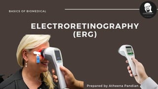
- 1. ELECTRORETINOGRAPHY (ERG) Prepared by Atheena Pandian BASICS OF BIOMEDICAL
- 2. Today's Coverage Points We'll Discuss What is ERG? Photoreceptors RODS and Cones Measurements Eye Response Recordings ERG Waves Diagnose 1. 2. 3. 4. 5. 6. 7. 8. Basics of Biomedical
- 3. Electroretinography meas ures the electrical responses of various cell types in the retina What is ERG? Basics of Biomedical
- 4. Photoreceptors Photoreceptors are the cells in the retina that respond to light. Their distinguishing feature is the presence of large amounts of tightly packed membrane that contains the photopigment rhodopsin or a related molecule. Basics of Biomedical
- 5. RODS Rods are responsible for vision at low light levels (scotopic vision). They do not mediate color vision, and have a low spatial acuity. CONES Cones are active at higher light levels (photopic vision), are capable of color vision and are responsible for high spatial acuity. The central fovea is populated exclusively by cones. There are 3 types of cones which we will refer to as the short-wavelength sensitive cones, the middle-wavelength sensitive cones and the long-wavelength sensitive cones or S-cone, M-cones, and L-cones for short. There are two types of photoreceptors in the human retina, rods and cones.
- 7. Electrodes are placed on the surface of the cornea (DTL silver/nylon fiber string or ERG Jet) or on the skin beneath the eye (Sensor Strips) to measure retinal responses Measurement Basics of Biomedical
- 8. Retinal pigment epithelium (RPE) responses are measured with an EOG test with skin-contact electrodes placed near the canthi. Measurement Basics of Biomedical
- 9. RECORDING During a recording, the patient's eyes are exposed to standardized stimuli and the resulting signal is displayed showing the time course of the signal's amplitude (voltage) Signals are very small, and typically are measured in microvolts or nanovolts. Basics of Biomedical
- 10. Basics of Biomedical Eye Response The ERG is composed of electrical potentials contributed by different cell types within the retina, and the stimulus conditions (flash or pattern stimulus, whether a background light is present, and the colors of the stimulus and background) can elicit stronger response from certain components.
- 11. Basics of Biomedical Eye Response If a dim flash ERG is performed on a dark- adapted eye, the response is primarily from the rod system. Flash ERGs performed on a light adapted eye will reflect the activity of the cone system.
- 12. ERG Wave Sufficiently bright flashes will elicit ERGs containing an a-wave (initial negative deflection) followed by a b-wave (positive deflection). Basics of Biomedical
- 13. Retinitis pigmentosa and related hereditary degenerations Retinitis punctata albescens Leber's congenital amaurosis Choroideremia Gyrate atrophy of the retina and choroid Goldman-Favre syndrome etc the electroretinogram (ERG) is used for the diagnosis of various retinal diseases. Diagnose
- 14. GOOD LUCK Basics of Biomedical