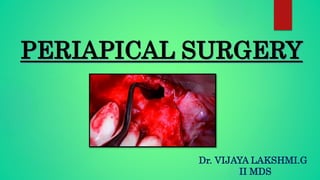
Periapical surgery viji
•Download as PPTX, PDF•
14 likes•2,648 views

Periapical surgery

Recommended
More Related Content
What's hot
What's hot (20)
Similar to Periapical surgery viji
Similar to Periapical surgery viji (20)
More from Dr. Vijaya Lakshmi
Recently uploaded
Recently uploaded (20)
Periapical surgery viji
- 1. Dr. VIJAYA LAKSHMI.G II MDS
- 2. • Introduction • Definition • History • Indication • Contraindication • Classification of endodontic surgeries • Recent advances • Conclusion • References CONTENTS
- 3. Surgical intervention is required where endodontic treatment has failed and tooth is to be retained rather than extracted. The percentage of success of endodontic treatment has been consistently high but failures may arise due to infection, poor access cavity preparation, inadequate instrumentation, obturation, missed canals and coronal leakage. So if this happens, Surgical endodontics is needed to save the tooth. INTRODUCTION
- 4. Surgical endodontics is defined as, “Removal of tissues other than the contents of root canal to retain a tooth with pulpal or periapical involvement” DEFINITION
- 5. • Surgical endodontics is not a recent innovation. • Trephination and incision and drainage are being done since ancient times. • In 11th century, first case of endodontic surgery was performed by Albucasis. • Root end resection (Apicectomy) was first documented in 1871 and apicectomy with retrograde cavity preparation and filling with amalgam was documented in 1890. HISTORY
- 6. • Root amputation was first introduced by Black and Inlitch in 1886 , then was dealt by Younger (1894) and Guerini (1909) • In 1930, indications for endodontic surgery were proposed. • In 1940, Triangular flap was first described by Fischer. • Neumann and Eikan described Trapezoidal flap in 1940. • Semilunar incision was first described by Partsch hence it is also known as Partsch incision.
- 7. INDICATIONS 1. Need for surgical drainage 2. Failed endodontic treatment a) Irretrievable root canal filling material b) Irretrievable intraradicular post
- 8. 3. Calcification of the pulp space 4. Procedural errors a) Instrument fragmentation b) Non-negotiable ledging c) Root perforation
- 9. 5. Symptomatic overfilling. 6. Anatomic variations. A. Root dilaceration. B. Apical root fenestration. 7. Exploratory surgery and Biopsy.
- 10. 8. Corrective surgery. a) Root resorptive defects b) Root caries Root resection Hemi-section Bi-cuspidization
- 11. Poor systemic health. Local anatomical considerations Poor periodontal status. Short root length. Acute infection. Non restorable teeth CONTRAINDICATIONS
- 12. Success of surgical treatment over non-surgical treatment. Medical history Periodontal evaluation Patient’s motivation Pre medications Informed consent PRE-SURGICAL CONSIDERATIONS
- 14. I. Surgical drainage 1. Incision and drainage 2. Cortical trephination (fistulative surgery) II. Periradicular surgery 1. Curettage 2. Biopsy 3. Root-end resection 4. Root-end preparation and filling CLASSIFICATION OF ENDODONTIC SURGICAL PROCEDURES
- 15. III. Corrective surgery 1. PERFORATION REPAIR a) Mechanical (iatrogenic) b) Resorptive (internal and external) 2. PERIODONTAL MANAGEMENT a) Root resection b) Hemisection & Bi-cuspidization 3. REPLACEMENT SURGERY a) Replantation 4. IMPLANT SURGERY a) Endodontic implants b) Root-form osseointegrated implants
- 16. In most cases drainage through the canal is all that is needed to treat the periradicular abcess of pulpal origin but there are times, when invasion of anatomic spaces has extended to a point that does not allow drainage through the tooth, and effectively remove the pus then It becomes mandatory to incise and drain the abcess. INCISION AND DRAINAGE
- 17. PRINCIPLES OF DESIGN- Principles and guidelines are applied to the location and extent of incision. Why should one follow the principles ??? “The adherence to these principles will ensure that the flapped 1.soft tissues will fit snugly in their original position 2.will properly cover the osseous wound site and 3.provide an adequate vascular bed for healing” INCISIONS AND FLAPS
- 18. PRINCIPLES: 1. Avoid severing vessels and nerves 2. Make incisions far away from the surgical area to ensure that the wound margins are over sound bone and there is room for adjustments when unexpected extensions are necessary. 3. Design the flap so that there is adequate visibility without overexposure of bone.
- 19. 4. The base of the flap should be the widest portion to maintain proper circulation. 5. There should be no sharp angles on the flap 6. Vertical or oblique incision should not be over root eminence. It is best to incise in the trough.
- 20. 7. Maintain the integrity of the interdental papillae. 8. Use sharp instruments to avoid tearing the mucoperiosteum. 9. Be gentle with the flap.
- 21. 10. Do not incise close to the gingival sulcus while using a horizontal or semilunar incision 11. Incise in the attached gingiva for semilunar flaps. NOTE “More trauma results from short incision rather than long incision”.
- 22. Types of incision Vertical incision Sulcular incision Semilunar incision Ochsenbein-Leubke incision
- 23. Classification of Flaps 1. Full mucoperiosteal flaps: (a) Triangular (one vertical releasing incision) (b) Rectangular (two vertical releasing incisions) (c) Trapezoidal (broad-based rectangular) 2. Limited mucoperiosteal flaps (a) Submarginal curved (semilunar) (b) Submarginal scalloped rectangular (Ochsenbein-Luebke)
- 25. FULL MUCOPERIOSTEAL FLAPS TRIANGULAR FLAP The triangular flap is formed by a intrasulcular incision and one vertical releasing incision.
- 26. ADVANTAGES: Good wound healing as there is minimal disruption of the vascular supply to the flapped tissue. Ease of flap reapproximation, with a minimal number of sutures required. DISADVANTAGES: It provides Limited surgical access because of the single vertical releasing incision. Difficult to expose the root apices of long teeth (eg, maxillary cuspids and mandibular incisors.)
- 27. Additional access can be easily obtained by placement of a distal releasing incision. It is recommended for maxillary incisors and posterior teeth. “It is the only recommended flap design for mandibular posterior teeth”.
- 28. RECTANGULAR FLAP: The rectangular flap is formed by an intrasulcular and two vertical releasing incisions.
- 29. ADVANTAGES: Increased surgical access to the root apex. This flap design is especially useful for mandibular anterior teeth, multiple teeth, and teeth with long roots, such as maxillary canines.
- 30. DISADVANTAGES: Difficulty in reapproximation of the flap margins and wound closure. Postsurgical stabilization is also more difficult as the flapped tissues are held in position solely by the sutures. This results in a greater potential for postsurgical flap dislodgment. This flap design is not recommended for posterior teeth.
- 31. TRAPEZOIDAL FLAP Similar to the rectangular flap with the exception that the two vertical releasing incisions meet intrasulcular incision at an obtuse angle.
- 32. The angled vertical releasing incisions are designed to create a broad-based flap with the vestibular portion being wider than the sulcular portion. Flap design is made on the assumption that it will provide a better blood supply to the flapped tissues.
- 33. Since the blood vessels and collagen fibers in the mucoperiosteal tissues are oriented in a vertical direction, the angled vertical releasing incisions will severe more of these structures. This will result in more bleeding, a disruption of the vascular supply to the unflapped tissues, and shrinkage of the flapped tissues.
- 34. Limited Mucoperiosteal Flaps: Submarginal Curved (Semilunar) Flap: The Submarginal or semilunar flap is formed by a curved incision in the alveolar mucosa and the attached gingiva.
- 35. The incision begins in the alveolar mucosa extending into the attached gingiva and then curves back into the alveolar mucosa. Advantages– No advantages Disadvantages- 1. Poor surgical access 2. Poor wound healing “This flap design is not recommended for periradicular surgery”.
- 36. Submarginal scalloped rectangular (Luebke-ochsenbein) flap: The Submarginal scalloped rectangular flap is a modification of the rectangular flap in which the horizontal incision is not placed in the gingival sulcus but in the buccal or labial attached gingiva.
- 37. ADVANTAGES: It does not involve the marginal or interdental gingiva and the crestal bone is not exposed. DISADVANTAGES: Vertically oriented blood vessels and collagen fibers are severed, resulting in more bleeding and a greater potential for flap shrinkage, delayed healing, and scar formation.
- 39. FLAP REFLECTION: Flap reflection is the process of separating the soft tissues (mucosa and periosteum) from the from the surface of the bone. The periosteal elevator is used gently to elevate the periosteum and its superficial tissues from the cortical plate.
- 40. After reflection of the attached gingival tissues, elevation is continued more apically lifting the alveolar mucosa along with periosteum until adequate surgical access is obtained. A thin gauze may be used for reflection to prevent tearing of the flap.
- 41. HARD TISSUE MANAGEMENT Hard tissue management in endodontic surgery involves 3 stages: 1.Trephination 2.Periradicular curretage 3. Periradicular surgery (i) Root end resection (Apicectomy) (ii) Root end preparation & filling
- 42. OSTEOTOMY: Osteotomy is the removal of some portion of the cortical plate to expose the root end. Clinician should precisely locate the root end. A number of factors should be considered to determine the location of the bony window. The angle of the crown to the root should be assessed.
- 43. When a root prominence or eminence in the cortical plate is present, the root angulation and position are more easily determined. Measurement of the entire tooth length on well- angled radiograph and transferred to the surgical site by the use of a sterile millimeter ruler.
- 44. When the cortical plate is intact, locate the body of the root coronal to the apex where the bone covering the root is thinner. Once the root has been located and identified, the bone covering the root is slowly and carefully removed with light brush strokes, working in an apical direction until the root apex is identified.
- 45. Barnes identified four ways by which the root surface can be distinguished from the surrounding osseous tissue: (1) Root structure generally has a yellowish color, (2) Roots do not bleed when probed, (3) Root texture is smooth and hard as compared to the granular and porous nature of bone, and (4) The root is surrounded by the periodontal ligament.
- 46. Definition- It is the perforation made through the cortical plate or apical foramen to accomplish the release of pressure in the periapical area from the accumulation of exudate within the alveolar bone. Indications- This technique is employed in cases of periapical abcess in which there is no swelling or drainage but much pain. TREPHINATION
- 47. Procedure- Small incision is made over the periapical region. Flap is reflected and bone is examined. Radiograph is taken with radio-opaque marker for confirmation. So that there is no chance of penetration in the wrong area.
- 48. CORTICAL TREPHENATION: Perforation of the cortical plate to accomplish the release of pressure from the accumulation of exudate within the alveolar bone.
- 49. The treatment of choice for these patients is drainage through the root canal system (apical trephination) whenever possible. Apical trephination involves penetration of the apical foramen with a small endodontic file and enlarging the apical opening to a size No. 20 or No. 25 file to allow drainage from the periradicular lesion into the canal space. The decision about whether to perform apical or cortical trephination is based primarily on clinical judgment regarding the urgency of obtaining drainage.
- 50. PERIRADICULAR CURETTAGE: Involves removal of the periradicular inflammatory tissue and is best accomplished by using various sizes and shapes of sharp surgical bone curettes and angled periodontal curettes.
- 51. Entire tissue mass is removed by inserting the bone curette, between the soft tissue mass and the lateral wall of the bony crypt with the concave surface of the curette facing the bone.
- 52. Once the soft tissue lesion has been freed along with the periphery, the bone curette should be turned with the concave portion toward the soft tissue and used in a scraping manner to free the tissue from the deep walls of the bony crypt.
- 53. ROOT-END RESECTION (APICOECTOMY) Historically, many authors have advocated periradicular curettage as the definitive treatment in endodontic surgery without root-end resection. Their rationale was to maintain a cemental covering on the root surface and to maintain as much root length as possible for tooth stability. PERIRADICULAR SURGERY
- 54. INDICATIONS: These indications may be classified as, 1) Biological 2) Technical. Biologic factors: Persistent symptoms, Persistent periradicular lesion.
- 55. Technical factors: Periapical infection in teeth with… Radicular posts, Crowned teeth without posts, Irretrievable root canal filling materials, Procedural accidents.
- 56. There are three important factors for the surgeon to consider before performing a root-end resection: (1) Instrumentation, (2) Extent of the root end resection, (3) Angle of the resection.
- 57. 1. Instrumentation: Ingle et al. recommended that root-end resection is best accomplished by the use of tapered fissure bur or round bur in a low-speed straight handpiece. Gutmann and Harrison, have suggested the use of a high-speed handpiece and a surgical length plain fissure bur.
- 58. 2. Extent of the Root-End Resection: Earlier, it was believed that it is necessary to resect the root at the level of healthy bone.
- 59. Average length of root resection is 3mm which is considered enough to eliminate the source of infection. However surgeon must evaluate the patient on an individual basis. 1. Visual and operative access to the surgical site 2. Anatomy of the root (shape, length, curvature). 3. Number of canals and their position in the root
- 60. 4. Need to place a root-end filling surrounded by solid dentin. 5. Presence and location of procedural error 6. Presence and extent of periodontal defects. NOTE: “Conservation of tooth structure during root-end resection is desirable; however, conservation conservation should not compromise the goals of the surgical procedure”.
- 61. 3. Angle of Root-End Resection It should be 30 ° -45 ° from the line perpendicular to the long axis of the tooth facing toward the buccal or facial aspect of the root. The purpose is to provide enhanced visibility to the root end and operative access to accomplish a root end preparation.
- 62. CLINICAL EVALUATION OF ROOT END RESECTION BEVEL IN PERIAPICAL SURGERY corresponding author: Marina Kacarska, Address: Vodnjanska b.b, Skopje, e-mail: marina_kacarska@yahoo.com Faculty of Dentistry, Oral Surgery, University Ss. Cyril and Methodius, Skopje The intention of this article was to clinically assess the root resections bevels and to estimate their relation to applied periapical surgeries. Thirty periapical surgeries were performed in a conventional manner, while thirty were contemporary ultrasonic surgeries. Following the completion of strictly planned and performed intraoperative procedures, the resection bevels were assessed. To obtain the real bevel angles a compass was used. Significantly lesser resection bevel associated with ultrasonic periapical surgery contributes to root preservation and favorable surgical outcome.
- 63. Root-End Preparation The purpose of a root-end preparation in periradicular surgery is to create a cavity to receive a root-end filling. It is performed by the use of small round or inverted cone burs and straight low- speed handpiece. It should be done parallel to the long axis of the root.
- 64. ROOT-END FILLING
- 65. The purpose of a root-end filling is to establish a seal between the root canal space and the periapical tissues. Suitable root-end filling material should be, (1) Able to prevent leakage of bacteria and their bioproducts into the periradicular tissues, (2) Nontoxic & Noncarcinogenic, (3) Biocompatible with the host tissues, (4) Insoluble in tissue fluids, (5) Dimensionally stable, (6) Unaffected by moisture during setting, (7) Easy to use
- 66. Root-End Filling Materials: Numerous materials have been suggested for use as root-end fillings, including: including: Amalgam, Gutta-percha, Glass ionomers, Composite resins, Carboxylate cements, Zinc phosphate cements, Zinc oxide–eugenol cements, Mineral tri-oxide aggregate (MTA).
- 67. Apical dentin permeability and microleakage associated with root end resection and retrograde filling Gilheany PA1, Figdor D, Tyas MJ. This study evaluated the apical leakage associated with various depths of retrograde fillings placed in root apices which had been resected at one of three different angles. Leakage was assessed with a hydraulic conductance apparatus. Teeth were divided into groups corresponding to the angle of apical resection (0, 30, and 45 degrees to the long axis of the root) and apical leakage was determined following incremental increases in the depth of the retrograde filling (Ketac Silver). Increasing the depth of the retrograde filling significantly decreased apical leakage; there was also a significant increase in leakage as the amount of bevel increased. Both the permeability of resected apical dentin and microleakage around the retrograde filling material had a significant influence on apical leakage.
- 68. • MTA showed significantly less microleakage when compared to Biodentine and light cure GIC. • No statistical difference between the ultrasonic retrotip preparation and conventional bur preparation. • MTA with ultrasonic preparation is the better material as root end filling material to prevent microleakage
- 70. ROOT AMPUTATION Removal of one or more roots of a multirooted tooth while other roots are retained.
- 74. CONTRAINDICATIONS FOR ROOT AMPUTATION
- 75. TECHNIQUE: 1. Vertical cut method 2. Pre-surgical crown contouring method 3. Horizontal preparation
- 77. BICUSPIDATION Bisection/bicuspidisation refers to division of crown that leaves the two halves along with their respective roots. Indications- Severe bone destruction in furcation with excellent bone support at other areas
- 79. Close proximity to mental foramen favours intention reimplantation REPLACEMENT SURGERY
- 80. Periradicular surgery with a retroseal is more advantageous for first molars when roots are more curved, widespread, and are more prone to fracture compared with second molars whose roots are more tapered and close together. ADVANTAGES- • Procedure is easier to perform • Less time consuming • Fewer complications of paresthesia, maxillary sinus penetration Inferior alveolar canal penetration pain swelling periodontal pocket formation • Greater chance to detect root fracture • More cost effective DISADVANTAGES- • Possible crown or root fracture • Greater likelihood of root resorption
- 82. Endodontic implants Rakesh K. Yadav, A. P. Tikku, Anil Chandra, K. K. Wadhwani, Ashutosh kr, and Mayank Singh Reamers were extruded to use as endodontic implantsPeriodontal involvement in retained E MTA based sealer fillapex was used to obturate and GIC was used for filling pulp chamber Six months post-op showed healing and better stability
- 83. Several authors have compared the effects of continuous and interrupted suture techniques. Their findings indicate that the interrupted suturing technique provides better flap adaptation than does the continuous technique and, therefore, is the recommended technique, and the most commonly used, for endodontic surgery. REPOSITIONING AND SUTURING
- 87. Micro-scalpels Rubinstein Mini-Molts retractor Minnesota retractor Micro mirrors Pluggers Stropko irrigator Impact-Air 45 handpiece MTA carrier systems
- 89. Root-end preparation with bur Root-end preparation with ultrasonic tip
- 90. Traditional Endodontic Surgery Versus Modern Technique: A 5-Year Controlled Clinical Trial Silvia Tortorici, MD,* Paolo Difalco, DDS, PhD,* Luigi Caradonna, MD,* and Stefano Tete`, MDÞ Compared traditional apicoectomy versus modern apicoectomy, method of osteotomy (type of instruments used), type of preparation of retrograde cavity (different apicoectomy angles and instruments used for root-end preparation), and root-end filling material used (gray mineral trioxide aggregate or silver amalgam). In conclusion, modern apicoectomy resulted in a probability of success more than 5 times higher (odds ratio, 5.20 [95% confidence interval, 3.94Y6.92]; P G 0.001) compared with the traditional technique.
- 91. An update in periapical surgery Eva Martí-Bowen 1, Miguel Peñarrocha 2 The Erbium-YAG laser has shown great potential in application to periapical surgery. The thermal damage induced by this laser in soft tissues, bone and other structures is comparatively less than with other laser systems, as a result of which postoperative discomfort is lessened. The main advantages with respect to rotary instrumentation comprise a reduction in tissue trauma and the risk of contamination LASER IN PERIAPICAL SURGERY
- 93. During the last 20 years, endodontics has encountered dramatic shift in the use of periradicular surgery. Previously, periradicular surgery was commonly considered as the treatment of choice when nonsurgical treatment had failed but nowadays periradicular surgery has become very selective in contemporary dental practice. CONCLUSION
- 94. Text book of endodontics, Ingle 5th edition. Textbook of oral & maxillofacial surgery By Daniel M. Laskin. Vol.2 Text book of endodontics, Nisha Garg. Text book of endodontics By Grossman. Text book of Surgical endodontics, Guttman REFERENCES
- 95. Thank you…
Editor's Notes
- Exploratory surgery & Biopsy- it is a rare indication in teeth where a fracture is suspected Teeth with a vital pulp with a radicular radiolucency or previous history of malignancy
- Anatomical considerations such as mandibular second molar area where roots are lingually inclined , apices are close to mandibular canal, too thick buccal plate. Proximity to nasal floor and maxillary sinus. Deep pockets , Poor bony support , tooth mobility and furcation involvement .
- .
- 2. This avoids possibility of collapse of flap into the bony defect with subsequent depression 3. Coz size doesn’t affect the rate of healing. 4.
- 5. There should be a gentle curve because sharp corners tend to slough because of poor circulation causing excessive scarring.\ 6. because in the Trough between the adjacent teeth, mucosa and attached gingiva are thicker , so it has better circulation ,it can afford stronger needle entry and exit points and are more extensible during edema.
- 7. Papilla at the incision line has to be either excluded or included. 8. Incision should be made in one pass. So repositioning is easy and there is less tendency for dehiscence an scar formation 9. Forklike retractors or tweezers should not be used . 10. Because lack of blood supply can cause loss of gingiva there should be 2-3 mm of attached gingiva around each tooth.
- dept
- The horizontal incision is scalloped and follows the contour of the marginal gingiva above the free gingival groove.
- .can be identified using metheylene blue dye
- Marker is small sterilised foil of x ray film which is burnished into bone concavity.
- According to “Gutmann” and “Harrison”, no studies are available to support this and it is questionable, especially if the source of periradicular infection is still within the root canal system.
- Procedural accidents such as fragmentation of instruments , perforation in apical 1/3 rd of root.
- NOTE- “Plain fissure burs, at high and low speed, produce the smoothest resected root surface”.
- 1. (example: root end resection of buccal root of maxillary first premolar to gain access to the palatal root). 3. (example: mesiobuccal root of maxillary molars, mesial root of mandibular molars, two canals in central incisors).
- 4. Example – when there is infected dentin then length of root end resection will be more 5. (example: perforation, ledge, broken instrument, apical extent of root canal filling). 6.- example – level of bone loss.
- NOTE: Recent literature states that beveling of root end results in opening of dentinal tubules on the resected root surface that may communicate with the root canal space and result in apical leakage, even when a root end filling has been placed.
- To eliminate a weak diseased root to allow the stronger roots to survive which, if retained together would collectively fail.
- 2 nd pic- canal obstruction-(separated instrument)
- Lack of necessary osseous support to the remaining root/ roots Fused roots Remaining root or roots are endodontically inoperable Lack of patient motivation for homecare remedies.
- Grossman- this is an act of deliberately removing a tooth and –after examination, diagnosis, endodontic manipulation, repair and returning the tooth to its original socket.
- Utilising the existing tooth by placing a metal post down into one of the root canals of the tooth and extending into the bone. Indications- Poor periodontal support Good alveolar bone support and can be maintaine dby implants Pulpoperiodontal therapy
- The irrigator fits over a triflow syringe and allows for the directional micro control of air and water. Air pressure can be regulated down to 4 psi.
- Endodontic microsurgery- micromirrors, isthmus, ultrasonic KiS tip,
- Root-end preparation with bur resulted in lingual preparation while the integrity of the root apex is preserved because of the co-axial preparation within the root canal when performed with ultrasonic