
Recommended
More Related Content
What's hot
What's hot (20)
Similar to caudal devation correction.pptx
Similar to caudal devation correction.pptx (20)
More from EmanZayed17
More from EmanZayed17 (16)
Recently uploaded
Recently uploaded (20)
caudal devation correction.pptx
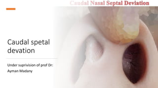
- 1. Caudal spetal devation Under suprivision of prof Dr: Ayman Madany
- 2. Introduction Caudal septal deviation is defined as deviation of the anterior most portion of the nasal septum. In addition to functional symptoms, caudal septal deviation can cause significant cosmetic deformities, including lobule deviation, tip ptosis, and deformity of the middle one-third of the nose. Caudal septal deviation differs from traditional septal deviation in that it involves a portion of the septum that contributes to the nasal valve area and the support of the nasal tip
- 3. Anantomy The nasal septum divides the left and right nasal cavities. It is lined by mucoperichondrium anteriorly (covering the quadrangular cartilage) and mucoperiosteum posteriorly (covering the bony septum), and superiorly becomes continuous with the cribriform plate mucosa, and inferiorly with the nasal floor mucosa. the posterior aspect of the quadrangular cartilage articulates neatly with the bony septum at the bony- cartilaginous junction. The bony septum consists of the perpendicular plate of the ethmoid bone superiorly, extending to the cribriform plate, and the vomer inferiorly, which borders the choana. The most inferior aspect of the nasal septum is the bony maxillary crest, which consists of the maxillary bone anteriorly and the palatine bone posteriorly.
- 4. CLASSIFICATIONS One common classification for caudal devation : Type A> Dislocation of caudal length with excess length coming out of one of the nostrils or excess length bending on itself, causing visible external deformity. Type B >“C- shaped” deviation of the caudal end because of excess anterior posterior length causing bending of the septum in one or both nostrils. Type C >Swing door caudal septal deviation, where the internal septum is in midline or mild deviation and the caudal septum is angulated causing deformity and obstruction. Type D >Complex caudal septal deviations including “S-Shaped” deviation.
- 5. Surgical Techniques: • The deviated caudal septum has a significant impact on nasal base specially the Tip position and symmetry. • Treatment of caudal septum can be the most significant element in treatment of many crocked noses. • The caudal septal deviation is classified into mild, moderate, and severe. No single method of correction showed efficacy in all cases. • In the time of Freer and Killian, a caudal and dorsal strip was always left. Frequently the most obvious portion of septal deformity, the caudal end was undisturbed and deformity persisted. • The earliest attempts to correct caudal septal deflections involved resection of the deformed segment which resulted in columellar retraction and ptosis of the nasal tip. By resecting caudal segment and implanting it separately in the columellar pocket, surgeons have attempted to eliminate the effects of mucosal scar contracture on membranous septum.
- 6. 1) Swinging Door • Method Metzenbaum was one of the first to describe a procedure for correction of the caudal septum. The caudal septum is dislocated from the attachment of the anterior nasal spine by wedge resection of the excessive vertical cartilage along the maxillary crest and fixed with an absorbable suture to the periosteum on the opposite side of the nasal spine using a figure-of-eight suture. • This method has been modifed by Pastorek and Becker, who introduced the “doorstop” technique. It involves the transposition of the deviated caudal septum over the anterior nasal spine to the opposite n asal cavity without further cartilage resection. However, the septum may slip from the midline of the maxillary crest when the suture loosens or when the soft tissue stretches out, which can lead to undercorrection of the caudal septum. Furthermore, there is a possibility of saddle nose deformity when excessive resection is made.
- 7. 2) Cross-hatching Incision • The cross-hatching incision based on the theory of interlocked stress was demonstrated by Gibson and Davis in 1957 and by Fry in 1966. • Multiple crossing incisions are performed on the concave side of the septal cartilage preserving intact contralateral cartilage alignment. • Although incisional technique is more conservative than cartilage wedge resection or cutting, it could be inefective and induce cartilage weakness or overcorrection. Furthermore, it is difcult to predict the efect of this technique because the eventual straightening of the septum is completed by a secondary healing process. 3) Scoring Incision • A partial-thickness scoring incision is made on the concave cartilage surface, which afects the interlocked cartilaginous stress and bends the tissue to the opposite side. • Because the scoring incision alone does not provide sufcient correction and the cartilage returns to its original deviation, applying 2-octylcyanoacrylate (2- OCA) tissue adhesive onto scoring incision may increase efcacy and prevent concavity recurrence. The percentage of straight septum by postoperative anterior rhinoscopy and postoperative symptom score for nasal obstruction were signifcantly better in the scoring+OCA group than scoring alone group. However, a temporary foreign body reaction characterized by septal swelling occurred in 12.5% of the scoring+OCA group.
- 8. 4) Septal Batten Graft • Batten graft has been introduced for correction caudal deviation due to the weakening of the caudal septal support. T • he graft is inserted submucosally on the concave side of the nasal septum and fxed to the septum to correct the curvature. • Caudal septal batten grafting using septal cartilage or bone has been reported to straighten and strengthen the deviated caudal septum. Furthermore, several alloplastic implants have been introduced to replace autologous implants. Silicone, Gore-Tex, Medpor, and polycaprolactone are currently available and have been used with variable success rates. • Bony batten grafting improved subjective nasal obstruction evaluated by the Nasal Obstruction Symptom Evaluation (NOSE) scale in all patients. • the use of the cartilage or bone on the caudal septum can make the caudal septum and nose too thick and stif, which can be anatomically unnatural and lead to nasal obstruction
- 9. 5) Tongue in groove • Kridel described the “tongue in groove” technique for the management of caudal deviations. The procedure involves placement of the caudal septum into the groove between the medial crura to hold it in place, the tongue in groove is a useful technique in a limited number of cases, specially hanging columella, but it causes stiffness of the nose and may shorten the nose.
- 10. • Steps • A full transfixion incision is created and the mucoperichondrium is elevated from the septum bilaterally in a posterior direction for at least 4 mm to expose both sides of the caudal end of the cartilaginous septum. • Caudal septal deviations often require partial separation of the posterior junction of the cartilaginous septum and the bony vomer; or a minimal resection of cartilage along the nasal floor at the maxillary crest to allow the septum to swing back to a midline position. • retrograde dissection is performed between the medial crura using fine forceps and tenotomy scissors to create a pocket. The medial crura are then advanced cephaloposteriorly, placing the denuded caudal septum into the potential space created between them. If there was excessive width to the columella preoperatively, soft tissue from the dissected pocket may be removed to help in the narrowing. The results of the initial trial placement will determine if any caudal septal cartilage excision is necessary
- 11. • Once any required cartilage trimming is completed, the caudal septum is again placed into the groove between the medial crura, and the nose is examined while the columella is gently held in place . • Once the precise desired relationship of the medial crura to the septum is obtained, these structures are fixed with a series of sutures between the medial crura and the caudal septum. • Typically 3 or 4 chromic sutures are placed in a through-and-through fashion using a straight needle. Alternatively, permanent (Prolene polypropylene; Ethicon Inc, Somerville, NJ) or semipermanent (PDS polydioxanone; Ethicon Inc) sutures may be placed in a buried fashion prior to membranous closure. • An external approach with dissection between the caudal aspect of the medial crura offers more complete exposure to facilitate buried suture placement
- 12. 6) marionette septoplasty • For cases of severe caudal septal deviation, Kayabasoglu developed a modified closed technique for extracorporeal septoplasty and called this type of endonasal caudal septal replacement grafting a “marionette septoplasty” because of the appearance of the sutures when the graft is fixed and the procedure’s resemblance to marionette art.
- 13. • Steps • a hemitransfixation incision is made on the side of the deviation, and submucoperichondrial flaps are elevated bilaterally • A 1-cm vertical chondrotomy is made 1 to 2 mm from the cephalic point of the dorsal aspect of the deviation. A horizontal chondrotomy is made from this point posteriorly, approximately 8 mm below and parallel to the dorsum. All septal structures in front of and below these incisions are removed. • A dorsal strut of cartilage is retained to facilitate stabilization of the cartilage grafts used to reconstruct the caudal septum. The harvested septum is used to create a straight L-shaped strut. • The dimensions of the L strut are such that the length of the nose is not shortened. The L strut is then sutured with 4-0 Vicryl Rapide at three points: the point corresponding to the K point, the point corresponding to the middle of the medial crura, and the point corresponding to the anterior nasal spine. These three temporary fixation points aid in positioning and stabilizing the graft during surgery.
- 14. • In order to place the L strut, limited dissection is performed between the remnant dorsal septal cartilage and the caudal part of the upper lateral cartilage to avoid postoperative dorsal depression. The dissection is then performed widely between the medial crura. The first point is fixed at the K point by passing the suture through the remnant dorsal cartilage and then through the L strut, and then tying a single knot. The knot is tightened carefully, and the L strut is moved into place overlying the remnant dorsal cartilage. Additional knots are tied for fixation • The sutures from the three points on the L strut are then placed through the corresponding positions on the nose and brought out of the skin overlying those points. With the three sutures attached to the L strut, the graft can now be positioned and stabilized from all three vectors until a satisfactory position is attained. Once the ideal position is found, the cephalic tip of the L strut is fixed to the remnant cartilage with 4-0 Prolene. It is the overlap of the cartilage in this step that can shorten the length of the nose, and this must be considered and avoided when constructing the L strut. The caudal portion of the L strut is fixed with 4-0 Vicryl Rapide between the medial crura
- 16. 7) Jang septoplasty • preserved the naturally strong junction between the maxillary crest and the septal cartilage and involved cutting the convex most part of the caudal septum and then reconnecting it with slight overlapping of the cut ends of the caudal cartilage strut and called this approach as the cutting and suture technique of the caudal L-strut • The surgical procedure includes a division of the deviated caudal L-strut preserved after resection of the deviated quadrangular septal cartilage at the central portion. A batten graft made of septal cartilage or bone is interposed between the cut ends of the caudal L-strut, the upper part of which mobilized toward the more concave side of the nasal cavity, and then sutured.
- 17. 8) Septal Cartilage Traction Suture Technique • Technique • Modified Killian incision was performed using the no. 15 blade at the concave site of the nasal cavity. After elevation of the mucoperichondrial flap, the deviated septal bone and cartilage were selectively removed, preserving an L-strut of dorsal and caudal cartilaginous septum at least 1.5 cm long. If the caudal septal deviation was not sufficiently corrected using that procedure, the vertical caudal cartilage excess was resected at the bottom, without disarticulation • from the anterior nasal spine, to make a flexible relationship between caudal septum and nasal spine. After removal of surplus caudal cartilage, the caudal septum was sutured on the modified Killian incision site at two or more points using 5-0 Vicryl (Ethicon, Somerville, NJ). The needle penetrating through the ipsilateral mucosa of incision site was passed through the most convex part of the caudal cartilage and then sutured through the opposite mucosa of incision site to pull into the concave side of the nasal cavity. Should the needle not penetrate the septal mucosa fully but not the opposite septal mucosa. Straightening of the caudal cartilage can be verified immediately after the septal cartilage traction suture technique.
- 19. Septal Extension Graft • The septal extension graft, first introduced by Byrd et al. • in 1997, achieved fixing a graft on the caudal or dorsal septum between both of the lower lateral cartilages while controlling nasal lengthening and tip projection, rotation, and shape. Compared to the columellar strut in patients with weak midvault or lower lateral cartilage, the septal extension graft has been found to be more favorable for maintaining tip projection through stronger support
- 20. • The pivot locking suture • Oh et al. demonstrated a pivot locking suture in which, in addition to the conventional anchoring suture, a figure-of-eight suture is performed on the cephalic and caudal margin where the graft and L-strut meet. This limits the up-and-down movement of the graft, and thereby improves the vertical stability. The vertical figure-of-eight suture • Unlike the direct extension procedure of Byrd et al., which presents difficulty in tight fixation, the vertical figure-of-eight suture instead of horizontal figure-of-eight suture yields more stability. Han et al. measured the shearing, buckling, and tensile force, after using a butt junction type through two simple interrupted sutures, two horizontal figure-of-eight locking sutures, and one vertical figure-of-eight suture. One vertical figure- of-eight suture was the most powerful method. Compared to the butt junction type, methods contacting broadly with the cartilage such as the overlapping type may produce a more powerful fixation, but swallowing of septal mucosa and wastage of cartilage need to be taken into account. Thus, the butt junction type with the vertical figure-of-eight suture may be a good substitution in those who lack available cartilage.
- 21. Thank you