SlideShare a Scribd company logo
1 of 17
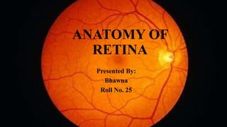
ANATOMY OF
RETINA
Presented By:
Bhawna
Roll No. 25
ANATOMY
• Retina- the innermost tunic of the eyeball
• thin, delicate and transparent membrane, which is the most
developed tissue of the eye.
Retina extends from the optic disc to the ora serrata .
 Retina is the thickest in the peripapillary (0.56mm) and thinnest
at ora serrate (0.1 mm).
GROSS DIVISION OF RETINA:
• Fundus refers to the dome shaped interior of the eyeball. Grossly,
fundus can be divided into two distinct regions:
 Posterior pole
Peripheral retina
I)Posterior pole: area of fundus posterior to the retinal
equator(imaginary line which is considered to lie in line with the
exist of the four vena vorticose )
• best examined : slit- lamp indirect bio microscopy using + 78 D
and + 90 D lens and by direct ophthalmoscopy.
• The posterior pole includes two distinct areas: the optic disc and
macula lutea.
1) Optic disc : also known as optic nerve head
• pink coloured, well defined vertically oval area with average
diameter of 1.5mm equal to 1DD (disc diameter).
• The optic disc thus represents the beginning of the optic nerve and
so is also referred to as optic nerve head.
• A depression seen in the disc is called the physiological cup.
• The central retinal artery and vein emerges through the
center of this cup.
• Because of absence of photoreceptors (rods and cones), the
optic disc produces an absolute scotoma in the visual field
called as physiological blind spot
2. Macula lutea: also known as yellow spot
• Itis comparatively darker than the surrounding fundus and is situated
at the posterior pole temporal to the optic disc.
• Fovea centralis is the central depressed part of the macula. the most
sensitive part of the retina.
• With lowest threshold for light and highest visual acuity, because it
contains only cones, in its center is a shining pit called foveola
• The tiny depression in the center of foveola is called umbo
• An area about 0.8mm in diameter (including foveola and some
surrounding area) does not contain any retinal capillaries and is
called foveal avascular zone (FAZ).
• Surrounding the fovea are the parafoveal and perifoveal areas.
II)Peripheral retina :area bounded posteriorly by the retinal equator and
anteriorly by the ora serrata
peripheral retina is best examined with indirect ophthalmoscopy and goldman
three mirror contact lens
MICROSCOPIC STRUCTURE: retina consists of 10 layers ,arranged in
two distinct functional components the pigment epithelium and the
neurosensory retina
1)Pigment epithelium: outermost layer of retina.
• It consists of a single layer of hexagonal cells containing melanin
pigment.
• It is firmly adherent to the underlying basal lamina of the choroid.
• Pigment epithelium provides metabolic support to the neurosensory
retina and also acts as an antireflective layer.
Interphotoreceptor matrix (IPM): present in the potential space
between pigment epithelium and the neurosensory retina
2). Layers of rods and cones: Rods and cones are the end organs of vision and
are also known as photoreceptors.
Layers of rods and cones contains only the outer segments of
photoreceptors cells arranged in a palisade manner.
There are about 120 million rods and 6.5 million cones.
Cones also contains a photosensitive substance and are primarily
responsible for highly discriminatory central vision (photopic vision) and
colour vision.
Rods contains a photosensitive substance visual purple (rhodopsin) and
subserve the peripheral vision and vision of low illumination (scotopic
vision).
3) External limiting membrane :it is a fenestrated membrane ,through which
pass processes of the rods and cones
4) OUTER NUCLEAR LAYER : it consists of nuclei of cones and rods
5) OUTER PLEXIFORM LAYER: it consists of connection of rods spherules
and cones pedicles with the dendrites of bipolar cells and horizontal cells
6) INNER NUCLEAR LAYER : it mainly consists of cell bodies of bipolar
cells.
• it also contain cell bodies of horizontal ,amacrine and muller’s cells and
capillaries of central artery of retina .
• The bipolar cells constitute the first order neurons .
7)INNER PLEXIFORM LAYER :it essentially consists of connections
between the axons of bipolar cells and dendrites of the ganglion cells ,and
processes of amacrine cells
8)GANGLION CELL LAYER : It mainly contains the cell bodies of ganglionic
cells (the second order neurons of visual pathway )
There are two types of ganglion cells :
Midget ganglion cells: present in the macular region and the dendrite
of each such cell synapses with the axon of single bipolar cell
Polysynaptic ganglion cells : lie predominantly in peripheral retina and
each such cell may synapse with up to a hundred bipolar cells
9) NERVE FIBRE LAYER (stratum opticum):consists of axons of the
ganglion cells ,which pass through the lamina cribrosa to form the optic
nerve .
10) Internal limiting membrane : it is the innermost layer and
separates the retina from vitreous .
Midget
ganglion
Polysynaptic
ganglionic
• It is formed by the union of terminal expansions of the muller’s fibres
,and is essentially a basement membrane
STRUCTURE OF FOVEA CENTRALIS :In this area ,there are no rods ,
cones are tightly packed and other layer of retina are very thin .
• Its central part (foveola)largely consists of cones and their nuclei
covered by a thin internal limiting membranes .all other retinal layers
are absent in this region
Functional division of retina :retina is divided into temporal
retina and nasal retina by a line drawn vertically through the
center of fovea
• Nerve fibre arising from temporal retina pass through the
optic nerve and optic tract of the same side to terminate in
the ipsilateral geniculate body
• Nerve fibres originating from the nasal retina after passing
through the optic nerve cross in the optic chiasma and travel
through the contralateral optic tract to terminate in the
contralateral geniculate body
BLOOD SUPPLY
• Outer four layer of the retina pigment epithelium , layer of rods and
cones , external limiting membrane and outer nuclear layer are
AVASCULAR
• they get their nutrition from the anterior ciliary arteries and
posterior ciliary arteries
• Inner six layer of retina are VASCULAR , get their supply from the
central retinal artery , which is a branch of ophthalmic artery
• Macular region supply by cilioretinal artery
• CENTRAL RETINAL ARTERY : emerges from the centre of the
physiological cup of the optic disc
• Divides into 4 branches :
1)Superior nasal 3) Inferior nasal
2)Superior temporal 4) Inferior temporal
• These are the end arteries, i.e they do not anastomosis with each other
RETINAL VEIN :
• Central retinal vein drains into cavernous sinus directly or through the
superior ophthalmic vein
• Only place where the retinal system anastomosis with ciliary system is in
the region of lamina cribrosa
BLOOD SUPPLY OF OPTIC NERVE HEAD :
 Surface nerve fibres layer: from retinal arterioles
 Prelaminar region : vessels of ciliary region
 Lamina cribrosa region : short posterior ciliary arteries
 Retrolaminar region :supplied by both ciliary and retinal circulation
ANATOMY OF RETINA.pptx

More Related Content

Similar to ANATOMY OF RETINA.pptx

Posterior segment of eye.pdf
Posterior segment of eye.pdfPosterior segment of eye.pdf
Posterior segment of eye.pdfSaroj Panthi
 
Anatomy of Uvea track
Anatomy of Uvea trackAnatomy of Uvea track
Anatomy of Uvea trackLhacha
 
385bceaa-1128-4537-b6a1-feee42cce7be.pdf
385bceaa-1128-4537-b6a1-feee42cce7be.pdf385bceaa-1128-4537-b6a1-feee42cce7be.pdf
385bceaa-1128-4537-b6a1-feee42cce7be.pdfdevb110695
 
Anatomy and Physiology of retina
Anatomy and Physiology of retinaAnatomy and Physiology of retina
Anatomy and Physiology of retinaVivek Adwe
 
Anatomy of optic nerve and its clinical significance
Anatomy of optic nerve and its clinical significanceAnatomy of optic nerve and its clinical significance
Anatomy of optic nerve and its clinical significancePabita Dhungel
 
vision.pptx
vision.pptxvision.pptx
vision.pptxLuzSan3
 
Anatomy and Physiology of Optic Nerve Dr.PrabhatDevkota.pptx
Anatomy and Physiology of Optic Nerve Dr.PrabhatDevkota.pptxAnatomy and Physiology of Optic Nerve Dr.PrabhatDevkota.pptx
Anatomy and Physiology of Optic Nerve Dr.PrabhatDevkota.pptxDr. Prabhat Devkota, MD
 
THE EYEBALL &conjuctiva anatomy in very easy way
THE EYEBALL &conjuctiva anatomy in very easy wayTHE EYEBALL &conjuctiva anatomy in very easy way
THE EYEBALL &conjuctiva anatomy in very easy wayABIDOFFICIALCHANNEL
 
RETINA - anatomy & physiology
RETINA - anatomy & physiologyRETINA - anatomy & physiology
RETINA - anatomy & physiologymonika pallan
 
Suraj chhetri retina
Suraj chhetri retinaSuraj chhetri retina
Suraj chhetri retinaSuraj Chhetri
 

Similar to ANATOMY OF RETINA.pptx (20)

Retina
RetinaRetina
Retina
 
Anatomy of the retina
Anatomy of the retinaAnatomy of the retina
Anatomy of the retina
 
Anatomy and Applied aspects of Uvea
Anatomy and Applied aspects of UveaAnatomy and Applied aspects of Uvea
Anatomy and Applied aspects of Uvea
 
Posterior segment of eye.pdf
Posterior segment of eye.pdfPosterior segment of eye.pdf
Posterior segment of eye.pdf
 
Anatomy of Uvea track
Anatomy of Uvea trackAnatomy of Uvea track
Anatomy of Uvea track
 
Retina
RetinaRetina
Retina
 
eye ball anat.pptx
eye ball anat.pptxeye ball anat.pptx
eye ball anat.pptx
 
385bceaa-1128-4537-b6a1-feee42cce7be.pdf
385bceaa-1128-4537-b6a1-feee42cce7be.pdf385bceaa-1128-4537-b6a1-feee42cce7be.pdf
385bceaa-1128-4537-b6a1-feee42cce7be.pdf
 
Anatomy and Physiology of retina
Anatomy and Physiology of retinaAnatomy and Physiology of retina
Anatomy and Physiology of retina
 
Anatomy of optic nerve and its clinical significance
Anatomy of optic nerve and its clinical significanceAnatomy of optic nerve and its clinical significance
Anatomy of optic nerve and its clinical significance
 
Retina
RetinaRetina
Retina
 
vision.pptx
vision.pptxvision.pptx
vision.pptx
 
Anatomy and Physiology of Optic Nerve Dr.PrabhatDevkota.pptx
Anatomy and Physiology of Optic Nerve Dr.PrabhatDevkota.pptxAnatomy and Physiology of Optic Nerve Dr.PrabhatDevkota.pptx
Anatomy and Physiology of Optic Nerve Dr.PrabhatDevkota.pptx
 
anatomy of retina
anatomy of retinaanatomy of retina
anatomy of retina
 
Uveal tract anatomy
Uveal tract anatomyUveal tract anatomy
Uveal tract anatomy
 
Anatomy of retina
Anatomy of retinaAnatomy of retina
Anatomy of retina
 
THE EYEBALL &conjuctiva anatomy in very easy way
THE EYEBALL &conjuctiva anatomy in very easy wayTHE EYEBALL &conjuctiva anatomy in very easy way
THE EYEBALL &conjuctiva anatomy in very easy way
 
RETINA - anatomy & physiology
RETINA - anatomy & physiologyRETINA - anatomy & physiology
RETINA - anatomy & physiology
 
Suraj chhetri retina
Suraj chhetri retinaSuraj chhetri retina
Suraj chhetri retina
 
Retina : an overview
Retina : an overviewRetina : an overview
Retina : an overview
 

More from 9459654457

investigation in pediatrics for a case of .pptx
investigation in pediatrics for a case of .pptxinvestigation in pediatrics for a case of .pptx
investigation in pediatrics for a case of .pptx9459654457
 
hip joint anatomy physiology and injuries.pptx
hip joint anatomy physiology and injuries.pptxhip joint anatomy physiology and injuries.pptx
hip joint anatomy physiology and injuries.pptx9459654457
 
EXPLORATORY LAPROTOMY indications and procedure.pptx
EXPLORATORY LAPROTOMY indications and procedure.pptxEXPLORATORY LAPROTOMY indications and procedure.pptx
EXPLORATORY LAPROTOMY indications and procedure.pptx9459654457
 
Vasculitis- Small, medium, large vessel vasculitis.pptx
Vasculitis- Small, medium, large vessel vasculitis.pptxVasculitis- Small, medium, large vessel vasculitis.pptx
Vasculitis- Small, medium, large vessel vasculitis.pptx9459654457
 
Lichtenstein hernioplasty surgery ppt.pptx
Lichtenstein hernioplasty surgery ppt.pptxLichtenstein hernioplasty surgery ppt.pptx
Lichtenstein hernioplasty surgery ppt.pptx9459654457
 
Steroidal contraceptives.pptx
Steroidal contraceptives.pptxSteroidal contraceptives.pptx
Steroidal contraceptives.pptx9459654457
 
ANTINEUTROPHIL CYTOPLASMIC ANTIBODY ASSOCIATED VASCULITIS.pptx
ANTINEUTROPHIL CYTOPLASMIC ANTIBODY ASSOCIATED  VASCULITIS.pptxANTINEUTROPHIL CYTOPLASMIC ANTIBODY ASSOCIATED  VASCULITIS.pptx
ANTINEUTROPHIL CYTOPLASMIC ANTIBODY ASSOCIATED VASCULITIS.pptx9459654457
 
old-age-faculty---opmh-personality-disorders-in-older-adults-tier-2---2021394...
old-age-faculty---opmh-personality-disorders-in-older-adults-tier-2---2021394...old-age-faculty---opmh-personality-disorders-in-older-adults-tier-2---2021394...
old-age-faculty---opmh-personality-disorders-in-older-adults-tier-2---2021394...9459654457
 
Rh negative case.pptx
Rh negative case.pptxRh negative case.pptx
Rh negative case.pptx9459654457
 
Anatomy of larynx
Anatomy of larynxAnatomy of larynx
Anatomy of larynx9459654457
 
CRITERIA FOR SCREENING.pptx
CRITERIA FOR SCREENING.pptxCRITERIA FOR SCREENING.pptx
CRITERIA FOR SCREENING.pptx9459654457
 
ANTENATAL_SCREENING_IN_FIRST_AND_SECOND_TRIMESTER.ppt
ANTENATAL_SCREENING_IN_FIRST_AND_SECOND_TRIMESTER.pptANTENATAL_SCREENING_IN_FIRST_AND_SECOND_TRIMESTER.ppt
ANTENATAL_SCREENING_IN_FIRST_AND_SECOND_TRIMESTER.ppt9459654457
 
Antenatal care.pptx
Antenatal care.pptxAntenatal care.pptx
Antenatal care.pptx9459654457
 
hydatid cyst.pptx
hydatid cyst.pptxhydatid cyst.pptx
hydatid cyst.pptx9459654457
 
Presentation 1.pptx
Presentation 1.pptxPresentation 1.pptx
Presentation 1.pptx9459654457
 
ca gall bladder seminar.ppt
ca gall bladder seminar.pptca gall bladder seminar.ppt
ca gall bladder seminar.ppt9459654457
 
Metabolic Alkalosis .pptx
Metabolic Alkalosis .pptxMetabolic Alkalosis .pptx
Metabolic Alkalosis .pptx9459654457
 
friends Quiz.pptx
friends Quiz.pptxfriends Quiz.pptx
friends Quiz.pptx9459654457
 

More from 9459654457 (20)

investigation in pediatrics for a case of .pptx
investigation in pediatrics for a case of .pptxinvestigation in pediatrics for a case of .pptx
investigation in pediatrics for a case of .pptx
 
hip joint anatomy physiology and injuries.pptx
hip joint anatomy physiology and injuries.pptxhip joint anatomy physiology and injuries.pptx
hip joint anatomy physiology and injuries.pptx
 
EXPLORATORY LAPROTOMY indications and procedure.pptx
EXPLORATORY LAPROTOMY indications and procedure.pptxEXPLORATORY LAPROTOMY indications and procedure.pptx
EXPLORATORY LAPROTOMY indications and procedure.pptx
 
Vasculitis- Small, medium, large vessel vasculitis.pptx
Vasculitis- Small, medium, large vessel vasculitis.pptxVasculitis- Small, medium, large vessel vasculitis.pptx
Vasculitis- Small, medium, large vessel vasculitis.pptx
 
Lichtenstein hernioplasty surgery ppt.pptx
Lichtenstein hernioplasty surgery ppt.pptxLichtenstein hernioplasty surgery ppt.pptx
Lichtenstein hernioplasty surgery ppt.pptx
 
Steroidal contraceptives.pptx
Steroidal contraceptives.pptxSteroidal contraceptives.pptx
Steroidal contraceptives.pptx
 
ANTINEUTROPHIL CYTOPLASMIC ANTIBODY ASSOCIATED VASCULITIS.pptx
ANTINEUTROPHIL CYTOPLASMIC ANTIBODY ASSOCIATED  VASCULITIS.pptxANTINEUTROPHIL CYTOPLASMIC ANTIBODY ASSOCIATED  VASCULITIS.pptx
ANTINEUTROPHIL CYTOPLASMIC ANTIBODY ASSOCIATED VASCULITIS.pptx
 
old-age-faculty---opmh-personality-disorders-in-older-adults-tier-2---2021394...
old-age-faculty---opmh-personality-disorders-in-older-adults-tier-2---2021394...old-age-faculty---opmh-personality-disorders-in-older-adults-tier-2---2021394...
old-age-faculty---opmh-personality-disorders-in-older-adults-tier-2---2021394...
 
Rh negative case.pptx
Rh negative case.pptxRh negative case.pptx
Rh negative case.pptx
 
Anatomy of larynx
Anatomy of larynxAnatomy of larynx
Anatomy of larynx
 
IHD .pptx
IHD .pptxIHD .pptx
IHD .pptx
 
angina.pptx
angina.pptxangina.pptx
angina.pptx
 
CRITERIA FOR SCREENING.pptx
CRITERIA FOR SCREENING.pptxCRITERIA FOR SCREENING.pptx
CRITERIA FOR SCREENING.pptx
 
ANTENATAL_SCREENING_IN_FIRST_AND_SECOND_TRIMESTER.ppt
ANTENATAL_SCREENING_IN_FIRST_AND_SECOND_TRIMESTER.pptANTENATAL_SCREENING_IN_FIRST_AND_SECOND_TRIMESTER.ppt
ANTENATAL_SCREENING_IN_FIRST_AND_SECOND_TRIMESTER.ppt
 
Antenatal care.pptx
Antenatal care.pptxAntenatal care.pptx
Antenatal care.pptx
 
hydatid cyst.pptx
hydatid cyst.pptxhydatid cyst.pptx
hydatid cyst.pptx
 
Presentation 1.pptx
Presentation 1.pptxPresentation 1.pptx
Presentation 1.pptx
 
ca gall bladder seminar.ppt
ca gall bladder seminar.pptca gall bladder seminar.ppt
ca gall bladder seminar.ppt
 
Metabolic Alkalosis .pptx
Metabolic Alkalosis .pptxMetabolic Alkalosis .pptx
Metabolic Alkalosis .pptx
 
friends Quiz.pptx
friends Quiz.pptxfriends Quiz.pptx
friends Quiz.pptx
 

Recently uploaded

History Class XII Ch. 3 Kinship, Caste and Class (1).pptx
History Class XII Ch. 3 Kinship, Caste and Class (1).pptxHistory Class XII Ch. 3 Kinship, Caste and Class (1).pptx
History Class XII Ch. 3 Kinship, Caste and Class (1).pptxsocialsciencegdgrohi
 
BASLIQ CURRENT LOOKBOOK LOOKBOOK(1) (1).pdf
BASLIQ CURRENT LOOKBOOK  LOOKBOOK(1) (1).pdfBASLIQ CURRENT LOOKBOOK  LOOKBOOK(1) (1).pdf
BASLIQ CURRENT LOOKBOOK LOOKBOOK(1) (1).pdfSoniaTolstoy
 
How to Make a Pirate ship Primary Education.pptx
How to Make a Pirate ship Primary Education.pptxHow to Make a Pirate ship Primary Education.pptx
How to Make a Pirate ship Primary Education.pptxmanuelaromero2013
 
ENGLISH5 QUARTER4 MODULE1 WEEK1-3 How Visual and Multimedia Elements.pptx
ENGLISH5 QUARTER4 MODULE1 WEEK1-3 How Visual and Multimedia Elements.pptxENGLISH5 QUARTER4 MODULE1 WEEK1-3 How Visual and Multimedia Elements.pptx
ENGLISH5 QUARTER4 MODULE1 WEEK1-3 How Visual and Multimedia Elements.pptxAnaBeatriceAblay2
 
SOCIAL AND HISTORICAL CONTEXT - LFTVD.pptx
SOCIAL AND HISTORICAL CONTEXT - LFTVD.pptxSOCIAL AND HISTORICAL CONTEXT - LFTVD.pptx
SOCIAL AND HISTORICAL CONTEXT - LFTVD.pptxiammrhaywood
 
18-04-UA_REPORT_MEDIALITERAСY_INDEX-DM_23-1-final-eng.pdf
18-04-UA_REPORT_MEDIALITERAСY_INDEX-DM_23-1-final-eng.pdf18-04-UA_REPORT_MEDIALITERAСY_INDEX-DM_23-1-final-eng.pdf
18-04-UA_REPORT_MEDIALITERAСY_INDEX-DM_23-1-final-eng.pdfssuser54595a
 
Organic Name Reactions for the students and aspirants of Chemistry12th.pptx
Organic Name Reactions  for the students and aspirants of Chemistry12th.pptxOrganic Name Reactions  for the students and aspirants of Chemistry12th.pptx
Organic Name Reactions for the students and aspirants of Chemistry12th.pptxVS Mahajan Coaching Centre
 
Mastering the Unannounced Regulatory Inspection
Mastering the Unannounced Regulatory InspectionMastering the Unannounced Regulatory Inspection
Mastering the Unannounced Regulatory InspectionSafetyChain Software
 
Painted Grey Ware.pptx, PGW Culture of India
Painted Grey Ware.pptx, PGW Culture of IndiaPainted Grey Ware.pptx, PGW Culture of India
Painted Grey Ware.pptx, PGW Culture of IndiaVirag Sontakke
 
Computed Fields and api Depends in the Odoo 17
Computed Fields and api Depends in the Odoo 17Computed Fields and api Depends in the Odoo 17
Computed Fields and api Depends in the Odoo 17Celine George
 
Alper Gobel In Media Res Media Component
Alper Gobel In Media Res Media ComponentAlper Gobel In Media Res Media Component
Alper Gobel In Media Res Media ComponentInMediaRes1
 
Employee wellbeing at the workplace.pptx
Employee wellbeing at the workplace.pptxEmployee wellbeing at the workplace.pptx
Employee wellbeing at the workplace.pptxNirmalaLoungPoorunde1
 
Enzyme, Pharmaceutical Aids, Miscellaneous Last Part of Chapter no 5th.pdf
Enzyme, Pharmaceutical Aids, Miscellaneous Last Part of Chapter no 5th.pdfEnzyme, Pharmaceutical Aids, Miscellaneous Last Part of Chapter no 5th.pdf
Enzyme, Pharmaceutical Aids, Miscellaneous Last Part of Chapter no 5th.pdfSumit Tiwari
 
Pharmacognosy Flower 3. Compositae 2023.pdf
Pharmacognosy Flower 3. Compositae 2023.pdfPharmacognosy Flower 3. Compositae 2023.pdf
Pharmacognosy Flower 3. Compositae 2023.pdfMahmoud M. Sallam
 
Science lesson Moon for 4th quarter lesson
Science lesson Moon for 4th quarter lessonScience lesson Moon for 4th quarter lesson
Science lesson Moon for 4th quarter lessonJericReyAuditor
 
The Most Excellent Way | 1 Corinthians 13
The Most Excellent Way | 1 Corinthians 13The Most Excellent Way | 1 Corinthians 13
The Most Excellent Way | 1 Corinthians 13Steve Thomason
 
Crayon Activity Handout For the Crayon A
Crayon Activity Handout For the Crayon ACrayon Activity Handout For the Crayon A
Crayon Activity Handout For the Crayon AUnboundStockton
 
call girls in Kamla Market (DELHI) 🔝 >༒9953330565🔝 genuine Escort Service 🔝✔️✔️
call girls in Kamla Market (DELHI) 🔝 >༒9953330565🔝 genuine Escort Service 🔝✔️✔️call girls in Kamla Market (DELHI) 🔝 >༒9953330565🔝 genuine Escort Service 🔝✔️✔️
call girls in Kamla Market (DELHI) 🔝 >༒9953330565🔝 genuine Escort Service 🔝✔️✔️9953056974 Low Rate Call Girls In Saket, Delhi NCR
 
Software Engineering Methodologies (overview)
Software Engineering Methodologies (overview)Software Engineering Methodologies (overview)
Software Engineering Methodologies (overview)eniolaolutunde
 

Recently uploaded (20)

History Class XII Ch. 3 Kinship, Caste and Class (1).pptx
History Class XII Ch. 3 Kinship, Caste and Class (1).pptxHistory Class XII Ch. 3 Kinship, Caste and Class (1).pptx
History Class XII Ch. 3 Kinship, Caste and Class (1).pptx
 
BASLIQ CURRENT LOOKBOOK LOOKBOOK(1) (1).pdf
BASLIQ CURRENT LOOKBOOK  LOOKBOOK(1) (1).pdfBASLIQ CURRENT LOOKBOOK  LOOKBOOK(1) (1).pdf
BASLIQ CURRENT LOOKBOOK LOOKBOOK(1) (1).pdf
 
How to Make a Pirate ship Primary Education.pptx
How to Make a Pirate ship Primary Education.pptxHow to Make a Pirate ship Primary Education.pptx
How to Make a Pirate ship Primary Education.pptx
 
ENGLISH5 QUARTER4 MODULE1 WEEK1-3 How Visual and Multimedia Elements.pptx
ENGLISH5 QUARTER4 MODULE1 WEEK1-3 How Visual and Multimedia Elements.pptxENGLISH5 QUARTER4 MODULE1 WEEK1-3 How Visual and Multimedia Elements.pptx
ENGLISH5 QUARTER4 MODULE1 WEEK1-3 How Visual and Multimedia Elements.pptx
 
SOCIAL AND HISTORICAL CONTEXT - LFTVD.pptx
SOCIAL AND HISTORICAL CONTEXT - LFTVD.pptxSOCIAL AND HISTORICAL CONTEXT - LFTVD.pptx
SOCIAL AND HISTORICAL CONTEXT - LFTVD.pptx
 
18-04-UA_REPORT_MEDIALITERAСY_INDEX-DM_23-1-final-eng.pdf
18-04-UA_REPORT_MEDIALITERAСY_INDEX-DM_23-1-final-eng.pdf18-04-UA_REPORT_MEDIALITERAСY_INDEX-DM_23-1-final-eng.pdf
18-04-UA_REPORT_MEDIALITERAСY_INDEX-DM_23-1-final-eng.pdf
 
Organic Name Reactions for the students and aspirants of Chemistry12th.pptx
Organic Name Reactions  for the students and aspirants of Chemistry12th.pptxOrganic Name Reactions  for the students and aspirants of Chemistry12th.pptx
Organic Name Reactions for the students and aspirants of Chemistry12th.pptx
 
Mastering the Unannounced Regulatory Inspection
Mastering the Unannounced Regulatory InspectionMastering the Unannounced Regulatory Inspection
Mastering the Unannounced Regulatory Inspection
 
Painted Grey Ware.pptx, PGW Culture of India
Painted Grey Ware.pptx, PGW Culture of IndiaPainted Grey Ware.pptx, PGW Culture of India
Painted Grey Ware.pptx, PGW Culture of India
 
Computed Fields and api Depends in the Odoo 17
Computed Fields and api Depends in the Odoo 17Computed Fields and api Depends in the Odoo 17
Computed Fields and api Depends in the Odoo 17
 
Alper Gobel In Media Res Media Component
Alper Gobel In Media Res Media ComponentAlper Gobel In Media Res Media Component
Alper Gobel In Media Res Media Component
 
Employee wellbeing at the workplace.pptx
Employee wellbeing at the workplace.pptxEmployee wellbeing at the workplace.pptx
Employee wellbeing at the workplace.pptx
 
Enzyme, Pharmaceutical Aids, Miscellaneous Last Part of Chapter no 5th.pdf
Enzyme, Pharmaceutical Aids, Miscellaneous Last Part of Chapter no 5th.pdfEnzyme, Pharmaceutical Aids, Miscellaneous Last Part of Chapter no 5th.pdf
Enzyme, Pharmaceutical Aids, Miscellaneous Last Part of Chapter no 5th.pdf
 
Pharmacognosy Flower 3. Compositae 2023.pdf
Pharmacognosy Flower 3. Compositae 2023.pdfPharmacognosy Flower 3. Compositae 2023.pdf
Pharmacognosy Flower 3. Compositae 2023.pdf
 
9953330565 Low Rate Call Girls In Rohini Delhi NCR
9953330565 Low Rate Call Girls In Rohini  Delhi NCR9953330565 Low Rate Call Girls In Rohini  Delhi NCR
9953330565 Low Rate Call Girls In Rohini Delhi NCR
 
Science lesson Moon for 4th quarter lesson
Science lesson Moon for 4th quarter lessonScience lesson Moon for 4th quarter lesson
Science lesson Moon for 4th quarter lesson
 
The Most Excellent Way | 1 Corinthians 13
The Most Excellent Way | 1 Corinthians 13The Most Excellent Way | 1 Corinthians 13
The Most Excellent Way | 1 Corinthians 13
 
Crayon Activity Handout For the Crayon A
Crayon Activity Handout For the Crayon ACrayon Activity Handout For the Crayon A
Crayon Activity Handout For the Crayon A
 
call girls in Kamla Market (DELHI) 🔝 >༒9953330565🔝 genuine Escort Service 🔝✔️✔️
call girls in Kamla Market (DELHI) 🔝 >༒9953330565🔝 genuine Escort Service 🔝✔️✔️call girls in Kamla Market (DELHI) 🔝 >༒9953330565🔝 genuine Escort Service 🔝✔️✔️
call girls in Kamla Market (DELHI) 🔝 >༒9953330565🔝 genuine Escort Service 🔝✔️✔️
 
Software Engineering Methodologies (overview)
Software Engineering Methodologies (overview)Software Engineering Methodologies (overview)
Software Engineering Methodologies (overview)
 

ANATOMY OF RETINA.pptx

  • 2. ANATOMY • Retina- the innermost tunic of the eyeball • thin, delicate and transparent membrane, which is the most developed tissue of the eye. Retina extends from the optic disc to the ora serrata .  Retina is the thickest in the peripapillary (0.56mm) and thinnest at ora serrate (0.1 mm). GROSS DIVISION OF RETINA: • Fundus refers to the dome shaped interior of the eyeball. Grossly, fundus can be divided into two distinct regions:  Posterior pole Peripheral retina
  • 3. I)Posterior pole: area of fundus posterior to the retinal equator(imaginary line which is considered to lie in line with the exist of the four vena vorticose ) • best examined : slit- lamp indirect bio microscopy using + 78 D and + 90 D lens and by direct ophthalmoscopy. • The posterior pole includes two distinct areas: the optic disc and macula lutea. 1) Optic disc : also known as optic nerve head • pink coloured, well defined vertically oval area with average diameter of 1.5mm equal to 1DD (disc diameter). • The optic disc thus represents the beginning of the optic nerve and so is also referred to as optic nerve head.
  • 4. • A depression seen in the disc is called the physiological cup. • The central retinal artery and vein emerges through the center of this cup. • Because of absence of photoreceptors (rods and cones), the optic disc produces an absolute scotoma in the visual field called as physiological blind spot
  • 5. 2. Macula lutea: also known as yellow spot • Itis comparatively darker than the surrounding fundus and is situated at the posterior pole temporal to the optic disc. • Fovea centralis is the central depressed part of the macula. the most sensitive part of the retina. • With lowest threshold for light and highest visual acuity, because it contains only cones, in its center is a shining pit called foveola • The tiny depression in the center of foveola is called umbo • An area about 0.8mm in diameter (including foveola and some surrounding area) does not contain any retinal capillaries and is called foveal avascular zone (FAZ). • Surrounding the fovea are the parafoveal and perifoveal areas.
  • 6. II)Peripheral retina :area bounded posteriorly by the retinal equator and anteriorly by the ora serrata peripheral retina is best examined with indirect ophthalmoscopy and goldman three mirror contact lens
  • 7. MICROSCOPIC STRUCTURE: retina consists of 10 layers ,arranged in two distinct functional components the pigment epithelium and the neurosensory retina 1)Pigment epithelium: outermost layer of retina. • It consists of a single layer of hexagonal cells containing melanin pigment. • It is firmly adherent to the underlying basal lamina of the choroid. • Pigment epithelium provides metabolic support to the neurosensory retina and also acts as an antireflective layer. Interphotoreceptor matrix (IPM): present in the potential space between pigment epithelium and the neurosensory retina
  • 8. 2). Layers of rods and cones: Rods and cones are the end organs of vision and are also known as photoreceptors. Layers of rods and cones contains only the outer segments of photoreceptors cells arranged in a palisade manner. There are about 120 million rods and 6.5 million cones. Cones also contains a photosensitive substance and are primarily responsible for highly discriminatory central vision (photopic vision) and colour vision. Rods contains a photosensitive substance visual purple (rhodopsin) and subserve the peripheral vision and vision of low illumination (scotopic vision). 3) External limiting membrane :it is a fenestrated membrane ,through which pass processes of the rods and cones
  • 9. 4) OUTER NUCLEAR LAYER : it consists of nuclei of cones and rods 5) OUTER PLEXIFORM LAYER: it consists of connection of rods spherules and cones pedicles with the dendrites of bipolar cells and horizontal cells 6) INNER NUCLEAR LAYER : it mainly consists of cell bodies of bipolar cells. • it also contain cell bodies of horizontal ,amacrine and muller’s cells and capillaries of central artery of retina . • The bipolar cells constitute the first order neurons . 7)INNER PLEXIFORM LAYER :it essentially consists of connections between the axons of bipolar cells and dendrites of the ganglion cells ,and processes of amacrine cells 8)GANGLION CELL LAYER : It mainly contains the cell bodies of ganglionic cells (the second order neurons of visual pathway )
  • 10. There are two types of ganglion cells : Midget ganglion cells: present in the macular region and the dendrite of each such cell synapses with the axon of single bipolar cell Polysynaptic ganglion cells : lie predominantly in peripheral retina and each such cell may synapse with up to a hundred bipolar cells 9) NERVE FIBRE LAYER (stratum opticum):consists of axons of the ganglion cells ,which pass through the lamina cribrosa to form the optic nerve . 10) Internal limiting membrane : it is the innermost layer and separates the retina from vitreous . Midget ganglion Polysynaptic ganglionic
  • 11. • It is formed by the union of terminal expansions of the muller’s fibres ,and is essentially a basement membrane
  • 12. STRUCTURE OF FOVEA CENTRALIS :In this area ,there are no rods , cones are tightly packed and other layer of retina are very thin . • Its central part (foveola)largely consists of cones and their nuclei covered by a thin internal limiting membranes .all other retinal layers are absent in this region
  • 13. Functional division of retina :retina is divided into temporal retina and nasal retina by a line drawn vertically through the center of fovea • Nerve fibre arising from temporal retina pass through the optic nerve and optic tract of the same side to terminate in the ipsilateral geniculate body • Nerve fibres originating from the nasal retina after passing through the optic nerve cross in the optic chiasma and travel through the contralateral optic tract to terminate in the contralateral geniculate body
  • 14. BLOOD SUPPLY • Outer four layer of the retina pigment epithelium , layer of rods and cones , external limiting membrane and outer nuclear layer are AVASCULAR • they get their nutrition from the anterior ciliary arteries and posterior ciliary arteries • Inner six layer of retina are VASCULAR , get their supply from the central retinal artery , which is a branch of ophthalmic artery • Macular region supply by cilioretinal artery • CENTRAL RETINAL ARTERY : emerges from the centre of the physiological cup of the optic disc • Divides into 4 branches :
  • 15. 1)Superior nasal 3) Inferior nasal 2)Superior temporal 4) Inferior temporal • These are the end arteries, i.e they do not anastomosis with each other RETINAL VEIN : • Central retinal vein drains into cavernous sinus directly or through the superior ophthalmic vein • Only place where the retinal system anastomosis with ciliary system is in the region of lamina cribrosa BLOOD SUPPLY OF OPTIC NERVE HEAD :  Surface nerve fibres layer: from retinal arterioles  Prelaminar region : vessels of ciliary region  Lamina cribrosa region : short posterior ciliary arteries  Retrolaminar region :supplied by both ciliary and retinal circulation