SlideShare a Scribd company logo
1 of 5
Download to read offline
Short case publication... Version 2.10| Edited by professor Yasser Metwally | December 2008


                                                                                                     Short case

                                                                                                     Edited by
                                                                                             Professor Yasser Metwally
                                                                                               Professor of neurology
                                                                                          Ain Shams university school of medicine
                                                                                                     Cairo, Egypt

                                                                                                Visit my web site at:
                                                                                             http://yassermetwally.com


27 years old male patient presented clinically with bilateral pyramidal manifestations, more on the left side, bilateral
parkinsonian manifestations and bilateral cerebellar manifestations more on the left side. Slit lamb examination of the
cornea revealed Kayser-Fleischer rings. Serum ceruloplasmin was reduced and urinary excretion of copper was increased.
Liver biopsy revealed increased concentration of copper.

DIAGNOSIS: WILSON DISEASE
Figure 1. Postcontrast MRI T1 studies showing ventricular dilatation, bilateral more or less symmetrical
hypointensities affecting predominantly the putamen and the internal capsule (mainly the posterior limbs). Notice
the bilateral fronto-partial hypointensities with probable cystic changes in the subcortical region of the right
posterior fronto-parietal region, the hypointensity probably involves the motor strip in the posterior frontal area
(area number 4). Also noticed mild cortical atrophy predominantly affecting the bilateral frontal regions. Notice the
linear hypointensity involving the thalamus and extending to the upper midbrain in the presumed area of the
pyramidal tract in (C). The MRI T1 hypointense changes demonstrated in these MRI images correspond,
pathologically, to edema, cystic changes, astrogliosis and demyelination involving mainly the cortical motor strip
and the descending cortico-spinal tract in the internal capsule, thalamus and upper midbrain. The demonstrated
lesions did not have mass effect and did not show any evidence of postcontrast enhancement.


                                                                Figure 2. Postcontrast MRI T1 images showing
                                                                ventricular dilatation, bilateral more or less
                                                                symmetrical linear hypointensities in the
                                                                presumed area of the cortico-spinal tract. Notice
                                                                the bilateral fronto-partial hypointensities with
                                                                probable subcortical cystic changes. The MRI T1
                                                                hypointense changes demonstrated in these MRI
                                                                images correspond, pathologically, to edema,
                                                                cystic changes, astrogliosis and demyelination
                                                                involving mainly the cortical motor strip and the
                                                                descending cortico-spinal tract in the internal
                                                                capsule, thalamus and upper midbrain.
Figure 3. Postcontrast MRI T1 images
                                                                          showing ventricular dilatation, bilateral
                                                                          more or less symmetrical linear
                                                                          hypointensities in the presumed area of
                                                                          the cortico-spinal tract. Notice the
                                                                          bilateral fronto-partial hypointensities
                                                                          with probable subcortical cystic changes.
                                                                          The MRI T1 hypointense changes
                                                                          demonstrated in these MRI images
                                                                          correspond, pathologically, to edema,
                                                                          cystic    changes,     astrogliosis  and
                                                                          demyelination involving mainly the
cortical motor strip and the descending cortico-spinal tract in the internal capsule, thalamus and upper midbrain.


Figure 4. MRI T2 images showing bilateral, more of less symmetrical hyperintensities involving the thalamus,
putamen, head of caudate nucleus, and the globus pallidus. Notice the bilateral hypointensities in the posterior
limb of the internal capsule probably involving the cortico-spinal tract. The crus cerebri appears hypointense in
(B) probably due to concomitant iron deposition. The red nucleus appears hyperintense in (A) and is seen
surrounded by a hypointense rim, This pattern is probably due to involvement of the dentatorubrothalamic tract
which is the main efferent pathway of the cerebellum and originates in the dentate nucleus. The dentate nucleus
mainly gives rise to the superior cerebellar peduncle, which decussates in the dorsal mesencephalon. The
superior cerebellar peduncle terminates, surrounds, or traverses the red nucleus and involvement of this tract, by
demyelination, and astroglosis, might be resposible for the abnormal signal intensity in and around the red
nucleus. The dentatothalamic tract terminates in the ventrolateral nucleus of the thalamus.
Figure 5. MRI T2 images showing bifrontal hyperintensities, mainly involving the posterior regions of the frontal
lobes with slight right sided predominance. The MRI T2 bifrontal hyperintensities mainly involves the subcortical
white matter (cystic changes and astrogliosis), with some linear cortical MRI T2 hypointensities which could be
due to concomitant iron deposition. Notice the bilateral, more or less symmetrical, basal ganglionic
hyperintensities (cystic changes and astrogliosis) and the moderate dilatation of the frontal horns of the lateral
ventricles.


Figure 6. MRI FLAIR images showing bilateral, more or less symmetrical, basal ganglionic hyperintensities (cystic
changes and astrogliosis) and moderate dilatation of the frontal horns of the lateral ventricles.
Figure 7 . Slit lamb examination of the cornea revealed Kayser-Fleischer rings.


References

1. Metwally, MYM: Textbook of neurimaging, A CD-ROM publication, (Metwally, MYM editor) WEB-CD agency for
electronic publishing, version 9.4a October 2008


Addendum

   A new version of short case is uploaded in my web site every week (every Saturday and remains available till
   Friday.)
   To download the current version follow the link quot;http://pdf.yassermetwally.com/short.pdfquot;.
   You can download the long case version of this short case during the same week from: http://pdf.yassermetwally.com/case.pdf or
   visit web site: http://pdf.yassermetwally.com
   To download the software version of the publication (crow.exe) follow the link: http://neurology.yassermetwally.com/crow.zip
   At the end of each year, all the publications are compiled on a single CD-ROM, please contact the author to know
   more details.
   Screen resolution is better set at 1024*768 pixel screen area for optimum display
   For an archive of the previously reported cases go to www.yassermetwally.net, then under pages in the right panel,
   scroll down and click on the text entry quot;downloadable short cases in PDF formatquot;
   Also to view a list of the previously published case records follow the following link (http://wordpress.com/tag/
   case-record/) or click on it if it appears as a link in your PDF reader

More Related Content

What's hot

Spinal cord tumors
Spinal cord tumorsSpinal cord tumors
Spinal cord tumorsANILKUMAR BR
 
Presentation1, artifacts and pitfalls of the wrist and elbow joints.
Presentation1, artifacts and pitfalls of the wrist and elbow joints.Presentation1, artifacts and pitfalls of the wrist and elbow joints.
Presentation1, artifacts and pitfalls of the wrist and elbow joints.Abdellah Nazeer
 
Schmorl’s node case presentation
Schmorl’s node case presentationSchmorl’s node case presentation
Schmorl’s node case presentationREKHAKHARE
 
Presentation1, artifact and pitfalls of the knee, hip and ankle joints.
Presentation1, artifact and pitfalls of the knee, hip and ankle joints.Presentation1, artifact and pitfalls of the knee, hip and ankle joints.
Presentation1, artifact and pitfalls of the knee, hip and ankle joints.Abdellah Nazeer
 
Presentation1.pptx, radiological imaging of soft tissue masses of the hand an...
Presentation1.pptx, radiological imaging of soft tissue masses of the hand an...Presentation1.pptx, radiological imaging of soft tissue masses of the hand an...
Presentation1.pptx, radiological imaging of soft tissue masses of the hand an...Abdellah Nazeer
 
Presentation1, musculoskeletal anatomy.
Presentation1, musculoskeletal anatomy.Presentation1, musculoskeletal anatomy.
Presentation1, musculoskeletal anatomy.Abdellah Nazeer
 
Presentation1.pptx radio;ogical imaging of benign and malignant soft tissue t...
Presentation1.pptx radio;ogical imaging of benign and malignant soft tissue t...Presentation1.pptx radio;ogical imaging of benign and malignant soft tissue t...
Presentation1.pptx radio;ogical imaging of benign and malignant soft tissue t...Abdellah Nazeer
 
Temporomandibular joint disorders
Temporomandibular joint disorders Temporomandibular joint disorders
Temporomandibular joint disorders Eman Alsheikh
 
Spinal cord lesions and its radiological imaging finding.
Spinal cord lesions and its radiological imaging finding.Spinal cord lesions and its radiological imaging finding.
Spinal cord lesions and its radiological imaging finding.Navneet Ranjan
 
Heterotrophic ossification
Heterotrophic ossificationHeterotrophic ossification
Heterotrophic ossificationAnnnoble
 
Spinal tumors drsant91
Spinal tumors drsant91Spinal tumors drsant91
Spinal tumors drsant91Santosh Prasad
 
Presentation1, radiological imaging of anterior knee pain.
Presentation1, radiological imaging of anterior knee pain.Presentation1, radiological imaging of anterior knee pain.
Presentation1, radiological imaging of anterior knee pain.Abdellah Nazeer
 
Myositis ossificans
Myositis ossificans Myositis ossificans
Myositis ossificans orthoprince
 
Presentation1.pptx, radiological imaging of spinal dysraphism.
Presentation1.pptx, radiological imaging of spinal dysraphism.Presentation1.pptx, radiological imaging of spinal dysraphism.
Presentation1.pptx, radiological imaging of spinal dysraphism.Abdellah Nazeer
 
Temporomandibular Joint Disorders
Temporomandibular Joint DisordersTemporomandibular Joint Disorders
Temporomandibular Joint DisordersHadi Munib
 
Presentation1, radiological film reading of knee joint.
Presentation1, radiological film reading of knee joint.Presentation1, radiological film reading of knee joint.
Presentation1, radiological film reading of knee joint.Abdellah Nazeer
 

What's hot (20)

Spinal cord tumors
Spinal cord tumorsSpinal cord tumors
Spinal cord tumors
 
Presentation1, artifacts and pitfalls of the wrist and elbow joints.
Presentation1, artifacts and pitfalls of the wrist and elbow joints.Presentation1, artifacts and pitfalls of the wrist and elbow joints.
Presentation1, artifacts and pitfalls of the wrist and elbow joints.
 
Schmorl’s node case presentation
Schmorl’s node case presentationSchmorl’s node case presentation
Schmorl’s node case presentation
 
Presentation1, artifact and pitfalls of the knee, hip and ankle joints.
Presentation1, artifact and pitfalls of the knee, hip and ankle joints.Presentation1, artifact and pitfalls of the knee, hip and ankle joints.
Presentation1, artifact and pitfalls of the knee, hip and ankle joints.
 
Presentation1.pptx, radiological imaging of soft tissue masses of the hand an...
Presentation1.pptx, radiological imaging of soft tissue masses of the hand an...Presentation1.pptx, radiological imaging of soft tissue masses of the hand an...
Presentation1.pptx, radiological imaging of soft tissue masses of the hand an...
 
Presentation1, musculoskeletal anatomy.
Presentation1, musculoskeletal anatomy.Presentation1, musculoskeletal anatomy.
Presentation1, musculoskeletal anatomy.
 
Myositis ossificans
Myositis ossificansMyositis ossificans
Myositis ossificans
 
Myositis ossificans
Myositis ossificansMyositis ossificans
Myositis ossificans
 
Knee cystic lesions
Knee cystic lesionsKnee cystic lesions
Knee cystic lesions
 
Presentation1.pptx radio;ogical imaging of benign and malignant soft tissue t...
Presentation1.pptx radio;ogical imaging of benign and malignant soft tissue t...Presentation1.pptx radio;ogical imaging of benign and malignant soft tissue t...
Presentation1.pptx radio;ogical imaging of benign and malignant soft tissue t...
 
Temporomandibular joint disorders
Temporomandibular joint disorders Temporomandibular joint disorders
Temporomandibular joint disorders
 
Myositis ossificans
Myositis ossificans Myositis ossificans
Myositis ossificans
 
Spinal cord lesions and its radiological imaging finding.
Spinal cord lesions and its radiological imaging finding.Spinal cord lesions and its radiological imaging finding.
Spinal cord lesions and its radiological imaging finding.
 
Heterotrophic ossification
Heterotrophic ossificationHeterotrophic ossification
Heterotrophic ossification
 
Spinal tumors drsant91
Spinal tumors drsant91Spinal tumors drsant91
Spinal tumors drsant91
 
Presentation1, radiological imaging of anterior knee pain.
Presentation1, radiological imaging of anterior knee pain.Presentation1, radiological imaging of anterior knee pain.
Presentation1, radiological imaging of anterior knee pain.
 
Myositis ossificans
Myositis ossificans Myositis ossificans
Myositis ossificans
 
Presentation1.pptx, radiological imaging of spinal dysraphism.
Presentation1.pptx, radiological imaging of spinal dysraphism.Presentation1.pptx, radiological imaging of spinal dysraphism.
Presentation1.pptx, radiological imaging of spinal dysraphism.
 
Temporomandibular Joint Disorders
Temporomandibular Joint DisordersTemporomandibular Joint Disorders
Temporomandibular Joint Disorders
 
Presentation1, radiological film reading of knee joint.
Presentation1, radiological film reading of knee joint.Presentation1, radiological film reading of knee joint.
Presentation1, radiological film reading of knee joint.
 

Viewers also liked

Issues in brainmapping...Temporal lobe epilepsy
Issues in brainmapping...Temporal lobe epilepsyIssues in brainmapping...Temporal lobe epilepsy
Issues in brainmapping...Temporal lobe epilepsyProfessor Yasser Metwally
 
Undergraduate downloads...How to elicit clinical neurological signs
Undergraduate downloads...How to elicit clinical neurological signsUndergraduate downloads...How to elicit clinical neurological signs
Undergraduate downloads...How to elicit clinical neurological signsProfessor Yasser Metwally
 
Neurological differential diagnosis...Vertigo
Neurological differential diagnosis...VertigoNeurological differential diagnosis...Vertigo
Neurological differential diagnosis...VertigoProfessor Yasser Metwally
 
Management of cerebral hemorrhage...A quick guide
Management of cerebral hemorrhage...A quick guideManagement of cerebral hemorrhage...A quick guide
Management of cerebral hemorrhage...A quick guideProfessor Yasser Metwally
 
Short case...Cervical vascular spondylotic myelopathy
Short case...Cervical vascular spondylotic myelopathyShort case...Cervical vascular spondylotic myelopathy
Short case...Cervical vascular spondylotic myelopathyProfessor Yasser Metwally
 
Thesis section...nonsurgical management of brain tumors
Thesis section...nonsurgical management of brain tumorsThesis section...nonsurgical management of brain tumors
Thesis section...nonsurgical management of brain tumorsProfessor Yasser Metwally
 
Talking psychiatry...Psychosis in the elderly
Talking psychiatry...Psychosis in the elderlyTalking psychiatry...Psychosis in the elderly
Talking psychiatry...Psychosis in the elderlyProfessor Yasser Metwally
 
Case record...Multiple dural arteriovenous fistulas
Case record...Multiple dural arteriovenous fistulasCase record...Multiple dural arteriovenous fistulas
Case record...Multiple dural arteriovenous fistulasProfessor Yasser Metwally
 
Issues in brainmapping...The secrets of conventional EEG
Issues in brainmapping...The secrets of conventional EEGIssues in brainmapping...The secrets of conventional EEG
Issues in brainmapping...The secrets of conventional EEGProfessor Yasser Metwally
 
Radiological pathology of microvascular brain disease
Radiological pathology of microvascular brain diseaseRadiological pathology of microvascular brain disease
Radiological pathology of microvascular brain diseaseProfessor Yasser Metwally
 
Radiological pathology of brain developmental disorders
Radiological pathology of brain developmental disordersRadiological pathology of brain developmental disorders
Radiological pathology of brain developmental disordersProfessor Yasser Metwally
 
Case record...Arterial ectasia of the vertebro-basilar system
Case record...Arterial ectasia of the vertebro-basilar systemCase record...Arterial ectasia of the vertebro-basilar system
Case record...Arterial ectasia of the vertebro-basilar systemProfessor Yasser Metwally
 
Topic of the month: Radiological pathology of behcet disease
Topic of the month: Radiological pathology of behcet diseaseTopic of the month: Radiological pathology of behcet disease
Topic of the month: Radiological pathology of behcet diseaseProfessor Yasser Metwally
 

Viewers also liked (20)

Issues in brainmapping...Temporal lobe epilepsy
Issues in brainmapping...Temporal lobe epilepsyIssues in brainmapping...Temporal lobe epilepsy
Issues in brainmapping...Temporal lobe epilepsy
 
Short case...Cortical dysplasia
Short case...Cortical dysplasiaShort case...Cortical dysplasia
Short case...Cortical dysplasia
 
Undergraduate downloads...How to elicit clinical neurological signs
Undergraduate downloads...How to elicit clinical neurological signsUndergraduate downloads...How to elicit clinical neurological signs
Undergraduate downloads...How to elicit clinical neurological signs
 
Neurological differential diagnosis...Vertigo
Neurological differential diagnosis...VertigoNeurological differential diagnosis...Vertigo
Neurological differential diagnosis...Vertigo
 
Management of cerebral hemorrhage...A quick guide
Management of cerebral hemorrhage...A quick guideManagement of cerebral hemorrhage...A quick guide
Management of cerebral hemorrhage...A quick guide
 
Short case...Cervical vascular spondylotic myelopathy
Short case...Cervical vascular spondylotic myelopathyShort case...Cervical vascular spondylotic myelopathy
Short case...Cervical vascular spondylotic myelopathy
 
Thesis section...nonsurgical management of brain tumors
Thesis section...nonsurgical management of brain tumorsThesis section...nonsurgical management of brain tumors
Thesis section...nonsurgical management of brain tumors
 
Talking psychiatry...Psychosis in the elderly
Talking psychiatry...Psychosis in the elderlyTalking psychiatry...Psychosis in the elderly
Talking psychiatry...Psychosis in the elderly
 
Case record...Multiple dural arteriovenous fistulas
Case record...Multiple dural arteriovenous fistulasCase record...Multiple dural arteriovenous fistulas
Case record...Multiple dural arteriovenous fistulas
 
Issues in brainmapping...The secrets of conventional EEG
Issues in brainmapping...The secrets of conventional EEGIssues in brainmapping...The secrets of conventional EEG
Issues in brainmapping...The secrets of conventional EEG
 
Case record...Cortical dysplasia
Case record...Cortical dysplasiaCase record...Cortical dysplasia
Case record...Cortical dysplasia
 
Case record...Postinfectious cerebellitis
Case record...Postinfectious cerebellitisCase record...Postinfectious cerebellitis
Case record...Postinfectious cerebellitis
 
Radiological pathology of microvascular brain disease
Radiological pathology of microvascular brain diseaseRadiological pathology of microvascular brain disease
Radiological pathology of microvascular brain disease
 
Radiological pathology of brain developmental disorders
Radiological pathology of brain developmental disordersRadiological pathology of brain developmental disorders
Radiological pathology of brain developmental disorders
 
Case record...Cerebral palsy
Case record...Cerebral palsyCase record...Cerebral palsy
Case record...Cerebral palsy
 
Case record...Arterial ectasia of the vertebro-basilar system
Case record...Arterial ectasia of the vertebro-basilar systemCase record...Arterial ectasia of the vertebro-basilar system
Case record...Arterial ectasia of the vertebro-basilar system
 
Topic of the month: Radiological pathology of behcet disease
Topic of the month: Radiological pathology of behcet diseaseTopic of the month: Radiological pathology of behcet disease
Topic of the month: Radiological pathology of behcet disease
 
Case record... Behcet disease
Case record... Behcet diseaseCase record... Behcet disease
Case record... Behcet disease
 
Case record... Septo-optic dysplasia
Case record... Septo-optic dysplasiaCase record... Septo-optic dysplasia
Case record... Septo-optic dysplasia
 
Short case...Wilson disease
Short case...Wilson diseaseShort case...Wilson disease
Short case...Wilson disease
 

Similar to Short case...Wilson disease

Short case...Multiple craniocervical anomalies
Short case...Multiple craniocervical anomaliesShort case...Multiple craniocervical anomalies
Short case...Multiple craniocervical anomaliesProfessor Yasser Metwally
 
Short case...Spinal dural arteriovenous fistula
Short case...Spinal dural arteriovenous fistulaShort case...Spinal dural arteriovenous fistula
Short case...Spinal dural arteriovenous fistulaProfessor Yasser Metwally
 
Short case...Postinfectious transverse myelitis
Short case...Postinfectious transverse myelitisShort case...Postinfectious transverse myelitis
Short case...Postinfectious transverse myelitisProfessor Yasser Metwally
 
Short case...Acute postinfectious transverse myelitis
Short case...Acute postinfectious transverse myelitisShort case...Acute postinfectious transverse myelitis
Short case...Acute postinfectious transverse myelitisProfessor Yasser Metwally
 
Short case...Dysembryoplastic neuroepithelial tumor
Short case...Dysembryoplastic neuroepithelial tumorShort case...Dysembryoplastic neuroepithelial tumor
Short case...Dysembryoplastic neuroepithelial tumorProfessor Yasser Metwally
 
Case record...Congenital craniocervical anomalies
Case record...Congenital craniocervical anomaliesCase record...Congenital craniocervical anomalies
Case record...Congenital craniocervical anomaliesProfessor Yasser Metwally
 
Case record...Dysembryoplastic neuroepithelial tumor
Case record...Dysembryoplastic neuroepithelial tumorCase record...Dysembryoplastic neuroepithelial tumor
Case record...Dysembryoplastic neuroepithelial tumorProfessor Yasser Metwally
 

Similar to Short case...Wilson disease (20)

Short case...Tuberous sclerosis
Short case...Tuberous sclerosisShort case...Tuberous sclerosis
Short case...Tuberous sclerosis
 
Short case...brain stem glioma
Short case...brain stem gliomaShort case...brain stem glioma
Short case...brain stem glioma
 
Short case...Multiple craniocervical anomalies
Short case...Multiple craniocervical anomaliesShort case...Multiple craniocervical anomalies
Short case...Multiple craniocervical anomalies
 
Short case...tectal plate glioma
Short case...tectal plate gliomaShort case...tectal plate glioma
Short case...tectal plate glioma
 
Short case...Herpes encephalitis
Short case...Herpes encephalitisShort case...Herpes encephalitis
Short case...Herpes encephalitis
 
Short case...Focal midbrain glioma
Short case...Focal midbrain gliomaShort case...Focal midbrain glioma
Short case...Focal midbrain glioma
 
Short case...Spinal dysraphism
Short case...Spinal dysraphismShort case...Spinal dysraphism
Short case...Spinal dysraphism
 
Short case...Aicardi syndrome
Short case...Aicardi syndromeShort case...Aicardi syndrome
Short case...Aicardi syndrome
 
Short case...Neuro-Behcet
Short case...Neuro-BehcetShort case...Neuro-Behcet
Short case...Neuro-Behcet
 
Short case...Friedreich ataxia
Short case...Friedreich ataxiaShort case...Friedreich ataxia
Short case...Friedreich ataxia
 
Short case...Multiple meningiomas
Short case...Multiple meningiomasShort case...Multiple meningiomas
Short case...Multiple meningiomas
 
Short case...Spinal dural arteriovenous fistula
Short case...Spinal dural arteriovenous fistulaShort case...Spinal dural arteriovenous fistula
Short case...Spinal dural arteriovenous fistula
 
Short case...Postinfectious transverse myelitis
Short case...Postinfectious transverse myelitisShort case...Postinfectious transverse myelitis
Short case...Postinfectious transverse myelitis
 
Short case...Cerebral palsy
Short case...Cerebral palsyShort case...Cerebral palsy
Short case...Cerebral palsy
 
Short case...Acute postinfectious transverse myelitis
Short case...Acute postinfectious transverse myelitisShort case...Acute postinfectious transverse myelitis
Short case...Acute postinfectious transverse myelitis
 
Short case...Intramedullary cavernoma
Short case...Intramedullary cavernomaShort case...Intramedullary cavernoma
Short case...Intramedullary cavernoma
 
Short case...Dysembryoplastic neuroepithelial tumor
Short case...Dysembryoplastic neuroepithelial tumorShort case...Dysembryoplastic neuroepithelial tumor
Short case...Dysembryoplastic neuroepithelial tumor
 
Short case...Epidural empyema
Short case...Epidural empyemaShort case...Epidural empyema
Short case...Epidural empyema
 
Case record...Congenital craniocervical anomalies
Case record...Congenital craniocervical anomaliesCase record...Congenital craniocervical anomalies
Case record...Congenital craniocervical anomalies
 
Case record...Dysembryoplastic neuroepithelial tumor
Case record...Dysembryoplastic neuroepithelial tumorCase record...Dysembryoplastic neuroepithelial tumor
Case record...Dysembryoplastic neuroepithelial tumor
 

More from Professor Yasser Metwally

The Snake, the Scorpion, the turtle in Egypt
The Snake, the Scorpion, the turtle in EgyptThe Snake, the Scorpion, the turtle in Egypt
The Snake, the Scorpion, the turtle in EgyptProfessor Yasser Metwally
 
Radiological pathology of epileptic disorders
Radiological pathology of epileptic disordersRadiological pathology of epileptic disorders
Radiological pathology of epileptic disordersProfessor Yasser Metwally
 
Radiological pathology of cerebrovascular disorders
Radiological pathology of cerebrovascular disordersRadiological pathology of cerebrovascular disorders
Radiological pathology of cerebrovascular disordersProfessor Yasser Metwally
 
Radiological pathology of spontaneous cerebral hemorrhage
Radiological pathology of spontaneous cerebral hemorrhageRadiological pathology of spontaneous cerebral hemorrhage
Radiological pathology of spontaneous cerebral hemorrhageProfessor Yasser Metwally
 
Radiological pathology of cerebral amyloid angiography
Radiological pathology of cerebral amyloid angiographyRadiological pathology of cerebral amyloid angiography
Radiological pathology of cerebral amyloid angiographyProfessor Yasser Metwally
 
Radiological pathology of cerebral microbleeds
Radiological pathology of cerebral microbleedsRadiological pathology of cerebral microbleeds
Radiological pathology of cerebral microbleedsProfessor Yasser Metwally
 
Issues in radiological pathology: Radiological pathology of watershed infarct...
Issues in radiological pathology: Radiological pathology of watershed infarct...Issues in radiological pathology: Radiological pathology of watershed infarct...
Issues in radiological pathology: Radiological pathology of watershed infarct...Professor Yasser Metwally
 
Radiological pathology of cortical laminar necrosis
Radiological pathology of cortical laminar necrosisRadiological pathology of cortical laminar necrosis
Radiological pathology of cortical laminar necrosisProfessor Yasser Metwally
 

More from Professor Yasser Metwally (20)

The Egyptian Zoo in Cairo 2015
The Egyptian Zoo in Cairo 2015The Egyptian Zoo in Cairo 2015
The Egyptian Zoo in Cairo 2015
 
End of the great nile river in Ras Elbar
End of the great nile river in Ras ElbarEnd of the great nile river in Ras Elbar
End of the great nile river in Ras Elbar
 
The Lion and The tiger in Egypt
The Lion and The tiger in EgyptThe Lion and The tiger in Egypt
The Lion and The tiger in Egypt
 
The monkeys in Egypt
The monkeys in EgyptThe monkeys in Egypt
The monkeys in Egypt
 
The Snake, the Scorpion, the turtle in Egypt
The Snake, the Scorpion, the turtle in EgyptThe Snake, the Scorpion, the turtle in Egypt
The Snake, the Scorpion, the turtle in Egypt
 
The Egyptian Parrot
The Egyptian ParrotThe Egyptian Parrot
The Egyptian Parrot
 
The Egyptian Deer
The Egyptian DeerThe Egyptian Deer
The Egyptian Deer
 
The Egyptian Pelican
The Egyptian PelicanThe Egyptian Pelican
The Egyptian Pelican
 
The Flamingo bird in Egypt
The Flamingo bird in EgyptThe Flamingo bird in Egypt
The Flamingo bird in Egypt
 
Egyptian Cats
Egyptian CatsEgyptian Cats
Egyptian Cats
 
Radiological pathology of epileptic disorders
Radiological pathology of epileptic disordersRadiological pathology of epileptic disorders
Radiological pathology of epileptic disorders
 
Radiological pathology of cerebrovascular disorders
Radiological pathology of cerebrovascular disordersRadiological pathology of cerebrovascular disorders
Radiological pathology of cerebrovascular disorders
 
Radiological pathology of spontaneous cerebral hemorrhage
Radiological pathology of spontaneous cerebral hemorrhageRadiological pathology of spontaneous cerebral hemorrhage
Radiological pathology of spontaneous cerebral hemorrhage
 
Radiological pathology of cerebral amyloid angiography
Radiological pathology of cerebral amyloid angiographyRadiological pathology of cerebral amyloid angiography
Radiological pathology of cerebral amyloid angiography
 
Radiological pathology of cerebral microbleeds
Radiological pathology of cerebral microbleedsRadiological pathology of cerebral microbleeds
Radiological pathology of cerebral microbleeds
 
The Egyptian Zoo in Cairo
The Egyptian Zoo in CairoThe Egyptian Zoo in Cairo
The Egyptian Zoo in Cairo
 
Progressive multifocal leukoencephalopathy
Progressive multifocal leukoencephalopathyProgressive multifocal leukoencephalopathy
Progressive multifocal leukoencephalopathy
 
Progressive multifocal leukoencephalopathy
Progressive multifocal leukoencephalopathyProgressive multifocal leukoencephalopathy
Progressive multifocal leukoencephalopathy
 
Issues in radiological pathology: Radiological pathology of watershed infarct...
Issues in radiological pathology: Radiological pathology of watershed infarct...Issues in radiological pathology: Radiological pathology of watershed infarct...
Issues in radiological pathology: Radiological pathology of watershed infarct...
 
Radiological pathology of cortical laminar necrosis
Radiological pathology of cortical laminar necrosisRadiological pathology of cortical laminar necrosis
Radiological pathology of cortical laminar necrosis
 

Recently uploaded

General Principles of Intellectual Property: Concepts of Intellectual Proper...
General Principles of Intellectual Property: Concepts of Intellectual  Proper...General Principles of Intellectual Property: Concepts of Intellectual  Proper...
General Principles of Intellectual Property: Concepts of Intellectual Proper...Poonam Aher Patil
 
Measures of Central Tendency: Mean, Median and Mode
Measures of Central Tendency: Mean, Median and ModeMeasures of Central Tendency: Mean, Median and Mode
Measures of Central Tendency: Mean, Median and ModeThiyagu K
 
Beyond the EU: DORA and NIS 2 Directive's Global Impact
Beyond the EU: DORA and NIS 2 Directive's Global ImpactBeyond the EU: DORA and NIS 2 Directive's Global Impact
Beyond the EU: DORA and NIS 2 Directive's Global ImpactPECB
 
How to Give a Domain for a Field in Odoo 17
How to Give a Domain for a Field in Odoo 17How to Give a Domain for a Field in Odoo 17
How to Give a Domain for a Field in Odoo 17Celine George
 
TỔNG ÔN TẬP THI VÀO LỚP 10 MÔN TIẾNG ANH NĂM HỌC 2023 - 2024 CÓ ĐÁP ÁN (NGỮ Â...
TỔNG ÔN TẬP THI VÀO LỚP 10 MÔN TIẾNG ANH NĂM HỌC 2023 - 2024 CÓ ĐÁP ÁN (NGỮ Â...TỔNG ÔN TẬP THI VÀO LỚP 10 MÔN TIẾNG ANH NĂM HỌC 2023 - 2024 CÓ ĐÁP ÁN (NGỮ Â...
TỔNG ÔN TẬP THI VÀO LỚP 10 MÔN TIẾNG ANH NĂM HỌC 2023 - 2024 CÓ ĐÁP ÁN (NGỮ Â...Nguyen Thanh Tu Collection
 
Mixin Classes in Odoo 17 How to Extend Models Using Mixin Classes
Mixin Classes in Odoo 17  How to Extend Models Using Mixin ClassesMixin Classes in Odoo 17  How to Extend Models Using Mixin Classes
Mixin Classes in Odoo 17 How to Extend Models Using Mixin ClassesCeline George
 
Explore beautiful and ugly buildings. Mathematics helps us create beautiful d...
Explore beautiful and ugly buildings. Mathematics helps us create beautiful d...Explore beautiful and ugly buildings. Mathematics helps us create beautiful d...
Explore beautiful and ugly buildings. Mathematics helps us create beautiful d...christianmathematics
 
psychiatric nursing HISTORY COLLECTION .docx
psychiatric  nursing HISTORY  COLLECTION  .docxpsychiatric  nursing HISTORY  COLLECTION  .docx
psychiatric nursing HISTORY COLLECTION .docxPoojaSen20
 
Activity 01 - Artificial Culture (1).pdf
Activity 01 - Artificial Culture (1).pdfActivity 01 - Artificial Culture (1).pdf
Activity 01 - Artificial Culture (1).pdfciinovamais
 
Application orientated numerical on hev.ppt
Application orientated numerical on hev.pptApplication orientated numerical on hev.ppt
Application orientated numerical on hev.pptRamjanShidvankar
 
Holdier Curriculum Vitae (April 2024).pdf
Holdier Curriculum Vitae (April 2024).pdfHoldier Curriculum Vitae (April 2024).pdf
Holdier Curriculum Vitae (April 2024).pdfagholdier
 
Advanced Views - Calendar View in Odoo 17
Advanced Views - Calendar View in Odoo 17Advanced Views - Calendar View in Odoo 17
Advanced Views - Calendar View in Odoo 17Celine George
 
Presentation by Andreas Schleicher Tackling the School Absenteeism Crisis 30 ...
Presentation by Andreas Schleicher Tackling the School Absenteeism Crisis 30 ...Presentation by Andreas Schleicher Tackling the School Absenteeism Crisis 30 ...
Presentation by Andreas Schleicher Tackling the School Absenteeism Crisis 30 ...EduSkills OECD
 
This PowerPoint helps students to consider the concept of infinity.
This PowerPoint helps students to consider the concept of infinity.This PowerPoint helps students to consider the concept of infinity.
This PowerPoint helps students to consider the concept of infinity.christianmathematics
 
Unit-IV- Pharma. Marketing Channels.pptx
Unit-IV- Pharma. Marketing Channels.pptxUnit-IV- Pharma. Marketing Channels.pptx
Unit-IV- Pharma. Marketing Channels.pptxVishalSingh1417
 
Web & Social Media Analytics Previous Year Question Paper.pdf
Web & Social Media Analytics Previous Year Question Paper.pdfWeb & Social Media Analytics Previous Year Question Paper.pdf
Web & Social Media Analytics Previous Year Question Paper.pdfJayanti Pande
 
microwave assisted reaction. General introduction
microwave assisted reaction. General introductionmicrowave assisted reaction. General introduction
microwave assisted reaction. General introductionMaksud Ahmed
 
ICT Role in 21st Century Education & its Challenges.pptx
ICT Role in 21st Century Education & its Challenges.pptxICT Role in 21st Century Education & its Challenges.pptx
ICT Role in 21st Century Education & its Challenges.pptxAreebaZafar22
 
Energy Resources. ( B. Pharmacy, 1st Year, Sem-II) Natural Resources
Energy Resources. ( B. Pharmacy, 1st Year, Sem-II) Natural ResourcesEnergy Resources. ( B. Pharmacy, 1st Year, Sem-II) Natural Resources
Energy Resources. ( B. Pharmacy, 1st Year, Sem-II) Natural ResourcesShubhangi Sonawane
 
Nutritional Needs Presentation - HLTH 104
Nutritional Needs Presentation - HLTH 104Nutritional Needs Presentation - HLTH 104
Nutritional Needs Presentation - HLTH 104misteraugie
 

Recently uploaded (20)

General Principles of Intellectual Property: Concepts of Intellectual Proper...
General Principles of Intellectual Property: Concepts of Intellectual  Proper...General Principles of Intellectual Property: Concepts of Intellectual  Proper...
General Principles of Intellectual Property: Concepts of Intellectual Proper...
 
Measures of Central Tendency: Mean, Median and Mode
Measures of Central Tendency: Mean, Median and ModeMeasures of Central Tendency: Mean, Median and Mode
Measures of Central Tendency: Mean, Median and Mode
 
Beyond the EU: DORA and NIS 2 Directive's Global Impact
Beyond the EU: DORA and NIS 2 Directive's Global ImpactBeyond the EU: DORA and NIS 2 Directive's Global Impact
Beyond the EU: DORA and NIS 2 Directive's Global Impact
 
How to Give a Domain for a Field in Odoo 17
How to Give a Domain for a Field in Odoo 17How to Give a Domain for a Field in Odoo 17
How to Give a Domain for a Field in Odoo 17
 
TỔNG ÔN TẬP THI VÀO LỚP 10 MÔN TIẾNG ANH NĂM HỌC 2023 - 2024 CÓ ĐÁP ÁN (NGỮ Â...
TỔNG ÔN TẬP THI VÀO LỚP 10 MÔN TIẾNG ANH NĂM HỌC 2023 - 2024 CÓ ĐÁP ÁN (NGỮ Â...TỔNG ÔN TẬP THI VÀO LỚP 10 MÔN TIẾNG ANH NĂM HỌC 2023 - 2024 CÓ ĐÁP ÁN (NGỮ Â...
TỔNG ÔN TẬP THI VÀO LỚP 10 MÔN TIẾNG ANH NĂM HỌC 2023 - 2024 CÓ ĐÁP ÁN (NGỮ Â...
 
Mixin Classes in Odoo 17 How to Extend Models Using Mixin Classes
Mixin Classes in Odoo 17  How to Extend Models Using Mixin ClassesMixin Classes in Odoo 17  How to Extend Models Using Mixin Classes
Mixin Classes in Odoo 17 How to Extend Models Using Mixin Classes
 
Explore beautiful and ugly buildings. Mathematics helps us create beautiful d...
Explore beautiful and ugly buildings. Mathematics helps us create beautiful d...Explore beautiful and ugly buildings. Mathematics helps us create beautiful d...
Explore beautiful and ugly buildings. Mathematics helps us create beautiful d...
 
psychiatric nursing HISTORY COLLECTION .docx
psychiatric  nursing HISTORY  COLLECTION  .docxpsychiatric  nursing HISTORY  COLLECTION  .docx
psychiatric nursing HISTORY COLLECTION .docx
 
Activity 01 - Artificial Culture (1).pdf
Activity 01 - Artificial Culture (1).pdfActivity 01 - Artificial Culture (1).pdf
Activity 01 - Artificial Culture (1).pdf
 
Application orientated numerical on hev.ppt
Application orientated numerical on hev.pptApplication orientated numerical on hev.ppt
Application orientated numerical on hev.ppt
 
Holdier Curriculum Vitae (April 2024).pdf
Holdier Curriculum Vitae (April 2024).pdfHoldier Curriculum Vitae (April 2024).pdf
Holdier Curriculum Vitae (April 2024).pdf
 
Advanced Views - Calendar View in Odoo 17
Advanced Views - Calendar View in Odoo 17Advanced Views - Calendar View in Odoo 17
Advanced Views - Calendar View in Odoo 17
 
Presentation by Andreas Schleicher Tackling the School Absenteeism Crisis 30 ...
Presentation by Andreas Schleicher Tackling the School Absenteeism Crisis 30 ...Presentation by Andreas Schleicher Tackling the School Absenteeism Crisis 30 ...
Presentation by Andreas Schleicher Tackling the School Absenteeism Crisis 30 ...
 
This PowerPoint helps students to consider the concept of infinity.
This PowerPoint helps students to consider the concept of infinity.This PowerPoint helps students to consider the concept of infinity.
This PowerPoint helps students to consider the concept of infinity.
 
Unit-IV- Pharma. Marketing Channels.pptx
Unit-IV- Pharma. Marketing Channels.pptxUnit-IV- Pharma. Marketing Channels.pptx
Unit-IV- Pharma. Marketing Channels.pptx
 
Web & Social Media Analytics Previous Year Question Paper.pdf
Web & Social Media Analytics Previous Year Question Paper.pdfWeb & Social Media Analytics Previous Year Question Paper.pdf
Web & Social Media Analytics Previous Year Question Paper.pdf
 
microwave assisted reaction. General introduction
microwave assisted reaction. General introductionmicrowave assisted reaction. General introduction
microwave assisted reaction. General introduction
 
ICT Role in 21st Century Education & its Challenges.pptx
ICT Role in 21st Century Education & its Challenges.pptxICT Role in 21st Century Education & its Challenges.pptx
ICT Role in 21st Century Education & its Challenges.pptx
 
Energy Resources. ( B. Pharmacy, 1st Year, Sem-II) Natural Resources
Energy Resources. ( B. Pharmacy, 1st Year, Sem-II) Natural ResourcesEnergy Resources. ( B. Pharmacy, 1st Year, Sem-II) Natural Resources
Energy Resources. ( B. Pharmacy, 1st Year, Sem-II) Natural Resources
 
Nutritional Needs Presentation - HLTH 104
Nutritional Needs Presentation - HLTH 104Nutritional Needs Presentation - HLTH 104
Nutritional Needs Presentation - HLTH 104
 

Short case...Wilson disease

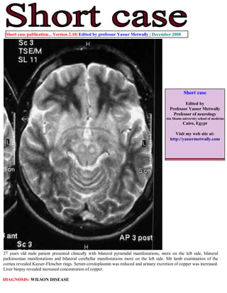
  • 1. Short case publication... Version 2.10| Edited by professor Yasser Metwally | December 2008 Short case Edited by Professor Yasser Metwally Professor of neurology Ain Shams university school of medicine Cairo, Egypt Visit my web site at: http://yassermetwally.com 27 years old male patient presented clinically with bilateral pyramidal manifestations, more on the left side, bilateral parkinsonian manifestations and bilateral cerebellar manifestations more on the left side. Slit lamb examination of the cornea revealed Kayser-Fleischer rings. Serum ceruloplasmin was reduced and urinary excretion of copper was increased. Liver biopsy revealed increased concentration of copper. DIAGNOSIS: WILSON DISEASE
  • 2. Figure 1. Postcontrast MRI T1 studies showing ventricular dilatation, bilateral more or less symmetrical hypointensities affecting predominantly the putamen and the internal capsule (mainly the posterior limbs). Notice the bilateral fronto-partial hypointensities with probable cystic changes in the subcortical region of the right posterior fronto-parietal region, the hypointensity probably involves the motor strip in the posterior frontal area (area number 4). Also noticed mild cortical atrophy predominantly affecting the bilateral frontal regions. Notice the linear hypointensity involving the thalamus and extending to the upper midbrain in the presumed area of the pyramidal tract in (C). The MRI T1 hypointense changes demonstrated in these MRI images correspond, pathologically, to edema, cystic changes, astrogliosis and demyelination involving mainly the cortical motor strip and the descending cortico-spinal tract in the internal capsule, thalamus and upper midbrain. The demonstrated lesions did not have mass effect and did not show any evidence of postcontrast enhancement. Figure 2. Postcontrast MRI T1 images showing ventricular dilatation, bilateral more or less symmetrical linear hypointensities in the presumed area of the cortico-spinal tract. Notice the bilateral fronto-partial hypointensities with probable subcortical cystic changes. The MRI T1 hypointense changes demonstrated in these MRI images correspond, pathologically, to edema, cystic changes, astrogliosis and demyelination involving mainly the cortical motor strip and the descending cortico-spinal tract in the internal capsule, thalamus and upper midbrain.
  • 3. Figure 3. Postcontrast MRI T1 images showing ventricular dilatation, bilateral more or less symmetrical linear hypointensities in the presumed area of the cortico-spinal tract. Notice the bilateral fronto-partial hypointensities with probable subcortical cystic changes. The MRI T1 hypointense changes demonstrated in these MRI images correspond, pathologically, to edema, cystic changes, astrogliosis and demyelination involving mainly the cortical motor strip and the descending cortico-spinal tract in the internal capsule, thalamus and upper midbrain. Figure 4. MRI T2 images showing bilateral, more of less symmetrical hyperintensities involving the thalamus, putamen, head of caudate nucleus, and the globus pallidus. Notice the bilateral hypointensities in the posterior limb of the internal capsule probably involving the cortico-spinal tract. The crus cerebri appears hypointense in (B) probably due to concomitant iron deposition. The red nucleus appears hyperintense in (A) and is seen surrounded by a hypointense rim, This pattern is probably due to involvement of the dentatorubrothalamic tract which is the main efferent pathway of the cerebellum and originates in the dentate nucleus. The dentate nucleus mainly gives rise to the superior cerebellar peduncle, which decussates in the dorsal mesencephalon. The superior cerebellar peduncle terminates, surrounds, or traverses the red nucleus and involvement of this tract, by demyelination, and astroglosis, might be resposible for the abnormal signal intensity in and around the red nucleus. The dentatothalamic tract terminates in the ventrolateral nucleus of the thalamus.
  • 4. Figure 5. MRI T2 images showing bifrontal hyperintensities, mainly involving the posterior regions of the frontal lobes with slight right sided predominance. The MRI T2 bifrontal hyperintensities mainly involves the subcortical white matter (cystic changes and astrogliosis), with some linear cortical MRI T2 hypointensities which could be due to concomitant iron deposition. Notice the bilateral, more or less symmetrical, basal ganglionic hyperintensities (cystic changes and astrogliosis) and the moderate dilatation of the frontal horns of the lateral ventricles. Figure 6. MRI FLAIR images showing bilateral, more or less symmetrical, basal ganglionic hyperintensities (cystic changes and astrogliosis) and moderate dilatation of the frontal horns of the lateral ventricles.
  • 5. Figure 7 . Slit lamb examination of the cornea revealed Kayser-Fleischer rings. References 1. Metwally, MYM: Textbook of neurimaging, A CD-ROM publication, (Metwally, MYM editor) WEB-CD agency for electronic publishing, version 9.4a October 2008 Addendum A new version of short case is uploaded in my web site every week (every Saturday and remains available till Friday.) To download the current version follow the link quot;http://pdf.yassermetwally.com/short.pdfquot;. You can download the long case version of this short case during the same week from: http://pdf.yassermetwally.com/case.pdf or visit web site: http://pdf.yassermetwally.com To download the software version of the publication (crow.exe) follow the link: http://neurology.yassermetwally.com/crow.zip At the end of each year, all the publications are compiled on a single CD-ROM, please contact the author to know more details. Screen resolution is better set at 1024*768 pixel screen area for optimum display For an archive of the previously reported cases go to www.yassermetwally.net, then under pages in the right panel, scroll down and click on the text entry quot;downloadable short cases in PDF formatquot; Also to view a list of the previously published case records follow the following link (http://wordpress.com/tag/ case-record/) or click on it if it appears as a link in your PDF reader