2. Summary
The nervous system and hormones enable us
to respond to external changes. They also help
us to control conditions inside our bodies.
Hormones are used in some forms of
contraception and in fertility treatments.
3. You should be able to:
■ evaluate the benefits of, and the problems that
may arise from, the use of hormones to control
fertility, including In Vitro Fertilisation (IVF)
4. Key terms
Central nervous system
Brain
Spinal cord
Stimulus / stimuli (plural)
Receptor
Impulse
Neurones
Nerve
Sensory neurone
Relay neurone
Motor neurone
Effector
Synapse
Reflex action
Muscle
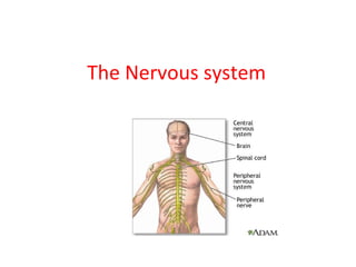
5. The Nervous System
What are its component
parts?
• Brain
• Spinal cord
• Nerves
What does it do?
• Enables you to control
your body
• Gives you feedback
about the world
6. Look away if you’re squeamish…
A dissected
brain, spinal
cord and
sections of
major
nerves.
9. Receptors in the eyes are sensitive to
___________________
Receptors in the ears are sensitive to
___________________
Receptors in the ____________ are sensitive to
changes in position
Receptors in the _____________ and
___________ are sensitive to chemicals that
enable us to taste and smell
Receptors in the skin are sensitive to
t________________, p_______________ and
p_______
10. The route impulses take
Sensory
neurone
Relay
neurones
(in brain or
spinal cord)
Effectors –
Motor neurone
11. The Spine
You don’t need
to know any of
these little
details – just
remember that
the spinal cord
carries messages
to and from the
brain.
22. Reflex Reactions
Can you think of any?
• Startle reflex – moving away, contraction of
arm and leg muscles, blinking, breathing
changes
• Withdrawal reflex – moving away from
potentially harmful influences (e.g. high
temperature)
• Iris reflex – pupil becomes smaller in bright
light
24. Why the knee jerk reflex?
• This reflex is quite useful for walking. Every
time you put weight on your foot, your
muscles contract to support you. Without this
reflex, we would all look silly staggering
around, having to consciously think about
working our muscles for each step, but with
the muscles reacting too hopelessly late to be
useful. Chewing gum at the same time
would be out of the question.
Editor's Notes
(I don’t know why this image shows the pelvis – it isn’t part of the nervous system!) Ask students to discuss in groups what they think are the component parts of the nervous system. The brain and spinal cord make up the ‘central nervous system’ while the nerves comprise the ‘peripheral nervous system’. Students can then discuss what they think are the main functions of the nervous system before you reveal them.
The nervous system – brain, spine and nerves - is made up of very specialised cells called nerve cells or neurones (also spelt ‘neurons’). They have all the main features of animal cells: cell membrane, cytoplasm, nucleus, etc. – but also some very specialised features which allow them to do their particular job.
The large blob towards the lower left seems to be the cell body of a neurone, with an axon branching off to the right.
Overview of the route signals take through the nervous system. The brain or spinal cord is sometimes referred to as the ‘co-ordinator’ in this process.
Students must be clear about the types of neurones, in particular sensory neurones (from receptors to spine/brain, giving feedback on the outside world) and motor neurones (from brain/spine to effectors, which are usually muscles, enabling the brain/spine to control the body). These neurones are one-way only. For non-reflex reactions, there would be many relay neurones involved on the right, as the impulse from the sensory neurone enters the brain and the brain then makes decisions on how to respond, and sends impulses to the appropriate muscles.
The key component here is the spinal cord, protected by the vertebrae of the spine. Nerves connect the spinal cord to all parts of the body.
Not clear enough for students to copy, but gives them an idea of the two neurone types. Students could be asked to pick out similarities and differences between the two.
More detailed – you may or may not wish to show this, as it shows more than students need to know at GCSE. For most syllabuses, they do not need to know the terms ‘synaptic endings’, ‘node of Ranvier’, ‘myelin sheath’ and ‘Schwann cell’.
These are all images of motor neurones (except perhaps for the photo – I don’t know what type of neurones these two are…) This is to show students that there are many different ways of representing the same thing. Colours are not always realistic, but may be used in different ways for clarity.
Again, notice that the colours do not necessarily reflect reality.
Individual neurones are bundled together in nerves. The neurones do not interfere with each other, but all act independently, and each neurone is one-way only. A single nerve contains both motor neurones and sensory neurones. A nerve is a bit like a bundle of optical fibres used in telecommunications.
Like all cells, nerve cells need energy to function. So the nerves also contain tiny blood vessels (capillaries) to carry oxygen and glucose to the cells.
Magnified image showing neurones in a nerve.
The axon (long part) of a neurone is surrounded by a fatty layer (called the myelin sheath). This acts as an electrical insulator. (Motor neurone disease is a disease which causes this insulator to break down. Signals leak out of the neurones and do not reach their destination muscles, so people with the disease gradually lose the ability to control their muscles.)
The green bands in the image are the fatty insulation layer, highly magnified.
The synapse is a tiny gap between one neurone and the next. The electrical signal reaches the end of the first neurone, and this triggers the release of chemicals. The arrival of these chemicals at the next neurone triggers the start of an electrical impulse. This is how the impulse gets from one neurone to the next. For most GCSE syllabuses, students do not need to know the terms ‘axon terminal’, ‘synaptic vesicles’ (‘sacs containing chemicals’ will do) or ‘neurotransmitter’ (‘chemicals’ is enough).
Incidentally, you may be wondering why there should be synapses at all – why not have the neurones directly connected to one another, without a gap? One reason is to ensure that the flow of impulses is in one direction only. At a synapse, only one of the neurones contains sacs of chemicals for release, while the other is the only one with receptors for those chemicals.
Not in GCSE, so you may want to hide this slide, but interesting to know that neurones can connect to each other in different ways. Taken from ‘Neuroscience For Kids’ at http://faculty.washington.edu/chudler/synapse.html
It is just now becoming possible to take a single neurone and incorporate it into a microchip, in which the neurone acts as a tiny electrical connector. This may lead to some interesting possibilities in the future…mind-reading?...downloading your memories?
Reflex reactions do not involve the brain; the brain is simply informed of them afterwards. The receptor causes an impulse along a sensory neurone, which connects to a relay neurone in the spine. Rather than taking the impulse up to the brain, it passes it directly to a motor neurone which causes a reaction in an appropriate muscle. Reflex reactions are usually there to protect the body from sources of danger, e.g. heat or injury. The body is full of ‘reflex arcs’ like this, ready for action should the need arise.
Students can test out the iris reflex.
Students could (carefully!) try this on themselves or each other. A hammer is not needed – the side of the hand works just as well. Gently tap just below the knee, and if you hit the right spot, the leg will jerk forwards. This movement is involuntary.