2. • Skin is the largest organ in the body with a
surface area of about 1.5 to 2m2
in adults.
• Performs many functions.
• Skin completely cover the body and is
continuous with the membranes lining the
body orifices.
3. Vocabulary
• Derma = Skin
– Dermatology
• Study of skin
– Dermatitis
• Inflammation of skin
• Epi = upon
– Epidermis
• Top layer of skin
• Vascular= pertaining to blood or lots of blood
supply
4. Function of Skin
• Cover- protects from germs, dehydration,
injury. First line of defense
• Regulates body temperature
• Manufactures vitamin D
• Site of many nerve endings
• Temporary storage of glucose, fat, water and
salt.
• Protects from UV radiation
• Can absorb chemical substances
– Nitroglycerin patch
– Ointment for rashes
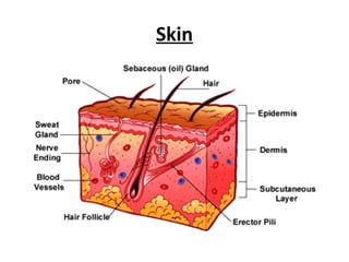
5. Skin- 3 basic layers
• Epidermis- epithelial cells with no blood vessel
– Avascular
• Dermis- True skin made of connective tissue and is
vascular
• Hypodermis- subcutaneous. Attaches integument to
muscle-
6. One Square Centimeter of Skin Contains
• 3,000,000 cells
• 10 hairs.
• 1 yard of blood vessels.
• 4 yards of nerves.
• 700 sweat glands.
• 200 nerve endings to record pain.
• 3000 sensory cells at the end of nerve fibers
7. The Epidermis
The epidermis is the outer or top layer and
is made up of five sub layers. From the
inner most layer they are called
Stratum Germinativum
Stratum Mucosum
Stratum Granulosum
Stratum Lucidum
Stratum Corneum
8. Epidermis- the layer on top
• Even the epidermis has layers!
– Very top layer is dead skin cells. Called Stratum
Corneum
– Protects you
• Every minute of the day we lose about 30,000 to
40,000 dead skin cells off the surface of our skin.
– Very bottom layer of the epidermis produces
more cells by undergoing continuous cell division.
Called Stratum Germinativum
9. Germinativum layer
The Germinativum layer is the bottom layer
and here the cells are constantly reproducing.
The melanocyte cells are also located in this
layer.
As new cells are formed and mitosis takes
place, the old cells are pushed towards the
surface of the skin.
10. Mucosum layer
The Mucosum layer is where tissue fluid is
stored.
The Granulosum layer is where the cells are
found with small granules in them, thought to
make the skin tough. Your lips and skin under
fingernails do not have this layer in them.
Granulosum layer
11. Lucidum layer
The Lucidum layer is only found on the
palms of your hands and soles of your feet.
This is the layer that thickens to fight
mechanical attack.
The Corneum layer is the top layer of your skin.
This is the layer you can see.
Here the cells are dead and continually flake
off the surface.
Corneum layer
12. The Dermis Layer
This layer is under the epidermis layer and is
sometimes called the true skin.
This layer contains the blood vessels.
These divide into a network of smaller vessels
called capillaries.
The blood supplies essential materials for growth,
nourishment and repair of the skin.
Dermis is tough and elastic and formed from
connective tissue and the matrix contains
collagen and elastic fibers.
Fibroblasts, macrophages and mast cells are the
main cells found in dermis.
13. Dermis-Thicker Inner Layer of Skin.
Matted masses of
• Connective tissue.
• Elastic fibers.
• Nerve endings.
• Muscles.
• Hair follicles.
• Oil and sweat glands
14. Dermis contains
• nerve endings
– Sensory receptors sensitive to Heat, cold, pain and
pressure
– Incoming stimuli activate these receptors and nerve
impulses generated in the sensory receptors are
conveyed by sensory nerves to the spinal cord first and
then to cerebrum.
• Blood vessels regulate body temperature
– Expand or contract
• Sebaceous glands-sebum
– Sebaceous glands present in the skin of all parts of the
body except palms of the hand and soles of the feet.
– Lubricated, protected, waterproof
15. • Sweat glands- sweat
– Cools, protects.
– Regulates body temperature
• Collagen and elastin-
• Errector pili:
little bundles of smooth muscle fibers attached to
hair follicles. Contraction makes the hair erect
• Immune cells
16. Subcutaneous aka hypodermis
• Loose connective
tissue and FAT-½ of
body’s stored fat.
• Connects the
integumentary
system to muscle
• Insulates
• Absorbs shock
17. • Fat cells do not multiply after puberty -- as your body stores
more fat, the number of fat cells remains the same. Each fat
cell simply gets bigger!
• Fat cells are large cells have very little cytoplasm, only 15
percent cell volume, a small nucleus and one large fat droplet
that makes up 85 percent of cell volume.
18. • Cross-section view of
your skin. The fat is
in the subcutaneous
layer, which is richly
supplied with blood
vessels.
19. Diseases of the skin
• Acne. A common and chronic disorder of the
sebaceous glands.
• Athlete’s foot. A contagious fungal infection
of the epidermis.
• Dermatitis. A nonspecific inflammation of the
skin.
• Psoriasis. The chronic inflammatory skin
disease. Cause unknown. No definitive
treatment.
20. Acne vulgaris
• Common in adolescent males
and is thought to be caused
by increased levels of
testosterone after puberty.
• Sebaceous glands in hair
follicles become blocked and
then infected, leading to
inflammation and pustule
formation.
• In severe cases permanent
scarring may result.
• Most common sites are :
face, chest and upper back.
•
21. Ahtlete’s foot
• Athlete’s foot, or tinea pedis, is
a fungal infection that can
grow and multiply on human
skin, especially the feet. It
grows best in a dark, moist,
and warm environment. A foot
inside a shoe is the perfect
place for the fungus. The same
fungus may also cause “jock
itch” in the groin.
22. Dermatitis (Eczema)
• Common inflammatory skin condition that may be
acute or chronic.
Acute dermatitis
• It is characterized by redness, swelling and there is
exudation of serous fluid usually accompanied by
pruritis (itching) and often followed by crusting and
scaling.
Chronic dermatitis
• If the condition becomes chronic the skin thickens
and may become leathery due to long term
scratching.
23. Atopic dermatitis
• Atopic dermatitis is associated with allergy and commonly
affects atopic individuals i.e. Those prone to hypersensitivity
disorders. Children who may suffer from hayfever or asthma
are often affected.
Contact dermatitis
Contact dermatitis may be caused by :
• Direct contact with irritants e.g. Cosmetics, soaps, detergent,
strong acids or alkalies, industrial chemicals.
• A hypersensitivity reaction to e.g. Latex, nickel, dyes and other
chemicals.
24. Psoriasis
• It is genetically determined inflammatory
skin condition and common in 15 to 40
years of age. More than 4.5 million adults
in the United States have been diagnosed
with psoriasis
• Cells of the basal layers of the epidermis
proliferate and the more rapid upward
progress of these cells through the
epidermis results in the incomplete
maturation of the upper layer.
• Patches of raised, reddish skin covered by
silvery-white scale. The skin often itches,
and it may crack and bleed when scales
are scratched or rubbed off.
• Trigger factors that lead to exacerbation
of the condition include trauma, infection
25. Skin cancer
• Most common type of cancer.
• Associated with exposure to ultraviolet light.
• Other factors.
– Hereditary
– Chemical exposure
26. Basal cell carcinoma.
• Most common, least malignant type of skin
cancer.
• Associated with long term exposure to sun light
and therefore occur mostly on sun exposed sites,
usually head and neck.
• Starts in the epidermis and extends to the dermis
or subcutaneous layer. 99% recovery.
• It appears as a shiny nodule and later rthis breaks
down, becoming an ulcer with irregular edges
commonly called a rodent ulcer.
• Locally invasive but seldom metastasises.
27. Malignant melanoma.
• Occurs in pigmented cells of the skin
called melanocytes.
• This is malignant proliferation of
melanocytes, usually originating in a
mole that may have an irregular outline.
It may ulcerate and bleed.
• Predisposing factors are a fair skin and
recurrent episodes of intensive
exposure to sunlight including repeated
exposure to sun burn, especially in
childhood.
• Spreads quickly to other areas. Most
deadly. Treatment is surgical removal
and chemotherapy.
• Metastasis usually develop early and
found in lymph nodes. Most common
sites of blood spread metastasis are
liver, lungs, bowel and bone marrow
(pet scan of patient whose skin
cancer has spread to other
organs)
28. Burns
• These may be caused by many types of trauma
including:
heat, cold, electricity , ionising radiations
and
corrosive chemicals including strong acids and
alkalies.
29. Burns are classified according to their depth:
First degree- when only epidermis is involved, the
surface is moist and there are signs of inflammation
including redness, swelling and pain. There are no
blisters.
Second degree- when epidermis and upper dermis
are affected. In addition to the above symptoms
blistering is usually present.
Third degree (deep)- when the epidermis and dermis
are destroyed. These burns are usually painless as
the sensory nerve endings in the dermis are
destroyed.