SlideShare a Scribd company logo
1 of 61
Anatomy of
Chest
Dr. Muhammad Bin Zulfiqar
PGR-1 FCPS SHL
Structures to identify
• Heart
• Lungs
• Mediastinum
• Pleural space
• Chest wall
• …Everything else!
– Bones, soft-tissues
IMAGING MODALITIES
1. Plain chest Radiograph
2. Fluoroscopy
3. Computerized tomography
4. Radionuclide lung scan
5. MRI
6. Ultrasound
7. Pulmonary angiography
Plain chest radiograph
• Diagnostic in 80% cases
• Standard views
1. Postero-anterior(P/A)
2. Lateral (right/left)
• Additional views
1. Oblique view(ribs)
2. Apical lordotic view
3. Expiration view
4. Decubitus view
CXR Interpretation
Normal structures visible
1. Tracheal air column.
2. Carina.
3. First rib.
4. Peripheral lung fields have no markings except:
5. The minor fissure.
6. Top of the R diaphragm is usually between the
anterior 6th & 7th ribs, and overlying the
posterior 10th & 11th ribs.
7. Left diaphragm is lower (in 90-95%) by roughly
half an interspace.
8. Inferior margins of the posterior ribs.
9. Anterior mediastinal line.
10. Superior vena cava.
11. Azygos vein.
12. Right descending pulmonary artery.
13. Pulmonary arteries and veins.
14. Right atrium.
15. Inferior vena cava.
16. Aortic arch.
17. Left pulmonary artery.
18. Border of the left ventricle.
19. Descending aorta.
20. Fat density lines in the intermuscular fascial
layers
Chest radiograph with
superimposed
mediastinal stripes.
Yellow: right
paratracheal stripe.
Light blue: right and
left paraspinal stripes.
Red: azygoesophageal
stripe. Brown:
pleuroesophageal
stripe. Purple:
anterior junction line
complex. Pink: left
subclavian artery
border. Light green:
posterior-superior
junction line. Dark
green: para-aortic
line.
Lobes and Fissures
RUL
LUL
RLL
RML
LLL
Left Lateral View Right Lateral View
LUL
LLL
RUL
RML
RLL
http://www.wikiradiography.com/page/Chest+Radiographic+Anatomy
AORTIC ARCH
LT. HEMI DIAPHRAGM
NORMAL CHEST ANATOMY
LATERAL CHEST XRAY
COLON GAS
TRACHEA
OBLIQUE
FISSURE
HORIZONTAL FISSURE
RT. HEMI
DIAPHRAGM
10
1. A line is drawn from
anterior surface of the
body of 6th thoracic
vertebrae passing
through the apex of the
heart up to anterior
lower most part of
diaphragm.
2. Another straight line is
drawn from anterior
surface of the body of T-
6 vertebrae to the
sternum.
.Segmental
Anatomy of Lung
Segmental
Anatomy of
Lung
Lateral view
On a normal lateral view the contours of the
heart are visible and the IVC is seen entering
the right atrium.
The retrosternal space should be radiolucent,
since it only contains air. Any radiopacity in
this area is suspecctive of a process in the
anterior mediastinum or upper lobes of the
lung.
As you go from superior to inferior over the
vertebral bodies they should get darker,
because usually there will be less soft tissue
and more radiolucent lung tissue (red arrow).
If this is not the case, look carefully for
pathology in the lower lobes.
The contours of the left and right
diaphragm should be visible.
The right diaphragm should be visible
all the way to the anterior chest wall
(red arrow).
Actually we see the interface between
the air in the lungs and the soft tissue
structures in the abdomen.
The left diaphragm can only be seen to
a point where it borders the heart
(blue arrow).
Here the interface is lost, since the
heart has the same density as the
structures below the diaphragm.
The left main pulmonary artery (in purple)
passes over the left main bronchus and is higher
than the right pulmonary artery (in blue) which
passes in front of the right main bronchus.
Pectus excavatum
In patients with a pectus excavatum the right heart border can be ill-defined, but this is normal.
It produces a silhouette sign and thus simulating a consolidation or atelectasis of the right middle
lobe.
The lateral view is helpful in such cases.
Pectus excavatum is a congenital deformity of the ribs and the sternum producing a concave
appearance of the anterior chest wall.
Vena azygos lobe
A common normal variant is the azygos lobe.
The azygos lobe is created when a laterally displaced azygos vein makes a
deep fissure in the upper part of the lung.
On a chest film it is seen as a fine line that crosses the apex of the right lung.
Here another patient with an azygos lobe.
The azygos vein is seen as a thick structure within the azygos
fissure.
In some patients an extra joint is seen in the anterior part of the
first rib at the point where the bone meets the calcified
cartilaginous part (arrow).
This may simulate a lung mass.
Cervical Rib
Computed Tomography
• Role of CT
– Main further investigation
for most CXR abnormality
(eg nodule/mass) or to
exclude disease with
normal CXR
– Main investigation for
certain scenarios (PE,
dissection, trauma)
Computed Tomography
• Numerous
protocols/techniques
depending on clinical
history
• Helical/spiral versus high
resolution
• Contrast
– Renal failure
– Allergy
Secondary lobule
The secondary lobule is the basic anatomic unit of pulmonary structure and function. It is the
smallest lung unit that is surrounded by connective tissue septa. The secondary lobule is
supplied by a small bronchiole (terminal bronchiole) in the center, that is paralleled by the
centrilobular artery.
Pulmonary veins and lymphatics run in the periphery of the lobule within the interlobular septa.
Centrilobular area is the central part of the secondary
lobule.
Perilymphatic area is the peripheral part of the secondary
lobule.
CT angiogram.
Frontal or
coronal view of
chest-3D slab
image showing
pulmonary
vessels.
MRI
• Multiple planes
• No radiation
• Common Indication
– Pancoast tumour
– Brachial plexus
– Cardiac
– Vascular (aorta)
• Usually targeted
examination (unlike
CT)
Coronal
Indications for MRI
•A chest MRI provides detailed pictures of tissues within the chest area.
•A chest MRI may be done for the following reasons:
•As an alternative to angiography, or to avoid repeated exposure to radiation
•Clarify findings from previous x-rays or CT scans
•Diagnose abnormal growths in the chest
•Evaluate blood flow
•Show lymph nodes and blood vessels
•Show the structures of the chest from multiple angles
•See if cancer in the chest has spread to other areas of the body - this is called
staging; staging helps guide future treatment and follow-up and gives you
some idea of what to expect in the future
•Tell the difference between tumors and normal tissue
Normal 32-year-old
female. MR angiography
derived maximum
intensity projection
reconstruction shows
normal pulmonary artery
anatomy: pulmonary
artery main stem (black
arrow), right pulmonary
artery (white arrow) and
left pulmonary artery
(asterisk).
the 3D reconstruction of the MRA image (1) allows the visualization of the entire pulmonary vasculature
identifying the subsegmental pulmonary arteries.
Dynamic contrast-enhanced MRA image in the oblique axial plane (2, 3, 4) allow the evaluation of pulmonary
circulation, including perfusion of the lung fields,
which is homogeneous and symmetric.
1: Right lung 2:Left lung
3:Cardiac silhouette 4.Mediastinum
THANK YOU
Anatomy of chest
Anatomy of chest
Anatomy of chest
Anatomy of chest

More Related Content

What's hot

Interpretation of chest x ray
Interpretation of chest x rayInterpretation of chest x ray
Interpretation of chest x raygowri shanker
 
Chest x ray positioning
Chest x ray  positioningChest x ray  positioning
Chest x ray positioningairwave12
 
Cxr revised 24 11-91
Cxr revised 24 11-91Cxr revised 24 11-91
Cxr revised 24 11-91aalmasi1970
 
mediastinal imaging and masses
mediastinal imaging and massesmediastinal imaging and masses
mediastinal imaging and massesArun Singh
 
CT ANATOMY OF NORMAL MEDIASTINUM MADE EASY
CT ANATOMY OF NORMAL MEDIASTINUM MADE EASYCT ANATOMY OF NORMAL MEDIASTINUM MADE EASY
CT ANATOMY OF NORMAL MEDIASTINUM MADE EASYDrNikrish Hegde
 
CT Procedure of Thorax (CT Chest)
CT Procedure of Thorax (CT Chest)CT Procedure of Thorax (CT Chest)
CT Procedure of Thorax (CT Chest)Upakar Paudel
 
Emergency x ray films Dr Ahmed Esawy
Emergency x ray films Dr Ahmed EsawyEmergency x ray films Dr Ahmed Esawy
Emergency x ray films Dr Ahmed EsawyAHMED ESAWY
 
Presentation1.pptx. interpretation of x ray chest.
Presentation1.pptx. interpretation of x ray chest.Presentation1.pptx. interpretation of x ray chest.
Presentation1.pptx. interpretation of x ray chest.Abdellah Nazeer
 
Doppler ultrasound of the kidneys
Doppler ultrasound of the kidneysDoppler ultrasound of the kidneys
Doppler ultrasound of the kidneysSamir Haffar
 
Learn Chest X-Ray With Its Normal Positioning & Radio-Anatomy
Learn Chest X-Ray With Its Normal Positioning & Radio-AnatomyLearn Chest X-Ray With Its Normal Positioning & Radio-Anatomy
Learn Chest X-Ray With Its Normal Positioning & Radio-AnatomyDr.Santosh Atreya
 
HRCT Chest - By Dr. Tinku Joseph
HRCT Chest - By Dr. Tinku JosephHRCT Chest - By Dr. Tinku Joseph
HRCT Chest - By Dr. Tinku JosephDr.Tinku Joseph
 
Presentation1, ultrasound examination of the chest.
Presentation1, ultrasound examination of the chest.Presentation1, ultrasound examination of the chest.
Presentation1, ultrasound examination of the chest.Abdellah Nazeer
 

What's hot (20)

CT CHEST ANATOMY
CT CHEST ANATOMYCT CHEST ANATOMY
CT CHEST ANATOMY
 
Interpretation of chest x ray
Interpretation of chest x rayInterpretation of chest x ray
Interpretation of chest x ray
 
BASIC CHEST X-RAY INTERPRETATION
BASIC CHEST X-RAY INTERPRETATIONBASIC CHEST X-RAY INTERPRETATION
BASIC CHEST X-RAY INTERPRETATION
 
Chest imaging
Chest imagingChest imaging
Chest imaging
 
Chest x ray positioning
Chest x ray  positioningChest x ray  positioning
Chest x ray positioning
 
Chest x ray pathology
Chest x ray pathologyChest x ray pathology
Chest x ray pathology
 
CT ABDOMEN ANATOMY
 CT ABDOMEN ANATOMY CT ABDOMEN ANATOMY
CT ABDOMEN ANATOMY
 
Cxr revised 24 11-91
Cxr revised 24 11-91Cxr revised 24 11-91
Cxr revised 24 11-91
 
mediastinal imaging and masses
mediastinal imaging and massesmediastinal imaging and masses
mediastinal imaging and masses
 
CT ANATOMY OF NORMAL MEDIASTINUM MADE EASY
CT ANATOMY OF NORMAL MEDIASTINUM MADE EASYCT ANATOMY OF NORMAL MEDIASTINUM MADE EASY
CT ANATOMY OF NORMAL MEDIASTINUM MADE EASY
 
CT Procedure of Thorax (CT Chest)
CT Procedure of Thorax (CT Chest)CT Procedure of Thorax (CT Chest)
CT Procedure of Thorax (CT Chest)
 
Emergency x ray films Dr Ahmed Esawy
Emergency x ray films Dr Ahmed EsawyEmergency x ray films Dr Ahmed Esawy
Emergency x ray films Dr Ahmed Esawy
 
Presentation1.pptx. interpretation of x ray chest.
Presentation1.pptx. interpretation of x ray chest.Presentation1.pptx. interpretation of x ray chest.
Presentation1.pptx. interpretation of x ray chest.
 
CT Anatomy
CT AnatomyCT Anatomy
CT Anatomy
 
Doppler ultrasound of the kidneys
Doppler ultrasound of the kidneysDoppler ultrasound of the kidneys
Doppler ultrasound of the kidneys
 
Learn Chest X-Ray With Its Normal Positioning & Radio-Anatomy
Learn Chest X-Ray With Its Normal Positioning & Radio-AnatomyLearn Chest X-Ray With Its Normal Positioning & Radio-Anatomy
Learn Chest X-Ray With Its Normal Positioning & Radio-Anatomy
 
Chest X-ray: Basics
Chest X-ray: BasicsChest X-ray: Basics
Chest X-ray: Basics
 
HRCT Chest - By Dr. Tinku Joseph
HRCT Chest - By Dr. Tinku JosephHRCT Chest - By Dr. Tinku Joseph
HRCT Chest - By Dr. Tinku Joseph
 
Chest xray
Chest xray  Chest xray
Chest xray
 
Presentation1, ultrasound examination of the chest.
Presentation1, ultrasound examination of the chest.Presentation1, ultrasound examination of the chest.
Presentation1, ultrasound examination of the chest.
 

Viewers also liked

Approach to ct chest 578
Approach to ct chest  578Approach to ct chest  578
Approach to ct chest 578divitto1
 
How read chest ct 3
How  read  chest ct  3How  read  chest ct  3
How read chest ct 3ANAS ALSOHLE
 
Fundamentals of chest radiology
Fundamentals of chest radiologyFundamentals of chest radiology
Fundamentals of chest radiologyDr. Sreedhar Rao
 
Radiology: Chest Imaging
Radiology: Chest ImagingRadiology: Chest Imaging
Radiology: Chest ImagingSCGH ED CME
 
Technical aspect of hrct; normal lung anatomy & hrct findings of lung disease
Technical aspect of hrct; normal lung anatomy & hrct findings of lung diseaseTechnical aspect of hrct; normal lung anatomy & hrct findings of lung disease
Technical aspect of hrct; normal lung anatomy & hrct findings of lung diseaseSarbesh Tiwari
 
basics of chest X- ray interpretation
basics of chest X- ray interpretationbasics of chest X- ray interpretation
basics of chest X- ray interpretationMaha Yousif
 
Segmental anatomy of lungs , anatomy of mediastinum and secondary lobule
Segmental anatomy of lungs , anatomy of mediastinum and secondary lobuleSegmental anatomy of lungs , anatomy of mediastinum and secondary lobule
Segmental anatomy of lungs , anatomy of mediastinum and secondary lobuleGamal Agmy
 
How read chest ct 1
How  read  chest ct  1How  read  chest ct  1
How read chest ct 1ANAS ALSOHLE
 
Chest radiology part 1
Chest radiology part 1Chest radiology part 1
Chest radiology part 1Gamal Agmy
 
Mediastnum ppt
Mediastnum pptMediastnum ppt
Mediastnum pptjyomita
 
Regional anatomy of the human thorax rs-2011
Regional anatomy of the human thorax rs-2011Regional anatomy of the human thorax rs-2011
Regional anatomy of the human thorax rs-2011AHS_anatomy2
 

Viewers also liked (20)

Chest X-ray anatomy
Chest X-ray anatomyChest X-ray anatomy
Chest X-ray anatomy
 
Normal chest ct
Normal chest ctNormal chest ct
Normal chest ct
 
Approach to ct chest 578
Approach to ct chest  578Approach to ct chest  578
Approach to ct chest 578
 
Anatomy of thorax (2)
Anatomy of thorax (2)Anatomy of thorax (2)
Anatomy of thorax (2)
 
How read chest ct 3
How  read  chest ct  3How  read  chest ct  3
How read chest ct 3
 
Fundamentals of chest radiology
Fundamentals of chest radiologyFundamentals of chest radiology
Fundamentals of chest radiology
 
Radiology: Chest Imaging
Radiology: Chest ImagingRadiology: Chest Imaging
Radiology: Chest Imaging
 
Lung anatomy.
Lung anatomy.Lung anatomy.
Lung anatomy.
 
Chest x ray
Chest x rayChest x ray
Chest x ray
 
Chest trauma
Chest traumaChest trauma
Chest trauma
 
Anatomy of thorax
Anatomy of thoraxAnatomy of thorax
Anatomy of thorax
 
Technical aspect of hrct; normal lung anatomy & hrct findings of lung disease
Technical aspect of hrct; normal lung anatomy & hrct findings of lung diseaseTechnical aspect of hrct; normal lung anatomy & hrct findings of lung disease
Technical aspect of hrct; normal lung anatomy & hrct findings of lung disease
 
Chest Trauma
Chest TraumaChest Trauma
Chest Trauma
 
basics of chest X- ray interpretation
basics of chest X- ray interpretationbasics of chest X- ray interpretation
basics of chest X- ray interpretation
 
Segmental anatomy of lungs , anatomy of mediastinum and secondary lobule
Segmental anatomy of lungs , anatomy of mediastinum and secondary lobuleSegmental anatomy of lungs , anatomy of mediastinum and secondary lobule
Segmental anatomy of lungs , anatomy of mediastinum and secondary lobule
 
How read chest ct 1
How  read  chest ct  1How  read  chest ct  1
How read chest ct 1
 
Chest radiology part 1
Chest radiology part 1Chest radiology part 1
Chest radiology part 1
 
Mediastnum ppt
Mediastnum pptMediastnum ppt
Mediastnum ppt
 
Chest trauma
Chest traumaChest trauma
Chest trauma
 
Regional anatomy of the human thorax rs-2011
Regional anatomy of the human thorax rs-2011Regional anatomy of the human thorax rs-2011
Regional anatomy of the human thorax rs-2011
 

Similar to Anatomy of chest

Thorax-XRAY and CT
Thorax-XRAY and CTThorax-XRAY and CT
Thorax-XRAY and CTdypradio
 
Rad Seminar CHEST IMAGING By Dr Siraj.pptx
Rad Seminar CHEST IMAGING By Dr Siraj.pptxRad Seminar CHEST IMAGING By Dr Siraj.pptx
Rad Seminar CHEST IMAGING By Dr Siraj.pptxImanuIliyas
 
Chest x ray anatomy - how to interpret chest x-ray (2)
Chest x ray anatomy - how to interpret chest x-ray (2)Chest x ray anatomy - how to interpret chest x-ray (2)
Chest x ray anatomy - how to interpret chest x-ray (2)Yusuf Shieba Elhamd
 
pathology related topics and its complete focus on every aspect
pathology related topics and its complete focus on every aspectpathology related topics and its complete focus on every aspect
pathology related topics and its complete focus on every aspectNausheen57
 
Chest x ray interpretation
Chest x ray interpretationChest x ray interpretation
Chest x ray interpretationKamal Sharma
 
Basic chest x ray interpretation
Basic chest x ray interpretationBasic chest x ray interpretation
Basic chest x ray interpretationHiba Ashibany
 
CHEST X-RAY F.pptx
CHEST X-RAY F.pptxCHEST X-RAY F.pptx
CHEST X-RAY F.pptxdevanshi92
 
Chest X-ray radiology_Power Point Presentation
Chest X-ray radiology_Power Point PresentationChest X-ray radiology_Power Point Presentation
Chest X-ray radiology_Power Point PresentationAhyaAziz
 
Chest x-ray.zp162335 (1)
Chest x-ray.zp162335 (1)Chest x-ray.zp162335 (1)
Chest x-ray.zp162335 (1)AndrFares
 
chest-x-ray.zp162335.pptx
chest-x-ray.zp162335.pptxchest-x-ray.zp162335.pptx
chest-x-ray.zp162335.pptxHamdiAlaqal
 

Similar to Anatomy of chest (20)

chest-x-ray.pptx
chest-x-ray.pptxchest-x-ray.pptx
chest-x-ray.pptx
 
Thorax-XRAY and CT
Thorax-XRAY and CTThorax-XRAY and CT
Thorax-XRAY and CT
 
Rad Seminar CHEST IMAGING By Dr Siraj.pptx
Rad Seminar CHEST IMAGING By Dr Siraj.pptxRad Seminar CHEST IMAGING By Dr Siraj.pptx
Rad Seminar CHEST IMAGING By Dr Siraj.pptx
 
Chest X ray ppt.ppt
Chest X ray ppt.pptChest X ray ppt.ppt
Chest X ray ppt.ppt
 
Chest x ray anatomy - how to interpret chest x-ray (2)
Chest x ray anatomy - how to interpret chest x-ray (2)Chest x ray anatomy - how to interpret chest x-ray (2)
Chest x ray anatomy - how to interpret chest x-ray (2)
 
pathology related topics and its complete focus on every aspect
pathology related topics and its complete focus on every aspectpathology related topics and its complete focus on every aspect
pathology related topics and its complete focus on every aspect
 
Chest x ray
Chest x rayChest x ray
Chest x ray
 
Chest x ray interpretation
Chest x ray interpretationChest x ray interpretation
Chest x ray interpretation
 
Basic chest x ray interpretation
Basic chest x ray interpretationBasic chest x ray interpretation
Basic chest x ray interpretation
 
Reading chest-x-rays
Reading chest-x-rays Reading chest-x-rays
Reading chest-x-rays
 
CHEST X-RAY F.pptx
CHEST X-RAY F.pptxCHEST X-RAY F.pptx
CHEST X-RAY F.pptx
 
Lung y3 2018 19 tl
Lung y3 2018 19 tlLung y3 2018 19 tl
Lung y3 2018 19 tl
 
Chest X-ray radiology_Power Point Presentation
Chest X-ray radiology_Power Point PresentationChest X-ray radiology_Power Point Presentation
Chest X-ray radiology_Power Point Presentation
 
cxr.ppt
cxr.pptcxr.ppt
cxr.ppt
 
chest-x-ray.zp162335.ppt
chest-x-ray.zp162335.pptchest-x-ray.zp162335.ppt
chest-x-ray.zp162335.ppt
 
Chest imaging
Chest imagingChest imaging
Chest imaging
 
Chest x-ray.zp162335
Chest x-ray.zp162335Chest x-ray.zp162335
Chest x-ray.zp162335
 
Chest x-ray.zp162335 (1)
Chest x-ray.zp162335 (1)Chest x-ray.zp162335 (1)
Chest x-ray.zp162335 (1)
 
chest-x-ray.zp162335.ppt
chest-x-ray.zp162335.pptchest-x-ray.zp162335.ppt
chest-x-ray.zp162335.ppt
 
chest-x-ray.zp162335.pptx
chest-x-ray.zp162335.pptxchest-x-ray.zp162335.pptx
chest-x-ray.zp162335.pptx
 

More from Dr. Muhammad Bin Zulfiqar

Role of medical imaging in developemental dysplasia of Hip Dr muhammad Bin Zu...
Role of medical imaging in developemental dysplasia of Hip Dr muhammad Bin Zu...Role of medical imaging in developemental dysplasia of Hip Dr muhammad Bin Zu...
Role of medical imaging in developemental dysplasia of Hip Dr muhammad Bin Zu...Dr. Muhammad Bin Zulfiqar
 
Trauma axial skeleton Dr. Muhammad Bin Zulfiqar
Trauma axial skeleton Dr. Muhammad Bin ZulfiqarTrauma axial skeleton Dr. Muhammad Bin Zulfiqar
Trauma axial skeleton Dr. Muhammad Bin ZulfiqarDr. Muhammad Bin Zulfiqar
 
21 non ischaemic acquired Dr.Muhammad Bin Zulfiqar
21 non ischaemic acquired Dr.Muhammad Bin Zulfiqar21 non ischaemic acquired Dr.Muhammad Bin Zulfiqar
21 non ischaemic acquired Dr.Muhammad Bin ZulfiqarDr. Muhammad Bin Zulfiqar
 
Mri anatomy of knee Dr. Muhammad Bin Zulfiqar
Mri anatomy of knee Dr. Muhammad Bin ZulfiqarMri anatomy of knee Dr. Muhammad Bin Zulfiqar
Mri anatomy of knee Dr. Muhammad Bin ZulfiqarDr. Muhammad Bin Zulfiqar
 
20 congenital heart disease Dr. Muhammmad Bin Zulfiqar
20 congenital heart disease Dr. Muhammmad Bin Zulfiqar20 congenital heart disease Dr. Muhammmad Bin Zulfiqar
20 congenital heart disease Dr. Muhammmad Bin ZulfiqarDr. Muhammad Bin Zulfiqar
 
An approach to cardiac xray Dr. Muhammad Bin Zulfiqar
An approach to cardiac xray Dr. Muhammad Bin ZulfiqarAn approach to cardiac xray Dr. Muhammad Bin Zulfiqar
An approach to cardiac xray Dr. Muhammad Bin ZulfiqarDr. Muhammad Bin Zulfiqar
 
19 cardiac anatomy and Imaging techniques Dr. Muhammad Bin Zulfiqar
19 cardiac anatomy and Imaging techniques Dr. Muhammad Bin Zulfiqar19 cardiac anatomy and Imaging techniques Dr. Muhammad Bin Zulfiqar
19 cardiac anatomy and Imaging techniques Dr. Muhammad Bin ZulfiqarDr. Muhammad Bin Zulfiqar
 
Pediatric brain tumors Dr. Muhammad Bin Zulfiqar
Pediatric brain tumors Dr. Muhammad Bin Zulfiqar Pediatric brain tumors Dr. Muhammad Bin Zulfiqar
Pediatric brain tumors Dr. Muhammad Bin Zulfiqar Dr. Muhammad Bin Zulfiqar
 
Prostate Biopsy--New Emerging Trends, Dr. Muhammad Bin Zulfiqar
Prostate Biopsy--New Emerging Trends, Dr. Muhammad Bin Zulfiqar Prostate Biopsy--New Emerging Trends, Dr. Muhammad Bin Zulfiqar
Prostate Biopsy--New Emerging Trends, Dr. Muhammad Bin Zulfiqar Dr. Muhammad Bin Zulfiqar
 
Eponymous fractures name Dr. muhammad Bin Zulfiqar
Eponymous fractures name Dr. muhammad Bin ZulfiqarEponymous fractures name Dr. muhammad Bin Zulfiqar
Eponymous fractures name Dr. muhammad Bin ZulfiqarDr. Muhammad Bin Zulfiqar
 
18 Airspace Diseases Dr. Muhammad Bin Zulfiqar
18 Airspace Diseases Dr. Muhammad Bin Zulfiqar18 Airspace Diseases Dr. Muhammad Bin Zulfiqar
18 Airspace Diseases Dr. Muhammad Bin ZulfiqarDr. Muhammad Bin Zulfiqar
 
Basic approach to brain CT Dr. Muhammad Bin Zulfiqar
Basic approach to brain CT Dr. Muhammad Bin ZulfiqarBasic approach to brain CT Dr. Muhammad Bin Zulfiqar
Basic approach to brain CT Dr. Muhammad Bin ZulfiqarDr. Muhammad Bin Zulfiqar
 
16 High Resolution Computed Tomography of Interstitial and Occupational Lung ...
16 High Resolution Computed Tomography of Interstitial and Occupational Lung ...16 High Resolution Computed Tomography of Interstitial and Occupational Lung ...
16 High Resolution Computed Tomography of Interstitial and Occupational Lung ...Dr. Muhammad Bin Zulfiqar
 
15 Pulmonary Neoplasms Dr. Muhammad Bin Zulfiqar
15 Pulmonary Neoplasms Dr. Muhammad Bin Zulfiqar15 Pulmonary Neoplasms Dr. Muhammad Bin Zulfiqar
15 Pulmonary Neoplasms Dr. Muhammad Bin ZulfiqarDr. Muhammad Bin Zulfiqar
 
Ultrasound of spinal cord in neonates Dr. Muhammad Bin Zulfiqar
Ultrasound of spinal cord in neonates Dr. Muhammad Bin ZulfiqarUltrasound of spinal cord in neonates Dr. Muhammad Bin Zulfiqar
Ultrasound of spinal cord in neonates Dr. Muhammad Bin ZulfiqarDr. Muhammad Bin Zulfiqar
 
Intervention radiology— an introduction Dr. Muhammad Bin Zulfiqar
Intervention radiology— an introduction Dr. Muhammad Bin ZulfiqarIntervention radiology— an introduction Dr. Muhammad Bin Zulfiqar
Intervention radiology— an introduction Dr. Muhammad Bin ZulfiqarDr. Muhammad Bin Zulfiqar
 

More from Dr. Muhammad Bin Zulfiqar (20)

Dislocations of joint. Joint Dislocation
Dislocations of joint. Joint DislocationDislocations of joint. Joint Dislocation
Dislocations of joint. Joint Dislocation
 
Role of color doppler ultrasound in rvhtn
Role of color doppler ultrasound in rvhtnRole of color doppler ultrasound in rvhtn
Role of color doppler ultrasound in rvhtn
 
Bone age assessment
Bone age assessmentBone age assessment
Bone age assessment
 
Role of medical imaging in developemental dysplasia of Hip Dr muhammad Bin Zu...
Role of medical imaging in developemental dysplasia of Hip Dr muhammad Bin Zu...Role of medical imaging in developemental dysplasia of Hip Dr muhammad Bin Zu...
Role of medical imaging in developemental dysplasia of Hip Dr muhammad Bin Zu...
 
Trauma axial skeleton Dr. Muhammad Bin Zulfiqar
Trauma axial skeleton Dr. Muhammad Bin ZulfiqarTrauma axial skeleton Dr. Muhammad Bin Zulfiqar
Trauma axial skeleton Dr. Muhammad Bin Zulfiqar
 
21 non ischaemic acquired Dr.Muhammad Bin Zulfiqar
21 non ischaemic acquired Dr.Muhammad Bin Zulfiqar21 non ischaemic acquired Dr.Muhammad Bin Zulfiqar
21 non ischaemic acquired Dr.Muhammad Bin Zulfiqar
 
Mri anatomy of knee Dr. Muhammad Bin Zulfiqar
Mri anatomy of knee Dr. Muhammad Bin ZulfiqarMri anatomy of knee Dr. Muhammad Bin Zulfiqar
Mri anatomy of knee Dr. Muhammad Bin Zulfiqar
 
20 congenital heart disease Dr. Muhammmad Bin Zulfiqar
20 congenital heart disease Dr. Muhammmad Bin Zulfiqar20 congenital heart disease Dr. Muhammmad Bin Zulfiqar
20 congenital heart disease Dr. Muhammmad Bin Zulfiqar
 
An approach to cardiac xray Dr. Muhammad Bin Zulfiqar
An approach to cardiac xray Dr. Muhammad Bin ZulfiqarAn approach to cardiac xray Dr. Muhammad Bin Zulfiqar
An approach to cardiac xray Dr. Muhammad Bin Zulfiqar
 
19 cardiac anatomy and Imaging techniques Dr. Muhammad Bin Zulfiqar
19 cardiac anatomy and Imaging techniques Dr. Muhammad Bin Zulfiqar19 cardiac anatomy and Imaging techniques Dr. Muhammad Bin Zulfiqar
19 cardiac anatomy and Imaging techniques Dr. Muhammad Bin Zulfiqar
 
Pediatric brain tumors Dr. Muhammad Bin Zulfiqar
Pediatric brain tumors Dr. Muhammad Bin Zulfiqar Pediatric brain tumors Dr. Muhammad Bin Zulfiqar
Pediatric brain tumors Dr. Muhammad Bin Zulfiqar
 
Prostate Biopsy--New Emerging Trends, Dr. Muhammad Bin Zulfiqar
Prostate Biopsy--New Emerging Trends, Dr. Muhammad Bin Zulfiqar Prostate Biopsy--New Emerging Trends, Dr. Muhammad Bin Zulfiqar
Prostate Biopsy--New Emerging Trends, Dr. Muhammad Bin Zulfiqar
 
Eponymous fractures name Dr. muhammad Bin Zulfiqar
Eponymous fractures name Dr. muhammad Bin ZulfiqarEponymous fractures name Dr. muhammad Bin Zulfiqar
Eponymous fractures name Dr. muhammad Bin Zulfiqar
 
18 Airspace Diseases Dr. Muhammad Bin Zulfiqar
18 Airspace Diseases Dr. Muhammad Bin Zulfiqar18 Airspace Diseases Dr. Muhammad Bin Zulfiqar
18 Airspace Diseases Dr. Muhammad Bin Zulfiqar
 
17 Thoracic Trauma and Related Topics
17 Thoracic Trauma andRelated Topics17 Thoracic Trauma andRelated Topics
17 Thoracic Trauma and Related Topics
 
Basic approach to brain CT Dr. Muhammad Bin Zulfiqar
Basic approach to brain CT Dr. Muhammad Bin ZulfiqarBasic approach to brain CT Dr. Muhammad Bin Zulfiqar
Basic approach to brain CT Dr. Muhammad Bin Zulfiqar
 
16 High Resolution Computed Tomography of Interstitial and Occupational Lung ...
16 High Resolution Computed Tomography of Interstitial and Occupational Lung ...16 High Resolution Computed Tomography of Interstitial and Occupational Lung ...
16 High Resolution Computed Tomography of Interstitial and Occupational Lung ...
 
15 Pulmonary Neoplasms Dr. Muhammad Bin Zulfiqar
15 Pulmonary Neoplasms Dr. Muhammad Bin Zulfiqar15 Pulmonary Neoplasms Dr. Muhammad Bin Zulfiqar
15 Pulmonary Neoplasms Dr. Muhammad Bin Zulfiqar
 
Ultrasound of spinal cord in neonates Dr. Muhammad Bin Zulfiqar
Ultrasound of spinal cord in neonates Dr. Muhammad Bin ZulfiqarUltrasound of spinal cord in neonates Dr. Muhammad Bin Zulfiqar
Ultrasound of spinal cord in neonates Dr. Muhammad Bin Zulfiqar
 
Intervention radiology— an introduction Dr. Muhammad Bin Zulfiqar
Intervention radiology— an introduction Dr. Muhammad Bin ZulfiqarIntervention radiology— an introduction Dr. Muhammad Bin Zulfiqar
Intervention radiology— an introduction Dr. Muhammad Bin Zulfiqar
 

Recently uploaded

SOC 101 Demonstration of Learning Presentation
SOC 101 Demonstration of Learning PresentationSOC 101 Demonstration of Learning Presentation
SOC 101 Demonstration of Learning Presentationcamerronhm
 
How to Create and Manage Wizard in Odoo 17
How to Create and Manage Wizard in Odoo 17How to Create and Manage Wizard in Odoo 17
How to Create and Manage Wizard in Odoo 17Celine George
 
1029 - Danh muc Sach Giao Khoa 10 . pdf
1029 -  Danh muc Sach Giao Khoa 10 . pdf1029 -  Danh muc Sach Giao Khoa 10 . pdf
1029 - Danh muc Sach Giao Khoa 10 . pdfQucHHunhnh
 
Explore beautiful and ugly buildings. Mathematics helps us create beautiful d...
Explore beautiful and ugly buildings. Mathematics helps us create beautiful d...Explore beautiful and ugly buildings. Mathematics helps us create beautiful d...
Explore beautiful and ugly buildings. Mathematics helps us create beautiful d...christianmathematics
 
Unit-IV; Professional Sales Representative (PSR).pptx
Unit-IV; Professional Sales Representative (PSR).pptxUnit-IV; Professional Sales Representative (PSR).pptx
Unit-IV; Professional Sales Representative (PSR).pptxVishalSingh1417
 
Magic bus Group work1and 2 (Team 3).pptx
Magic bus Group work1and 2 (Team 3).pptxMagic bus Group work1and 2 (Team 3).pptx
Magic bus Group work1and 2 (Team 3).pptxdhanalakshmis0310
 
SKILL OF INTRODUCING THE LESSON MICRO SKILLS.pptx
SKILL OF INTRODUCING THE LESSON MICRO SKILLS.pptxSKILL OF INTRODUCING THE LESSON MICRO SKILLS.pptx
SKILL OF INTRODUCING THE LESSON MICRO SKILLS.pptxAmanpreet Kaur
 
Mixin Classes in Odoo 17 How to Extend Models Using Mixin Classes
Mixin Classes in Odoo 17  How to Extend Models Using Mixin ClassesMixin Classes in Odoo 17  How to Extend Models Using Mixin Classes
Mixin Classes in Odoo 17 How to Extend Models Using Mixin ClassesCeline George
 
Food safety_Challenges food safety laboratories_.pdf
Food safety_Challenges food safety laboratories_.pdfFood safety_Challenges food safety laboratories_.pdf
Food safety_Challenges food safety laboratories_.pdfSherif Taha
 
Third Battle of Panipat detailed notes.pptx
Third Battle of Panipat detailed notes.pptxThird Battle of Panipat detailed notes.pptx
Third Battle of Panipat detailed notes.pptxAmita Gupta
 
ICT role in 21st century education and it's challenges.
ICT role in 21st century education and it's challenges.ICT role in 21st century education and it's challenges.
ICT role in 21st century education and it's challenges.MaryamAhmad92
 
PROCESS RECORDING FORMAT.docx
PROCESS      RECORDING        FORMAT.docxPROCESS      RECORDING        FORMAT.docx
PROCESS RECORDING FORMAT.docxPoojaSen20
 
Unit-IV- Pharma. Marketing Channels.pptx
Unit-IV- Pharma. Marketing Channels.pptxUnit-IV- Pharma. Marketing Channels.pptx
Unit-IV- Pharma. Marketing Channels.pptxVishalSingh1417
 
How to Give a Domain for a Field in Odoo 17
How to Give a Domain for a Field in Odoo 17How to Give a Domain for a Field in Odoo 17
How to Give a Domain for a Field in Odoo 17Celine George
 
Making communications land - Are they received and understood as intended? we...
Making communications land - Are they received and understood as intended? we...Making communications land - Are they received and understood as intended? we...
Making communications land - Are they received and understood as intended? we...Association for Project Management
 
This PowerPoint helps students to consider the concept of infinity.
This PowerPoint helps students to consider the concept of infinity.This PowerPoint helps students to consider the concept of infinity.
This PowerPoint helps students to consider the concept of infinity.christianmathematics
 
psychiatric nursing HISTORY COLLECTION .docx
psychiatric  nursing HISTORY  COLLECTION  .docxpsychiatric  nursing HISTORY  COLLECTION  .docx
psychiatric nursing HISTORY COLLECTION .docxPoojaSen20
 
Unit-V; Pricing (Pharma Marketing Management).pptx
Unit-V; Pricing (Pharma Marketing Management).pptxUnit-V; Pricing (Pharma Marketing Management).pptx
Unit-V; Pricing (Pharma Marketing Management).pptxVishalSingh1417
 
Russian Escort Service in Delhi 11k Hotel Foreigner Russian Call Girls in Delhi
Russian Escort Service in Delhi 11k Hotel Foreigner Russian Call Girls in DelhiRussian Escort Service in Delhi 11k Hotel Foreigner Russian Call Girls in Delhi
Russian Escort Service in Delhi 11k Hotel Foreigner Russian Call Girls in Delhikauryashika82
 

Recently uploaded (20)

SOC 101 Demonstration of Learning Presentation
SOC 101 Demonstration of Learning PresentationSOC 101 Demonstration of Learning Presentation
SOC 101 Demonstration of Learning Presentation
 
How to Create and Manage Wizard in Odoo 17
How to Create and Manage Wizard in Odoo 17How to Create and Manage Wizard in Odoo 17
How to Create and Manage Wizard in Odoo 17
 
1029 - Danh muc Sach Giao Khoa 10 . pdf
1029 -  Danh muc Sach Giao Khoa 10 . pdf1029 -  Danh muc Sach Giao Khoa 10 . pdf
1029 - Danh muc Sach Giao Khoa 10 . pdf
 
Mehran University Newsletter Vol-X, Issue-I, 2024
Mehran University Newsletter Vol-X, Issue-I, 2024Mehran University Newsletter Vol-X, Issue-I, 2024
Mehran University Newsletter Vol-X, Issue-I, 2024
 
Explore beautiful and ugly buildings. Mathematics helps us create beautiful d...
Explore beautiful and ugly buildings. Mathematics helps us create beautiful d...Explore beautiful and ugly buildings. Mathematics helps us create beautiful d...
Explore beautiful and ugly buildings. Mathematics helps us create beautiful d...
 
Unit-IV; Professional Sales Representative (PSR).pptx
Unit-IV; Professional Sales Representative (PSR).pptxUnit-IV; Professional Sales Representative (PSR).pptx
Unit-IV; Professional Sales Representative (PSR).pptx
 
Magic bus Group work1and 2 (Team 3).pptx
Magic bus Group work1and 2 (Team 3).pptxMagic bus Group work1and 2 (Team 3).pptx
Magic bus Group work1and 2 (Team 3).pptx
 
SKILL OF INTRODUCING THE LESSON MICRO SKILLS.pptx
SKILL OF INTRODUCING THE LESSON MICRO SKILLS.pptxSKILL OF INTRODUCING THE LESSON MICRO SKILLS.pptx
SKILL OF INTRODUCING THE LESSON MICRO SKILLS.pptx
 
Mixin Classes in Odoo 17 How to Extend Models Using Mixin Classes
Mixin Classes in Odoo 17  How to Extend Models Using Mixin ClassesMixin Classes in Odoo 17  How to Extend Models Using Mixin Classes
Mixin Classes in Odoo 17 How to Extend Models Using Mixin Classes
 
Food safety_Challenges food safety laboratories_.pdf
Food safety_Challenges food safety laboratories_.pdfFood safety_Challenges food safety laboratories_.pdf
Food safety_Challenges food safety laboratories_.pdf
 
Third Battle of Panipat detailed notes.pptx
Third Battle of Panipat detailed notes.pptxThird Battle of Panipat detailed notes.pptx
Third Battle of Panipat detailed notes.pptx
 
ICT role in 21st century education and it's challenges.
ICT role in 21st century education and it's challenges.ICT role in 21st century education and it's challenges.
ICT role in 21st century education and it's challenges.
 
PROCESS RECORDING FORMAT.docx
PROCESS      RECORDING        FORMAT.docxPROCESS      RECORDING        FORMAT.docx
PROCESS RECORDING FORMAT.docx
 
Unit-IV- Pharma. Marketing Channels.pptx
Unit-IV- Pharma. Marketing Channels.pptxUnit-IV- Pharma. Marketing Channels.pptx
Unit-IV- Pharma. Marketing Channels.pptx
 
How to Give a Domain for a Field in Odoo 17
How to Give a Domain for a Field in Odoo 17How to Give a Domain for a Field in Odoo 17
How to Give a Domain for a Field in Odoo 17
 
Making communications land - Are they received and understood as intended? we...
Making communications land - Are they received and understood as intended? we...Making communications land - Are they received and understood as intended? we...
Making communications land - Are they received and understood as intended? we...
 
This PowerPoint helps students to consider the concept of infinity.
This PowerPoint helps students to consider the concept of infinity.This PowerPoint helps students to consider the concept of infinity.
This PowerPoint helps students to consider the concept of infinity.
 
psychiatric nursing HISTORY COLLECTION .docx
psychiatric  nursing HISTORY  COLLECTION  .docxpsychiatric  nursing HISTORY  COLLECTION  .docx
psychiatric nursing HISTORY COLLECTION .docx
 
Unit-V; Pricing (Pharma Marketing Management).pptx
Unit-V; Pricing (Pharma Marketing Management).pptxUnit-V; Pricing (Pharma Marketing Management).pptx
Unit-V; Pricing (Pharma Marketing Management).pptx
 
Russian Escort Service in Delhi 11k Hotel Foreigner Russian Call Girls in Delhi
Russian Escort Service in Delhi 11k Hotel Foreigner Russian Call Girls in DelhiRussian Escort Service in Delhi 11k Hotel Foreigner Russian Call Girls in Delhi
Russian Escort Service in Delhi 11k Hotel Foreigner Russian Call Girls in Delhi
 

Anatomy of chest

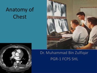
  • 1. Anatomy of Chest Dr. Muhammad Bin Zulfiqar PGR-1 FCPS SHL
  • 2. Structures to identify • Heart • Lungs • Mediastinum • Pleural space • Chest wall • …Everything else! – Bones, soft-tissues
  • 3. IMAGING MODALITIES 1. Plain chest Radiograph 2. Fluoroscopy 3. Computerized tomography 4. Radionuclide lung scan 5. MRI 6. Ultrasound 7. Pulmonary angiography
  • 4. Plain chest radiograph • Diagnostic in 80% cases • Standard views 1. Postero-anterior(P/A) 2. Lateral (right/left) • Additional views 1. Oblique view(ribs) 2. Apical lordotic view 3. Expiration view 4. Decubitus view
  • 5. CXR Interpretation Normal structures visible 1. Tracheal air column. 2. Carina. 3. First rib. 4. Peripheral lung fields have no markings except: 5. The minor fissure. 6. Top of the R diaphragm is usually between the anterior 6th & 7th ribs, and overlying the posterior 10th & 11th ribs. 7. Left diaphragm is lower (in 90-95%) by roughly half an interspace. 8. Inferior margins of the posterior ribs. 9. Anterior mediastinal line. 10. Superior vena cava. 11. Azygos vein. 12. Right descending pulmonary artery. 13. Pulmonary arteries and veins. 14. Right atrium. 15. Inferior vena cava. 16. Aortic arch. 17. Left pulmonary artery. 18. Border of the left ventricle. 19. Descending aorta. 20. Fat density lines in the intermuscular fascial layers
  • 6.
  • 7. Chest radiograph with superimposed mediastinal stripes. Yellow: right paratracheal stripe. Light blue: right and left paraspinal stripes. Red: azygoesophageal stripe. Brown: pleuroesophageal stripe. Purple: anterior junction line complex. Pink: left subclavian artery border. Light green: posterior-superior junction line. Dark green: para-aortic line.
  • 8.
  • 9. Lobes and Fissures RUL LUL RLL RML LLL Left Lateral View Right Lateral View LUL LLL RUL RML RLL http://www.wikiradiography.com/page/Chest+Radiographic+Anatomy
  • 10. AORTIC ARCH LT. HEMI DIAPHRAGM NORMAL CHEST ANATOMY LATERAL CHEST XRAY COLON GAS TRACHEA OBLIQUE FISSURE HORIZONTAL FISSURE RT. HEMI DIAPHRAGM 10 1. A line is drawn from anterior surface of the body of 6th thoracic vertebrae passing through the apex of the heart up to anterior lower most part of diaphragm. 2. Another straight line is drawn from anterior surface of the body of T- 6 vertebrae to the sternum.
  • 13. Lateral view On a normal lateral view the contours of the heart are visible and the IVC is seen entering the right atrium. The retrosternal space should be radiolucent, since it only contains air. Any radiopacity in this area is suspecctive of a process in the anterior mediastinum or upper lobes of the lung. As you go from superior to inferior over the vertebral bodies they should get darker, because usually there will be less soft tissue and more radiolucent lung tissue (red arrow). If this is not the case, look carefully for pathology in the lower lobes.
  • 14. The contours of the left and right diaphragm should be visible. The right diaphragm should be visible all the way to the anterior chest wall (red arrow). Actually we see the interface between the air in the lungs and the soft tissue structures in the abdomen. The left diaphragm can only be seen to a point where it borders the heart (blue arrow). Here the interface is lost, since the heart has the same density as the structures below the diaphragm.
  • 15. The left main pulmonary artery (in purple) passes over the left main bronchus and is higher than the right pulmonary artery (in blue) which passes in front of the right main bronchus.
  • 16. Pectus excavatum In patients with a pectus excavatum the right heart border can be ill-defined, but this is normal. It produces a silhouette sign and thus simulating a consolidation or atelectasis of the right middle lobe. The lateral view is helpful in such cases. Pectus excavatum is a congenital deformity of the ribs and the sternum producing a concave appearance of the anterior chest wall.
  • 17. Vena azygos lobe A common normal variant is the azygos lobe. The azygos lobe is created when a laterally displaced azygos vein makes a deep fissure in the upper part of the lung. On a chest film it is seen as a fine line that crosses the apex of the right lung.
  • 18. Here another patient with an azygos lobe. The azygos vein is seen as a thick structure within the azygos fissure.
  • 19. In some patients an extra joint is seen in the anterior part of the first rib at the point where the bone meets the calcified cartilaginous part (arrow). This may simulate a lung mass.
  • 21.
  • 22. Computed Tomography • Role of CT – Main further investigation for most CXR abnormality (eg nodule/mass) or to exclude disease with normal CXR – Main investigation for certain scenarios (PE, dissection, trauma)
  • 23. Computed Tomography • Numerous protocols/techniques depending on clinical history • Helical/spiral versus high resolution • Contrast – Renal failure – Allergy
  • 24.
  • 25.
  • 26.
  • 27.
  • 28.
  • 29.
  • 30.
  • 31.
  • 32.
  • 33.
  • 34.
  • 35.
  • 36.
  • 37.
  • 38.
  • 39.
  • 40.
  • 41.
  • 42.
  • 43.
  • 44.
  • 45.
  • 46.
  • 47. Secondary lobule The secondary lobule is the basic anatomic unit of pulmonary structure and function. It is the smallest lung unit that is surrounded by connective tissue septa. The secondary lobule is supplied by a small bronchiole (terminal bronchiole) in the center, that is paralleled by the centrilobular artery. Pulmonary veins and lymphatics run in the periphery of the lobule within the interlobular septa.
  • 48. Centrilobular area is the central part of the secondary lobule. Perilymphatic area is the peripheral part of the secondary lobule.
  • 49.
  • 50. CT angiogram. Frontal or coronal view of chest-3D slab image showing pulmonary vessels.
  • 51. MRI • Multiple planes • No radiation • Common Indication – Pancoast tumour – Brachial plexus – Cardiac – Vascular (aorta) • Usually targeted examination (unlike CT) Coronal
  • 52. Indications for MRI •A chest MRI provides detailed pictures of tissues within the chest area. •A chest MRI may be done for the following reasons: •As an alternative to angiography, or to avoid repeated exposure to radiation •Clarify findings from previous x-rays or CT scans •Diagnose abnormal growths in the chest •Evaluate blood flow •Show lymph nodes and blood vessels •Show the structures of the chest from multiple angles •See if cancer in the chest has spread to other areas of the body - this is called staging; staging helps guide future treatment and follow-up and gives you some idea of what to expect in the future •Tell the difference between tumors and normal tissue
  • 53. Normal 32-year-old female. MR angiography derived maximum intensity projection reconstruction shows normal pulmonary artery anatomy: pulmonary artery main stem (black arrow), right pulmonary artery (white arrow) and left pulmonary artery (asterisk).
  • 54. the 3D reconstruction of the MRA image (1) allows the visualization of the entire pulmonary vasculature identifying the subsegmental pulmonary arteries. Dynamic contrast-enhanced MRA image in the oblique axial plane (2, 3, 4) allow the evaluation of pulmonary circulation, including perfusion of the lung fields, which is homogeneous and symmetric.
  • 55.
  • 56. 1: Right lung 2:Left lung 3:Cardiac silhouette 4.Mediastinum