SlideShare a Scribd company logo
1 of 66
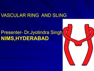
VASCULAR RING AND SLING

Presenter- Dr.Jyotindra Singh

NIMS,HYDERABAD
INTRODUCTION
Aortic arch anomalies producing tracheo esophageal
constriction account for 1 to 2% of all congenital heart disease.

Vascular ring is a congenital anomaly in which the aortic arch
and its branches completely or incompletely encircle and
compress the trachea or esophagus, or both.

Vascular sling is a congenital anomaly in which the left
pulmonary artery arises from the right pulmonary artery
extrapericardially (anomalous left pulmonary artery), courses
to the left behind the tracheal bifurcation and in front of the
esophagus to reach the left lung hilum, and forms a sling
around the trachea.
HISTORICAL ASPECTS
The condition of double aortic arch was apparently first described by
Hommel in 1737 (cited by Turner) and a century later by Von Siebold.
The complex development and regression of the aortic arches during fetal
development was elucidated by Congdon in 1922,

Wolman is credited with describing the syndrome of tracheal and
esophageal compression produced by a double arch in 1939.
Stimulus for modern interest in these anomalies was prompted by the first
surgical correction of a double aortic arch by Gross

in 1945.

The basis for radiologic diagnosis was initially described by Neuhauser.
HISTORICAL ASPECT
1948, Edwards introduced the hypothetical double aortic arch
scheme to conceptualize the numerous anomalies of the arch
complex.
This was further elaborated by Kirklin and Clagett in 1950 and
by Stewart, Kincaid, and Edwards in 1964.

In 1951, Barry provided a clear anatomic summary and review
of Congdon's basic work.
In 1999, Momma and colleagues followed by McElhinney and
colleagues identified chromosome 22ql l deletions associated
with isolated anomalies of laterality or branching of the aortic
arch
EMBRYOLOGY
By 5 weeks of fetal development, the
primordial heart tubes have fused and
six aortic (branchial) arches have
formed between the ventral roots and
dorsal aortae

During normal
development, persistence of the left
fourth aortic arch forms the arch of
the aorta and proximal left
subclavian artery.
The right fourth aortic arch forms the
innominate and right subclavian
arteries.
Involution of the distal right aorta
results in an unpaired single aortic
arch.

Aortic
--sac
Fourth Pair
RIGHT:
Becomes the
proximal part
of the right
subclavian
artery

LEFT: Forms
part of the arch
of aorta
Arch of Aorta
Derived as:
Proximal segment
from aortic sac

Middle segment from
the left 4th aortic arch

Distal segment from
the left dorsal aorta
Subclavian Artery
The right subclavian
artery formed from
the:
 Right 4th aortic
arch
 Right dorsal aorta
&
 Right 7th
intersegmental
artery
The left subclavian
artery formed from
the left 7th
intersegmental artery
Sixth Pair
RIGHT:
• Proximal part: persists as
the proximal part of the
right pulmonary artery
• Distal part: degenerates

LEFT:
• Proximal part: persists as
the proximal part of the left
pulmonary artery
• Distal part: forms ductus
arteriosus, a shunt
between pulmonary artery
Group I-Complete vascular ring
Double aortic arch
Right aortic arch with retroesophageal component
Mirror-image branching with retroesophageal ligamentum arteriosum
Retroesophageal left subclavian artery with ligamentum arteriosum
Retroesophageal left innominate artery
Right aortic arch with retroesophageal left subclavian artery
Left aortic arch and right descending aorta with right ligamentum
arteriosum or patent ductus arteriosus
Cervical aortic arch complex
Group II -Incomplete vascular ring
Left aortic arch and retroesophageal right subclavian
artery
Tracheal compression by innominate or left common
carotid artery
Ductus arteriosus sling

Malrotation of heart with patent ductus arteriosus

GROUP III- Pulmonary artery Sling
DOUBLE AORTIC ARCH
HOW IT IS FORMED ?

WHAT IS DOUBLE AORTIC
ARCH?
HOW IS VASCULAR RING
FORMED?
ORIGIN OF VESSELS?

RIGHT DOMINANT,LEFT
DOMINANT,BALANCED?
Double aortic arch
A double aortic arch is the most common complete vascular ring that causes
tracheoesophageal compression.
Patients typically present in the first months of life with symptoms of stridor,
respiratory distress, and a cough that sounds like a seal's bark. A simple cold may
precipitate severe respiratory difficulty.

In patients with double aortic arch, the ascending aorta arises normally, but as it
leaves the pericardium it divides into two branches, a left and a right aortic arch that
join posteriorly to form the descending aorta.

The left arch passes anteriorly and to the left of the trachea in the usual position and
is joined by the ductus arteriosus (or more often a ligamentum arteriosum), where it
becomes the descending aorta.
The right aortic arch passes to the right and then posterior to the esophagus to join
the left-sided descending aorta, thus completing the vascular ring .
DOUBLE AORTIC ARCH
Two-thirds of these infants, the right-sided (posterior) arch is dominant, and
in one-third, the left-sided (anterior) arch is dominant. Rarely, the arches are
of equal size (balanced arches).
The right arch gives origin to two vessels, the right common carotid and right
subclavian arteries,

The left arch gives origin to the left common carotid and left subclavian
arteries, in that order.
“Four artery sign.” - When the two dorsal subclavian arteries arise directly
from the aorta and not from a brachiocephalic artery.
2 dorsal subclavian arteries and 2 ventral carotid arteries spaced evenly
around the trachea.
Double Aortic Arch

Both arches patent
Symmetrical origin

Atretic L arch distal to
the origin of L SCA

Atretic segment
between L CCA
and L SCA
VARIANTS
The right aortic arch is often (50%) larger (right dominant) than the left arch, which usually
becomes narrow or atretic in its distal part beyond the origin of the left subclavian artery .

This portion may remain patent or be represented by a fibrous chord that joins the
descending aorta, often at the site of a diverticulum.

Less commonly (25%) the left aortic arch is larger (left dominant) than the right
arch, which, although smaller in its distal part after the origin of the right subclavian
artery, is rarely atretic .

Size of the right and left aortic arches is about equal (balanced) in about 25% of cases.

Associated cardiovascular anomalies are not common, but include tetralogy of Fallot and
transposition of the great arteries.
5. Double Aortic Arch
Clinical features
–

vascular ring – symtoms depend on tightness of ring
When both arches widely patent  tight ring  stridor in 1st wk
Atretic L arch  loose ring  present at 3-6/12 week or later
Rarely double AA present in adulthood - swallowing/resp. syms

21
DOUBLE AORTIC ARCH
CHILD WITH RECURRENT PNEUMONIA
Double Aortic Arch. Frontal chest shows impression on right-side of barium-filled esophagus from higher
right-sided arch and below it an impression on the left-side of the esophagus from left-sided arch.
Lateral film shows anterior displacement of both trachea and esophagus.
SURGICAL APPROACH & PRINCIPLE
Surgical approach is determined by which arch is dominant.

When the right arch is dominant (75%)- left thoracotomy.
When the left arch is dominant (18%)- right thoracotomy.

When the arches are balanced (7%), - left thoracotomy is preferred.

If the patient has a significant intracardiac lesion-median sternotomy.
MRI / CT scan shows Right posterior arch is smaller- Right thoracotomy
SURGICAL STEPS
What surgical approach taken?
Muscle-sparing technique,-elevating the serratus anterior and the latissimus
dorsi
Fourth intercostal space.
How is lung retracted?
Before opening pleura ,what structures to identify?
Relation of vagus nerve?
How is pleura opened?
Surgical steps
Ligamentum arteriosum identified?

Isolation of posterior arch
Which arch to preserve?
Site of division?--

Caution before clamp/vascular division?
Is mediatinal pleura sutured?
Post operative care
High humidity to loosen secretions;
oxygen therapy when needed, as
monitored by pulse oximetry;
chest physiotherapy;
and nasopharyngeal suctioning.
to achieve early extubation.
Nebulization.
Right Arch of Aorta
Occurs when the entire right aortic arch persists
&the segment of left dorsal aorta distal to the 7th
intersegmental artery involutes

TYPES:

 Without retropharyngeal component:
The DA passes from right pulmonary artery
to right arch of aorta. No effect on the
trachea & esophagus
 With retropharyngeal
component: The right arch lies
posterior to esophagus.
The attachment of DA to distal
part of the arch of aorta forms a
ring around the trachea &
esophagus and may lead to their
compression
Type I –RAA without retroesophageal component

• Mirror image of normal
• Interruption of embryonic left arch distal to ductus arteriosus.
Almost always ass. with congenital intracardiac disease

– Conotruncal anomalies – TOF, TA, TGA, DORV, LTGA, PA with RV aorta
– Other lesions – VSD, PA with IVS
Ductus is commonly L sided - attached to L innom. A. – no vascular ring

Echo/Angio -Distinctive branching pattern
CxR/ Ba oesophagography -R indentation of trachea/oesophagus
• Treatment
RAA only - No Rx needed

39
Type II vascular rings (right aortic arch with
retroesophageal ligamentum arteriosum)
account for 45% of complete vascular rings.
The majority of these defects have left
descending aortas.
A remnant or stump of the left fourth arch
(Kommerell's diverticulum) may persist.
The left subclavian artery arises aberrantly
behind the esophagus from the right-sided arch
or from Kommerell's diverticulum.
Near this point the left-sided ligamentum
emanates and joins the left pulmonary artery to
create a complete ring about the trachea and
esophagus.
40
Mirror-image branching and

retroesophageal ligamentum arteriosum.
Interruption of the left arch is proximal
(upstream) to the ductus arteriosus
The left-sided ligamentum arteriosum
extends from a diverticulum
(Kommerell) on the upper descending
thoracic aorta, behind the esophagus,
forward to the left pulmonary artery.
The vascular ring is formed by the
ascending portion of the right arch and
innominate artery anteriorly, by the
aortic diverticulum posteriorly, and by
the ligamentum arteriosum laterally.
This anomaly is rare.
KOMMERELL’s Diverticulum
identification of a Kommerell's
diverticulum as part of the pathology.

Remnant of the left fourth aortic arch
that did not undergo complete
involution.
The diverticulum may independently
compress the esophagus or trachea.

Complications of ruptured aneurysm
and aortic dissection.
This aneurysmal dilatation should be
resected if present and usually
necessitates transfer of the left
subclavian artery to the left carotid
artery
.

.

.
Representative 3D CTA showing Kommerell’s
diverticulum and aberrant left subclavian
artery

Sagittal view showing
compression of the trachea
by the arch and aberrant
subclavian artery
TYPE III
Type III-Retroesophageal left subclavian artery and ligamentum
(ductus) arteriosum.
Most common type of vascular ring
associated with right arch.

Interruption of the left arch occurs
between the left subclavian and left
common carotid arteries .
The first branch of the right arch
becomes the left common carotid
artery, and the descending aorta gives
origin to the left subclavian artery as
the fourth branch.
It is usually loose, so compression of
either the esophagus or trachea is
uncommon.
Associated cardiac anomalies are
rare.
Type IV-Retroesophageal left innominate artery

Here interruption occurs
between the left common
carotid and the right arch
A vascular ring is present, but
the anomaly is rare.
Surgical approach
The surgical approach is through a left
thoracotomy.
After careful dissection and identification
of the configuration of the aortic arch, the
ligamentum arteriosum is identified as
compressing the esophagus.
The ring is released by dividing the
ligamentum between vascular clamps
and oversewing the stumps with Prolene
suture.

Any adhesive bands around the
esophagus are lysed.
SURGICAL REPORT
11 children required additional surgery because their vascular ring
(ligamentum) was divided without addressing the associated Kommerell's
diverticulum.
All were symptomatic, with recurrent respiratory symptoms or recurrent
dysphagia.
All patients responded to reoperation with resection of the diverticulum and
transfer of the left subclavian artery to the left carotid artery with resolution of
airway symptoms
The primary strategy for patients with a right aortic arch, left ligamentum,
and Kommerell's diverticulum –
division of the ligamentum,
resection of Kommerell's diverticulum,
left subclavian artery transfer
CIRCUMFLEX AORTA
RAA + LEFT LIGAMENTUM
ARTERIOSUM + LEFT
DESCENDING THORACIC AORTA

UNCROSSING OPERATION

Median sternotomy+ CPB+ HCA

Aortic arch mobilized + divided
infront of tracheobronchial tree+
reanastomosed end to side to the
lateral aspect of ascending aorta.
LEFT AORTIC ARCH
Vascular rings are likely in the
uncommon combination of aortic arch
and right descending aorta.

The left arch crosses behind the
esophagus.

In combination with right patent
ductus arteriosus or ligamentum
arteriosum, a vascuLAR RING IS
FORMED.
CERVICAL AORTIC ARCH
Cervical aortic arch is a developmental entity
consisting of persistence of the right or left
third branchial arch and regression of the
fourth branchial arch.

The aorta is usually redundant and crosses to
the opposite side posterior to the esophagus.

A vascular ring is formed when there is an
aberrant subclavian artery on the side of the
aortic arch and a ligamentum arteriosum.

Abnormalities of brachiocephalic arterial
branching and arch laterality are common in
patients with cervical aortic arch.

Vascular ring is frequently present, usually
formed by the right aortic arch and aberrant
left subclavian artery, but occasionally by
double aortic arch.
2.1 L AA with retroesophageal R SCA

Most common arch anomaly – 0.5% population
Higher incidence in Downs with CHD – 38%
Mostly asymptomatic

Ba oesophagograph -Small filling defect slanting up and R

57
Innominate artery compression Syndrome
Anterior compression of the trachea by the innominate artery.
There is a “normal” left aortic arch- innominate artery originate more posteriorly
and leftward on the aortic arch than usual.

The artery then courses to the right, upward, and posterior to reach the thoracic
outlet, it compresses the trachea anteriorly.
Infants present with stridor, respiratory distress, cyanosis, and apnea with feeding.
The infant may hold its head hyperextended to splint the trachea and improve
breathing.

Rigid bronchoscopy -demonstrate a pulsatile anterior compression of the trachea

Anterior compression of the tracheal wall by the bronchoscope may compress the
innominate artery and temporarily obliterate the right radial pulse.
The diagnosis can be confirmed by CT scan, which will demonstrate flattening and
obliteration of the tracheal lumen by the contrast-filled innominate
PULMONARY ARTERY SLING
A pulmonary artery sling is a rare vascular
anomaly in which the left pulmonary artery
originates from the right pulmonary artery
and encircles the right mainstem bronchus
and distal trachea before coursing anterior
to the esophagus and descending aorta to
enter the hilum of the left lung
First reported by Glaevecke and Doehle.

Cosentino and associates- “ring-sling”
complex.
Nearly all infants present within the first
months of life with respiratory distress,
particularly if there are associated complete
tracheal rings.
complete tracheal rings were found in 58%
of patients with pulmonary artery sling.
A chest radiograph may show
unilateral hyperaeration of the right
lung field.
A barium esophagogram shows
anterior compression of the
esophagus on the lateral views.
Both CT and MRI will show the left
pulmonary artery originating from the
right pulmonary artery, encircling the
trachea, and coursing to the hilum of
the left lung .
Echocardiography is- diagnostic
procedure of choice for pulmonary
artery sling for the patient with an
unstable airway.
Bronchoscopy should be performed
in all these infants to check for
associated complete tracheal rings.
SURGICAL APPROACH
Surgical intervention should be
undertaken as soon as the diagnosis is
made because of the usual tenuous
respiratory status.
The first successful operation -by Potts
and coworkers69 at Children's Memorial
Hospital through a right thoracotomy.
Present approach with median
sternotomy and the use of
extracorporeal circulation.
This allows accurate division of the left
pulmonary artery with implantation into
the main pulmonary artery anterior to
the trachea

Over 50% will have associated complete
tracheal rings (the ring-sling
complex), the median sternotomy
approach allows simultaneous
VATS
Video-assisted thoracoscopic surgery (VATS)
has been successfully applied to some
patients with complete vascular rings.

The most suitable patients are those with a

right aortic arch with aberrant left
subclavian artery and left
ligamentum arteriosum (type IB) and
the occasional patient with a double aortic
arch with an atretic left arch.

In these patients a nonvascular segment of
the ring can be approached endoscopically
and safely divided.

MRI can accurately detect segmental patency
or atresia and thereby identify those vascular
ring patients suitable for the VATS approach.
THANK
YOU

More Related Content

What's hot

Antenatal Diagnosis of Fetal Heart Disease
Antenatal Diagnosis of Fetal Heart DiseaseAntenatal Diagnosis of Fetal Heart Disease
Antenatal Diagnosis of Fetal Heart DiseaseTarique Ajij
 
Av canal defect
Av canal defectAv canal defect
Av canal defectdrsrb
 
Chest xray for evaluation of cardiovascular system
Chest xray for evaluation of cardiovascular systemChest xray for evaluation of cardiovascular system
Chest xray for evaluation of cardiovascular systemPRAVEEN GUPTA
 
Congenitally corrected transposition of great arteries
Congenitally corrected transposition of great arteriesCongenitally corrected transposition of great arteries
Congenitally corrected transposition of great arteriesDheeraj Sharma
 
Ecg in congenital heart disease
Ecg in congenital heart diseaseEcg in congenital heart disease
Ecg in congenital heart diseaseRamachandra Barik
 
Presentation1, radiological imaging of scimitar syndrome
Presentation1, radiological imaging of scimitar syndromePresentation1, radiological imaging of scimitar syndrome
Presentation1, radiological imaging of scimitar syndromeAbdellah Nazeer
 
Peripheral pulmonary stenosis
Peripheral pulmonary stenosisPeripheral pulmonary stenosis
Peripheral pulmonary stenosisdrkvarun
 
Aortic arch anomalies
Aortic arch anomaliesAortic arch anomalies
Aortic arch anomaliesVarun Babu
 
IMAGING OF FETAL CVS AND ITS ANOMALIES
IMAGING OF FETAL CVS AND ITS ANOMALIESIMAGING OF FETAL CVS AND ITS ANOMALIES
IMAGING OF FETAL CVS AND ITS ANOMALIESAmeen Rageh
 
Congenital heart disease and vascular abnormality(x-ray findings)
Congenital heart disease and vascular abnormality(x-ray findings)Congenital heart disease and vascular abnormality(x-ray findings)
Congenital heart disease and vascular abnormality(x-ray findings)z2jeetendra
 
Imaging of Aortic Dissection
Imaging of Aortic DissectionImaging of Aortic Dissection
Imaging of Aortic DissectionSakher Alkhaderi
 
Total anomalous pulmonary venous connections seminar ppt.
Total anomalous pulmonary venous connections seminar ppt.Total anomalous pulmonary venous connections seminar ppt.
Total anomalous pulmonary venous connections seminar ppt.Pawan Ola
 
Presentation1 virbhan, TOF Dr Virbhan Balai
Presentation1  virbhan, TOF Dr Virbhan BalaiPresentation1  virbhan, TOF Dr Virbhan Balai
Presentation1 virbhan, TOF Dr Virbhan BalaiDr Virbhan Balai
 

What's hot (20)

Conotruncal anamolies
Conotruncal anamoliesConotruncal anamolies
Conotruncal anamolies
 
transposition of great arteries
transposition of great arteriestransposition of great arteries
transposition of great arteries
 
Antenatal Diagnosis of Fetal Heart Disease
Antenatal Diagnosis of Fetal Heart DiseaseAntenatal Diagnosis of Fetal Heart Disease
Antenatal Diagnosis of Fetal Heart Disease
 
Av canal defect
Av canal defectAv canal defect
Av canal defect
 
ATRIOVENTRICULAR SEPTAL DEFECT
ATRIOVENTRICULAR SEPTAL DEFECTATRIOVENTRICULAR SEPTAL DEFECT
ATRIOVENTRICULAR SEPTAL DEFECT
 
Chest xray for evaluation of cardiovascular system
Chest xray for evaluation of cardiovascular systemChest xray for evaluation of cardiovascular system
Chest xray for evaluation of cardiovascular system
 
Congenitally corrected transposition of great arteries
Congenitally corrected transposition of great arteriesCongenitally corrected transposition of great arteries
Congenitally corrected transposition of great arteries
 
Ecg in congenital heart disease
Ecg in congenital heart diseaseEcg in congenital heart disease
Ecg in congenital heart disease
 
Presentation1, radiological imaging of scimitar syndrome
Presentation1, radiological imaging of scimitar syndromePresentation1, radiological imaging of scimitar syndrome
Presentation1, radiological imaging of scimitar syndrome
 
EBSTEIN ANOMALY
EBSTEIN ANOMALYEBSTEIN ANOMALY
EBSTEIN ANOMALY
 
Cxr congenital
Cxr  congenitalCxr  congenital
Cxr congenital
 
Peripheral pulmonary stenosis
Peripheral pulmonary stenosisPeripheral pulmonary stenosis
Peripheral pulmonary stenosis
 
Aortic arch anomalies
Aortic arch anomaliesAortic arch anomalies
Aortic arch anomalies
 
Aortic aneurysm imaging
Aortic aneurysm imagingAortic aneurysm imaging
Aortic aneurysm imaging
 
IMAGING OF FETAL CVS AND ITS ANOMALIES
IMAGING OF FETAL CVS AND ITS ANOMALIESIMAGING OF FETAL CVS AND ITS ANOMALIES
IMAGING OF FETAL CVS AND ITS ANOMALIES
 
Congenital heart disease and vascular abnormality(x-ray findings)
Congenital heart disease and vascular abnormality(x-ray findings)Congenital heart disease and vascular abnormality(x-ray findings)
Congenital heart disease and vascular abnormality(x-ray findings)
 
Imaging of Aortic Dissection
Imaging of Aortic DissectionImaging of Aortic Dissection
Imaging of Aortic Dissection
 
Total anomalous pulmonary venous connections seminar ppt.
Total anomalous pulmonary venous connections seminar ppt.Total anomalous pulmonary venous connections seminar ppt.
Total anomalous pulmonary venous connections seminar ppt.
 
Presentation1 virbhan, TOF Dr Virbhan Balai
Presentation1  virbhan, TOF Dr Virbhan BalaiPresentation1  virbhan, TOF Dr Virbhan Balai
Presentation1 virbhan, TOF Dr Virbhan Balai
 
vsd
vsdvsd
vsd
 

Similar to Vascular ring and sling

Vascular anomalies chest
Vascular anomalies chestVascular anomalies chest
Vascular anomalies chestfenderhm
 
VASCULAR RINGS AND SLINGS TYPES HAEMODYNAMICS PRESENTATION AND DIAGNOSIS.pptx
VASCULAR RINGS AND SLINGS TYPES HAEMODYNAMICS PRESENTATION AND DIAGNOSIS.pptxVASCULAR RINGS AND SLINGS TYPES HAEMODYNAMICS PRESENTATION AND DIAGNOSIS.pptx
VASCULAR RINGS AND SLINGS TYPES HAEMODYNAMICS PRESENTATION AND DIAGNOSIS.pptxJaydeep Malakar
 
Aorta, SVC, PULMONARY TRUNK.pptx
Aorta, SVC, PULMONARY TRUNK.pptxAorta, SVC, PULMONARY TRUNK.pptx
Aorta, SVC, PULMONARY TRUNK.pptxMovieDownload83
 
AORTIC ARCH ANOMALIES 2.pptx
AORTIC ARCH ANOMALIES 2.pptxAORTIC ARCH ANOMALIES 2.pptx
AORTIC ARCH ANOMALIES 2.pptxVishnuDutt40
 
Cynotic heart disease plane radiography
Cynotic heart disease plane radiography Cynotic heart disease plane radiography
Cynotic heart disease plane radiography Dr-Girish Gunari
 
Complete transposition of great arteries
Complete transposition of great arteriesComplete transposition of great arteries
Complete transposition of great arteriesDheeraj Sharma
 
Development of heart ,,embryology,,virbhan
Development of heart ,,embryology,,virbhanDevelopment of heart ,,embryology,,virbhan
Development of heart ,,embryology,,virbhanDr Virbhan Balai
 
Single ventricle
Single ventricleSingle ventricle
Single ventricleNazmun Ara
 
Single ventricle
Single ventricleSingle ventricle
Single ventricleNazmun Ara
 
Single ventricle
Single ventricleSingle ventricle
Single ventricleNazmun Ara
 
Aortic arch anomalies
Aortic arch anomaliesAortic arch anomalies
Aortic arch anomaliesSuheil Dhanse
 
Vascular development
Vascular developmentVascular development
Vascular developmentRezaul hayat
 
Coronary circulation and fetal circulation
Coronary circulation and fetal circulationCoronary circulation and fetal circulation
Coronary circulation and fetal circulationPrincy Francis M
 
Aortic arch final.ppt
Aortic arch final.pptAortic arch final.ppt
Aortic arch final.pptHiralal Pawar
 
Pediatric Cardiology (1).pptx
Pediatric Cardiology (1).pptxPediatric Cardiology (1).pptx
Pediatric Cardiology (1).pptxelgieaulia
 
Development of heart and fetal circulation
Development of heart and fetal circulationDevelopment of heart and fetal circulation
Development of heart and fetal circulationDr. Mahesh Yadav
 

Similar to Vascular ring and sling (20)

Aortic arch anomalies
Aortic arch anomaliesAortic arch anomalies
Aortic arch anomalies
 
Vascular anomalies chest
Vascular anomalies chestVascular anomalies chest
Vascular anomalies chest
 
VASCULAR RINGS AND SLINGS TYPES HAEMODYNAMICS PRESENTATION AND DIAGNOSIS.pptx
VASCULAR RINGS AND SLINGS TYPES HAEMODYNAMICS PRESENTATION AND DIAGNOSIS.pptxVASCULAR RINGS AND SLINGS TYPES HAEMODYNAMICS PRESENTATION AND DIAGNOSIS.pptx
VASCULAR RINGS AND SLINGS TYPES HAEMODYNAMICS PRESENTATION AND DIAGNOSIS.pptx
 
Aorta, SVC, PULMONARY TRUNK.pptx
Aorta, SVC, PULMONARY TRUNK.pptxAorta, SVC, PULMONARY TRUNK.pptx
Aorta, SVC, PULMONARY TRUNK.pptx
 
AORTIC ARCH ANOMALIES 2.pptx
AORTIC ARCH ANOMALIES 2.pptxAORTIC ARCH ANOMALIES 2.pptx
AORTIC ARCH ANOMALIES 2.pptx
 
Cynotic heart disease plane radiography
Cynotic heart disease plane radiography Cynotic heart disease plane radiography
Cynotic heart disease plane radiography
 
Complete transposition of great arteries
Complete transposition of great arteriesComplete transposition of great arteries
Complete transposition of great arteries
 
Development of heart ,,embryology,,virbhan
Development of heart ,,embryology,,virbhanDevelopment of heart ,,embryology,,virbhan
Development of heart ,,embryology,,virbhan
 
Single ventricle
Single ventricleSingle ventricle
Single ventricle
 
Single ventricle
Single ventricleSingle ventricle
Single ventricle
 
Single ventricle
Single ventricleSingle ventricle
Single ventricle
 
Aortic arch anomalies
Aortic arch anomaliesAortic arch anomalies
Aortic arch anomalies
 
Vascular development
Vascular developmentVascular development
Vascular development
 
Coronary circulation and fetal circulation
Coronary circulation and fetal circulationCoronary circulation and fetal circulation
Coronary circulation and fetal circulation
 
Aortic arch final.ppt
Aortic arch final.pptAortic arch final.ppt
Aortic arch final.ppt
 
Cardiovascular adi
Cardiovascular adiCardiovascular adi
Cardiovascular adi
 
Pediatric Cardiology (1).pptx
Pediatric Cardiology (1).pptxPediatric Cardiology (1).pptx
Pediatric Cardiology (1).pptx
 
seminar ASD
seminar ASDseminar ASD
seminar ASD
 
Development of heart and fetal circulation
Development of heart and fetal circulationDevelopment of heart and fetal circulation
Development of heart and fetal circulation
 
Heart development 2
Heart development 2Heart development 2
Heart development 2
 

More from Jyotindra Singh

2020: Evidence based choices for AVR
2020: Evidence based choices for AVR 2020: Evidence based choices for AVR
2020: Evidence based choices for AVR Jyotindra Singh
 
Mitral Annular calcification
Mitral Annular calcificationMitral Annular calcification
Mitral Annular calcificationJyotindra Singh
 
Management-Severe Aortic stenosis with Low Transvalvular Gradient and Poor LV...
Management-Severe Aortic stenosis with Low Transvalvular Gradient and Poor LV...Management-Severe Aortic stenosis with Low Transvalvular Gradient and Poor LV...
Management-Severe Aortic stenosis with Low Transvalvular Gradient and Poor LV...Jyotindra Singh
 
Surgical closure Neonatal PDA
Surgical closure Neonatal PDA Surgical closure Neonatal PDA
Surgical closure Neonatal PDA Jyotindra Singh
 
MITRAL REPAIR - HOW DURABLE IS MITRAL VALVE REPAIR ??
MITRAL  REPAIR - HOW DURABLE IS MITRAL VALVE  REPAIR ??MITRAL  REPAIR - HOW DURABLE IS MITRAL VALVE  REPAIR ??
MITRAL REPAIR - HOW DURABLE IS MITRAL VALVE REPAIR ??Jyotindra Singh
 
Mitral surgery- When not to opearte
Mitral surgery- When not to opearte Mitral surgery- When not to opearte
Mitral surgery- When not to opearte Jyotindra Singh
 
HEART VALVES PROSTHESIS MISMATCH -FACTS OR FICTION
HEART VALVES PROSTHESIS MISMATCH -FACTS OR FICTIONHEART VALVES PROSTHESIS MISMATCH -FACTS OR FICTION
HEART VALVES PROSTHESIS MISMATCH -FACTS OR FICTIONJyotindra Singh
 
Mitral valve- Replacement/Repair- Lessons from literature
Mitral valve- Replacement/Repair- Lessons from literature Mitral valve- Replacement/Repair- Lessons from literature
Mitral valve- Replacement/Repair- Lessons from literature Jyotindra Singh
 
Immediate Intraoperative mitral valve repair complication ppt
Immediate Intraoperative  mitral valve repair complication  pptImmediate Intraoperative  mitral valve repair complication  ppt
Immediate Intraoperative mitral valve repair complication pptJyotindra Singh
 
Mitral valve current challenges
Mitral valve current challengesMitral valve current challenges
Mitral valve current challengesJyotindra Singh
 

More from Jyotindra Singh (20)

Bicuspid aortic valve
Bicuspid aortic valve Bicuspid aortic valve
Bicuspid aortic valve
 
Infective endocarditis
Infective endocarditis Infective endocarditis
Infective endocarditis
 
2020: Evidence based choices for AVR
2020: Evidence based choices for AVR 2020: Evidence based choices for AVR
2020: Evidence based choices for AVR
 
Cabg trial updates
Cabg trial updatesCabg trial updates
Cabg trial updates
 
Mitral Annular calcification
Mitral Annular calcificationMitral Annular calcification
Mitral Annular calcification
 
Ischaemic mr
Ischaemic mr Ischaemic mr
Ischaemic mr
 
Management-Severe Aortic stenosis with Low Transvalvular Gradient and Poor LV...
Management-Severe Aortic stenosis with Low Transvalvular Gradient and Poor LV...Management-Severe Aortic stenosis with Low Transvalvular Gradient and Poor LV...
Management-Severe Aortic stenosis with Low Transvalvular Gradient and Poor LV...
 
Surgical closure Neonatal PDA
Surgical closure Neonatal PDA Surgical closure Neonatal PDA
Surgical closure Neonatal PDA
 
MITRAL REPAIR - HOW DURABLE IS MITRAL VALVE REPAIR ??
MITRAL  REPAIR - HOW DURABLE IS MITRAL VALVE  REPAIR ??MITRAL  REPAIR - HOW DURABLE IS MITRAL VALVE  REPAIR ??
MITRAL REPAIR - HOW DURABLE IS MITRAL VALVE REPAIR ??
 
Mitral surgery- When not to opearte
Mitral surgery- When not to opearte Mitral surgery- When not to opearte
Mitral surgery- When not to opearte
 
TAVI Trials vs SAVR
TAVI Trials vs SAVRTAVI Trials vs SAVR
TAVI Trials vs SAVR
 
HEART VALVES PROSTHESIS MISMATCH -FACTS OR FICTION
HEART VALVES PROSTHESIS MISMATCH -FACTS OR FICTIONHEART VALVES PROSTHESIS MISMATCH -FACTS OR FICTION
HEART VALVES PROSTHESIS MISMATCH -FACTS OR FICTION
 
Mitral valve- Replacement/Repair- Lessons from literature
Mitral valve- Replacement/Repair- Lessons from literature Mitral valve- Replacement/Repair- Lessons from literature
Mitral valve- Replacement/Repair- Lessons from literature
 
Immediate Intraoperative mitral valve repair complication ppt
Immediate Intraoperative  mitral valve repair complication  pptImmediate Intraoperative  mitral valve repair complication  ppt
Immediate Intraoperative mitral valve repair complication ppt
 
Mitral valve current challenges
Mitral valve current challengesMitral valve current challenges
Mitral valve current challenges
 
Bicuspid aortic valve
Bicuspid aortic valveBicuspid aortic valve
Bicuspid aortic valve
 
Oral cancer
Oral cancerOral cancer
Oral cancer
 
Benign breast disease
Benign breast diseaseBenign breast disease
Benign breast disease
 
Aneurysm
AneurysmAneurysm
Aneurysm
 
Tumour markers
Tumour markersTumour markers
Tumour markers
 

Recently uploaded

1029-Danh muc Sach Giao Khoa khoi 6.pdf
1029-Danh muc Sach Giao Khoa khoi  6.pdf1029-Danh muc Sach Giao Khoa khoi  6.pdf
1029-Danh muc Sach Giao Khoa khoi 6.pdfQucHHunhnh
 
Ecosystem Interactions Class Discussion Presentation in Blue Green Lined Styl...
Ecosystem Interactions Class Discussion Presentation in Blue Green Lined Styl...Ecosystem Interactions Class Discussion Presentation in Blue Green Lined Styl...
Ecosystem Interactions Class Discussion Presentation in Blue Green Lined Styl...fonyou31
 
Key note speaker Neum_Admir Softic_ENG.pdf
Key note speaker Neum_Admir Softic_ENG.pdfKey note speaker Neum_Admir Softic_ENG.pdf
Key note speaker Neum_Admir Softic_ENG.pdfAdmir Softic
 
Arihant handbook biology for class 11 .pdf
Arihant handbook biology for class 11 .pdfArihant handbook biology for class 11 .pdf
Arihant handbook biology for class 11 .pdfchloefrazer622
 
Measures of Dispersion and Variability: Range, QD, AD and SD
Measures of Dispersion and Variability: Range, QD, AD and SDMeasures of Dispersion and Variability: Range, QD, AD and SD
Measures of Dispersion and Variability: Range, QD, AD and SDThiyagu K
 
Grant Readiness 101 TechSoup and Remy Consulting
Grant Readiness 101 TechSoup and Remy ConsultingGrant Readiness 101 TechSoup and Remy Consulting
Grant Readiness 101 TechSoup and Remy ConsultingTechSoup
 
1029 - Danh muc Sach Giao Khoa 10 . pdf
1029 -  Danh muc Sach Giao Khoa 10 . pdf1029 -  Danh muc Sach Giao Khoa 10 . pdf
1029 - Danh muc Sach Giao Khoa 10 . pdfQucHHunhnh
 
Presentation by Andreas Schleicher Tackling the School Absenteeism Crisis 30 ...
Presentation by Andreas Schleicher Tackling the School Absenteeism Crisis 30 ...Presentation by Andreas Schleicher Tackling the School Absenteeism Crisis 30 ...
Presentation by Andreas Schleicher Tackling the School Absenteeism Crisis 30 ...EduSkills OECD
 
Beyond the EU: DORA and NIS 2 Directive's Global Impact
Beyond the EU: DORA and NIS 2 Directive's Global ImpactBeyond the EU: DORA and NIS 2 Directive's Global Impact
Beyond the EU: DORA and NIS 2 Directive's Global ImpactPECB
 
Call Girls in Dwarka Mor Delhi Contact Us 9654467111
Call Girls in Dwarka Mor Delhi Contact Us 9654467111Call Girls in Dwarka Mor Delhi Contact Us 9654467111
Call Girls in Dwarka Mor Delhi Contact Us 9654467111Sapana Sha
 
Activity 01 - Artificial Culture (1).pdf
Activity 01 - Artificial Culture (1).pdfActivity 01 - Artificial Culture (1).pdf
Activity 01 - Artificial Culture (1).pdfciinovamais
 
Explore beautiful and ugly buildings. Mathematics helps us create beautiful d...
Explore beautiful and ugly buildings. Mathematics helps us create beautiful d...Explore beautiful and ugly buildings. Mathematics helps us create beautiful d...
Explore beautiful and ugly buildings. Mathematics helps us create beautiful d...christianmathematics
 
social pharmacy d-pharm 1st year by Pragati K. Mahajan
social pharmacy d-pharm 1st year by Pragati K. Mahajansocial pharmacy d-pharm 1st year by Pragati K. Mahajan
social pharmacy d-pharm 1st year by Pragati K. Mahajanpragatimahajan3
 
fourth grading exam for kindergarten in writing
fourth grading exam for kindergarten in writingfourth grading exam for kindergarten in writing
fourth grading exam for kindergarten in writingTeacherCyreneCayanan
 
Nutritional Needs Presentation - HLTH 104
Nutritional Needs Presentation - HLTH 104Nutritional Needs Presentation - HLTH 104
Nutritional Needs Presentation - HLTH 104misteraugie
 
Introduction to Nonprofit Accounting: The Basics
Introduction to Nonprofit Accounting: The BasicsIntroduction to Nonprofit Accounting: The Basics
Introduction to Nonprofit Accounting: The BasicsTechSoup
 
Russian Escort Service in Delhi 11k Hotel Foreigner Russian Call Girls in Delhi
Russian Escort Service in Delhi 11k Hotel Foreigner Russian Call Girls in DelhiRussian Escort Service in Delhi 11k Hotel Foreigner Russian Call Girls in Delhi
Russian Escort Service in Delhi 11k Hotel Foreigner Russian Call Girls in Delhikauryashika82
 

Recently uploaded (20)

Advance Mobile Application Development class 07
Advance Mobile Application Development class 07Advance Mobile Application Development class 07
Advance Mobile Application Development class 07
 
1029-Danh muc Sach Giao Khoa khoi 6.pdf
1029-Danh muc Sach Giao Khoa khoi  6.pdf1029-Danh muc Sach Giao Khoa khoi  6.pdf
1029-Danh muc Sach Giao Khoa khoi 6.pdf
 
Ecosystem Interactions Class Discussion Presentation in Blue Green Lined Styl...
Ecosystem Interactions Class Discussion Presentation in Blue Green Lined Styl...Ecosystem Interactions Class Discussion Presentation in Blue Green Lined Styl...
Ecosystem Interactions Class Discussion Presentation in Blue Green Lined Styl...
 
Key note speaker Neum_Admir Softic_ENG.pdf
Key note speaker Neum_Admir Softic_ENG.pdfKey note speaker Neum_Admir Softic_ENG.pdf
Key note speaker Neum_Admir Softic_ENG.pdf
 
Arihant handbook biology for class 11 .pdf
Arihant handbook biology for class 11 .pdfArihant handbook biology for class 11 .pdf
Arihant handbook biology for class 11 .pdf
 
Measures of Dispersion and Variability: Range, QD, AD and SD
Measures of Dispersion and Variability: Range, QD, AD and SDMeasures of Dispersion and Variability: Range, QD, AD and SD
Measures of Dispersion and Variability: Range, QD, AD and SD
 
INDIA QUIZ 2024 RLAC DELHI UNIVERSITY.pptx
INDIA QUIZ 2024 RLAC DELHI UNIVERSITY.pptxINDIA QUIZ 2024 RLAC DELHI UNIVERSITY.pptx
INDIA QUIZ 2024 RLAC DELHI UNIVERSITY.pptx
 
Grant Readiness 101 TechSoup and Remy Consulting
Grant Readiness 101 TechSoup and Remy ConsultingGrant Readiness 101 TechSoup and Remy Consulting
Grant Readiness 101 TechSoup and Remy Consulting
 
1029 - Danh muc Sach Giao Khoa 10 . pdf
1029 -  Danh muc Sach Giao Khoa 10 . pdf1029 -  Danh muc Sach Giao Khoa 10 . pdf
1029 - Danh muc Sach Giao Khoa 10 . pdf
 
Presentation by Andreas Schleicher Tackling the School Absenteeism Crisis 30 ...
Presentation by Andreas Schleicher Tackling the School Absenteeism Crisis 30 ...Presentation by Andreas Schleicher Tackling the School Absenteeism Crisis 30 ...
Presentation by Andreas Schleicher Tackling the School Absenteeism Crisis 30 ...
 
Beyond the EU: DORA and NIS 2 Directive's Global Impact
Beyond the EU: DORA and NIS 2 Directive's Global ImpactBeyond the EU: DORA and NIS 2 Directive's Global Impact
Beyond the EU: DORA and NIS 2 Directive's Global Impact
 
Call Girls in Dwarka Mor Delhi Contact Us 9654467111
Call Girls in Dwarka Mor Delhi Contact Us 9654467111Call Girls in Dwarka Mor Delhi Contact Us 9654467111
Call Girls in Dwarka Mor Delhi Contact Us 9654467111
 
Activity 01 - Artificial Culture (1).pdf
Activity 01 - Artificial Culture (1).pdfActivity 01 - Artificial Culture (1).pdf
Activity 01 - Artificial Culture (1).pdf
 
Explore beautiful and ugly buildings. Mathematics helps us create beautiful d...
Explore beautiful and ugly buildings. Mathematics helps us create beautiful d...Explore beautiful and ugly buildings. Mathematics helps us create beautiful d...
Explore beautiful and ugly buildings. Mathematics helps us create beautiful d...
 
social pharmacy d-pharm 1st year by Pragati K. Mahajan
social pharmacy d-pharm 1st year by Pragati K. Mahajansocial pharmacy d-pharm 1st year by Pragati K. Mahajan
social pharmacy d-pharm 1st year by Pragati K. Mahajan
 
fourth grading exam for kindergarten in writing
fourth grading exam for kindergarten in writingfourth grading exam for kindergarten in writing
fourth grading exam for kindergarten in writing
 
Nutritional Needs Presentation - HLTH 104
Nutritional Needs Presentation - HLTH 104Nutritional Needs Presentation - HLTH 104
Nutritional Needs Presentation - HLTH 104
 
Introduction to Nonprofit Accounting: The Basics
Introduction to Nonprofit Accounting: The BasicsIntroduction to Nonprofit Accounting: The Basics
Introduction to Nonprofit Accounting: The Basics
 
Russian Escort Service in Delhi 11k Hotel Foreigner Russian Call Girls in Delhi
Russian Escort Service in Delhi 11k Hotel Foreigner Russian Call Girls in DelhiRussian Escort Service in Delhi 11k Hotel Foreigner Russian Call Girls in Delhi
Russian Escort Service in Delhi 11k Hotel Foreigner Russian Call Girls in Delhi
 
Mattingly "AI & Prompt Design: Structured Data, Assistants, & RAG"
Mattingly "AI & Prompt Design: Structured Data, Assistants, & RAG"Mattingly "AI & Prompt Design: Structured Data, Assistants, & RAG"
Mattingly "AI & Prompt Design: Structured Data, Assistants, & RAG"
 

Vascular ring and sling

  • 1. VASCULAR RING AND SLING Presenter- Dr.Jyotindra Singh NIMS,HYDERABAD
  • 2. INTRODUCTION Aortic arch anomalies producing tracheo esophageal constriction account for 1 to 2% of all congenital heart disease. Vascular ring is a congenital anomaly in which the aortic arch and its branches completely or incompletely encircle and compress the trachea or esophagus, or both. Vascular sling is a congenital anomaly in which the left pulmonary artery arises from the right pulmonary artery extrapericardially (anomalous left pulmonary artery), courses to the left behind the tracheal bifurcation and in front of the esophagus to reach the left lung hilum, and forms a sling around the trachea.
  • 3.
  • 4. HISTORICAL ASPECTS The condition of double aortic arch was apparently first described by Hommel in 1737 (cited by Turner) and a century later by Von Siebold. The complex development and regression of the aortic arches during fetal development was elucidated by Congdon in 1922, Wolman is credited with describing the syndrome of tracheal and esophageal compression produced by a double arch in 1939. Stimulus for modern interest in these anomalies was prompted by the first surgical correction of a double aortic arch by Gross in 1945. The basis for radiologic diagnosis was initially described by Neuhauser.
  • 5. HISTORICAL ASPECT 1948, Edwards introduced the hypothetical double aortic arch scheme to conceptualize the numerous anomalies of the arch complex. This was further elaborated by Kirklin and Clagett in 1950 and by Stewart, Kincaid, and Edwards in 1964. In 1951, Barry provided a clear anatomic summary and review of Congdon's basic work. In 1999, Momma and colleagues followed by McElhinney and colleagues identified chromosome 22ql l deletions associated with isolated anomalies of laterality or branching of the aortic arch
  • 6.
  • 7. EMBRYOLOGY By 5 weeks of fetal development, the primordial heart tubes have fused and six aortic (branchial) arches have formed between the ventral roots and dorsal aortae During normal development, persistence of the left fourth aortic arch forms the arch of the aorta and proximal left subclavian artery. The right fourth aortic arch forms the innominate and right subclavian arteries. Involution of the distal right aorta results in an unpaired single aortic arch. Aortic --sac
  • 8. Fourth Pair RIGHT: Becomes the proximal part of the right subclavian artery LEFT: Forms part of the arch of aorta
  • 9. Arch of Aorta Derived as: Proximal segment from aortic sac Middle segment from the left 4th aortic arch Distal segment from the left dorsal aorta
  • 10. Subclavian Artery The right subclavian artery formed from the:  Right 4th aortic arch  Right dorsal aorta &  Right 7th intersegmental artery The left subclavian artery formed from the left 7th intersegmental artery
  • 11. Sixth Pair RIGHT: • Proximal part: persists as the proximal part of the right pulmonary artery • Distal part: degenerates LEFT: • Proximal part: persists as the proximal part of the left pulmonary artery • Distal part: forms ductus arteriosus, a shunt between pulmonary artery
  • 12. Group I-Complete vascular ring Double aortic arch Right aortic arch with retroesophageal component Mirror-image branching with retroesophageal ligamentum arteriosum Retroesophageal left subclavian artery with ligamentum arteriosum Retroesophageal left innominate artery Right aortic arch with retroesophageal left subclavian artery Left aortic arch and right descending aorta with right ligamentum arteriosum or patent ductus arteriosus Cervical aortic arch complex
  • 13. Group II -Incomplete vascular ring Left aortic arch and retroesophageal right subclavian artery Tracheal compression by innominate or left common carotid artery Ductus arteriosus sling Malrotation of heart with patent ductus arteriosus GROUP III- Pulmonary artery Sling
  • 14.
  • 15. DOUBLE AORTIC ARCH HOW IT IS FORMED ? WHAT IS DOUBLE AORTIC ARCH? HOW IS VASCULAR RING FORMED? ORIGIN OF VESSELS? RIGHT DOMINANT,LEFT DOMINANT,BALANCED?
  • 16.
  • 17. Double aortic arch A double aortic arch is the most common complete vascular ring that causes tracheoesophageal compression. Patients typically present in the first months of life with symptoms of stridor, respiratory distress, and a cough that sounds like a seal's bark. A simple cold may precipitate severe respiratory difficulty. In patients with double aortic arch, the ascending aorta arises normally, but as it leaves the pericardium it divides into two branches, a left and a right aortic arch that join posteriorly to form the descending aorta. The left arch passes anteriorly and to the left of the trachea in the usual position and is joined by the ductus arteriosus (or more often a ligamentum arteriosum), where it becomes the descending aorta. The right aortic arch passes to the right and then posterior to the esophagus to join the left-sided descending aorta, thus completing the vascular ring .
  • 18. DOUBLE AORTIC ARCH Two-thirds of these infants, the right-sided (posterior) arch is dominant, and in one-third, the left-sided (anterior) arch is dominant. Rarely, the arches are of equal size (balanced arches). The right arch gives origin to two vessels, the right common carotid and right subclavian arteries, The left arch gives origin to the left common carotid and left subclavian arteries, in that order. “Four artery sign.” - When the two dorsal subclavian arteries arise directly from the aorta and not from a brachiocephalic artery. 2 dorsal subclavian arteries and 2 ventral carotid arteries spaced evenly around the trachea.
  • 19. Double Aortic Arch Both arches patent Symmetrical origin Atretic L arch distal to the origin of L SCA Atretic segment between L CCA and L SCA
  • 20. VARIANTS The right aortic arch is often (50%) larger (right dominant) than the left arch, which usually becomes narrow or atretic in its distal part beyond the origin of the left subclavian artery . This portion may remain patent or be represented by a fibrous chord that joins the descending aorta, often at the site of a diverticulum. Less commonly (25%) the left aortic arch is larger (left dominant) than the right arch, which, although smaller in its distal part after the origin of the right subclavian artery, is rarely atretic . Size of the right and left aortic arches is about equal (balanced) in about 25% of cases. Associated cardiovascular anomalies are not common, but include tetralogy of Fallot and transposition of the great arteries.
  • 21. 5. Double Aortic Arch Clinical features – vascular ring – symtoms depend on tightness of ring When both arches widely patent  tight ring  stridor in 1st wk Atretic L arch  loose ring  present at 3-6/12 week or later Rarely double AA present in adulthood - swallowing/resp. syms 21
  • 22.
  • 23. DOUBLE AORTIC ARCH CHILD WITH RECURRENT PNEUMONIA
  • 24. Double Aortic Arch. Frontal chest shows impression on right-side of barium-filled esophagus from higher right-sided arch and below it an impression on the left-side of the esophagus from left-sided arch. Lateral film shows anterior displacement of both trachea and esophagus.
  • 25.
  • 26.
  • 27.
  • 28. SURGICAL APPROACH & PRINCIPLE Surgical approach is determined by which arch is dominant. When the right arch is dominant (75%)- left thoracotomy. When the left arch is dominant (18%)- right thoracotomy. When the arches are balanced (7%), - left thoracotomy is preferred. If the patient has a significant intracardiac lesion-median sternotomy. MRI / CT scan shows Right posterior arch is smaller- Right thoracotomy
  • 29.
  • 30.
  • 31.
  • 32.
  • 33.
  • 34. SURGICAL STEPS What surgical approach taken? Muscle-sparing technique,-elevating the serratus anterior and the latissimus dorsi Fourth intercostal space. How is lung retracted? Before opening pleura ,what structures to identify? Relation of vagus nerve? How is pleura opened?
  • 35. Surgical steps Ligamentum arteriosum identified? Isolation of posterior arch Which arch to preserve? Site of division?-- Caution before clamp/vascular division? Is mediatinal pleura sutured?
  • 36. Post operative care High humidity to loosen secretions; oxygen therapy when needed, as monitored by pulse oximetry; chest physiotherapy; and nasopharyngeal suctioning. to achieve early extubation. Nebulization.
  • 37.
  • 38. Right Arch of Aorta Occurs when the entire right aortic arch persists &the segment of left dorsal aorta distal to the 7th intersegmental artery involutes TYPES:  Without retropharyngeal component: The DA passes from right pulmonary artery to right arch of aorta. No effect on the trachea & esophagus  With retropharyngeal component: The right arch lies posterior to esophagus. The attachment of DA to distal part of the arch of aorta forms a ring around the trachea & esophagus and may lead to their compression
  • 39. Type I –RAA without retroesophageal component • Mirror image of normal • Interruption of embryonic left arch distal to ductus arteriosus. Almost always ass. with congenital intracardiac disease – Conotruncal anomalies – TOF, TA, TGA, DORV, LTGA, PA with RV aorta – Other lesions – VSD, PA with IVS Ductus is commonly L sided - attached to L innom. A. – no vascular ring Echo/Angio -Distinctive branching pattern CxR/ Ba oesophagography -R indentation of trachea/oesophagus • Treatment RAA only - No Rx needed 39
  • 40. Type II vascular rings (right aortic arch with retroesophageal ligamentum arteriosum) account for 45% of complete vascular rings. The majority of these defects have left descending aortas. A remnant or stump of the left fourth arch (Kommerell's diverticulum) may persist. The left subclavian artery arises aberrantly behind the esophagus from the right-sided arch or from Kommerell's diverticulum. Near this point the left-sided ligamentum emanates and joins the left pulmonary artery to create a complete ring about the trachea and esophagus. 40
  • 41. Mirror-image branching and retroesophageal ligamentum arteriosum. Interruption of the left arch is proximal (upstream) to the ductus arteriosus The left-sided ligamentum arteriosum extends from a diverticulum (Kommerell) on the upper descending thoracic aorta, behind the esophagus, forward to the left pulmonary artery. The vascular ring is formed by the ascending portion of the right arch and innominate artery anteriorly, by the aortic diverticulum posteriorly, and by the ligamentum arteriosum laterally. This anomaly is rare.
  • 42. KOMMERELL’s Diverticulum identification of a Kommerell's diverticulum as part of the pathology. Remnant of the left fourth aortic arch that did not undergo complete involution. The diverticulum may independently compress the esophagus or trachea. Complications of ruptured aneurysm and aortic dissection. This aneurysmal dilatation should be resected if present and usually necessitates transfer of the left subclavian artery to the left carotid artery
  • 43.
  • 44. . . .
  • 45. Representative 3D CTA showing Kommerell’s diverticulum and aberrant left subclavian artery Sagittal view showing compression of the trachea by the arch and aberrant subclavian artery
  • 47. Type III-Retroesophageal left subclavian artery and ligamentum (ductus) arteriosum. Most common type of vascular ring associated with right arch. Interruption of the left arch occurs between the left subclavian and left common carotid arteries . The first branch of the right arch becomes the left common carotid artery, and the descending aorta gives origin to the left subclavian artery as the fourth branch. It is usually loose, so compression of either the esophagus or trachea is uncommon. Associated cardiac anomalies are rare.
  • 48.
  • 49. Type IV-Retroesophageal left innominate artery Here interruption occurs between the left common carotid and the right arch A vascular ring is present, but the anomaly is rare.
  • 50.
  • 51. Surgical approach The surgical approach is through a left thoracotomy. After careful dissection and identification of the configuration of the aortic arch, the ligamentum arteriosum is identified as compressing the esophagus. The ring is released by dividing the ligamentum between vascular clamps and oversewing the stumps with Prolene suture. Any adhesive bands around the esophagus are lysed.
  • 52.
  • 53. SURGICAL REPORT 11 children required additional surgery because their vascular ring (ligamentum) was divided without addressing the associated Kommerell's diverticulum. All were symptomatic, with recurrent respiratory symptoms or recurrent dysphagia. All patients responded to reoperation with resection of the diverticulum and transfer of the left subclavian artery to the left carotid artery with resolution of airway symptoms The primary strategy for patients with a right aortic arch, left ligamentum, and Kommerell's diverticulum – division of the ligamentum, resection of Kommerell's diverticulum, left subclavian artery transfer
  • 54. CIRCUMFLEX AORTA RAA + LEFT LIGAMENTUM ARTERIOSUM + LEFT DESCENDING THORACIC AORTA UNCROSSING OPERATION Median sternotomy+ CPB+ HCA Aortic arch mobilized + divided infront of tracheobronchial tree+ reanastomosed end to side to the lateral aspect of ascending aorta.
  • 55. LEFT AORTIC ARCH Vascular rings are likely in the uncommon combination of aortic arch and right descending aorta. The left arch crosses behind the esophagus. In combination with right patent ductus arteriosus or ligamentum arteriosum, a vascuLAR RING IS FORMED.
  • 56. CERVICAL AORTIC ARCH Cervical aortic arch is a developmental entity consisting of persistence of the right or left third branchial arch and regression of the fourth branchial arch. The aorta is usually redundant and crosses to the opposite side posterior to the esophagus. A vascular ring is formed when there is an aberrant subclavian artery on the side of the aortic arch and a ligamentum arteriosum. Abnormalities of brachiocephalic arterial branching and arch laterality are common in patients with cervical aortic arch. Vascular ring is frequently present, usually formed by the right aortic arch and aberrant left subclavian artery, but occasionally by double aortic arch.
  • 57. 2.1 L AA with retroesophageal R SCA Most common arch anomaly – 0.5% population Higher incidence in Downs with CHD – 38% Mostly asymptomatic Ba oesophagograph -Small filling defect slanting up and R 57
  • 58.
  • 59. Innominate artery compression Syndrome Anterior compression of the trachea by the innominate artery. There is a “normal” left aortic arch- innominate artery originate more posteriorly and leftward on the aortic arch than usual. The artery then courses to the right, upward, and posterior to reach the thoracic outlet, it compresses the trachea anteriorly. Infants present with stridor, respiratory distress, cyanosis, and apnea with feeding. The infant may hold its head hyperextended to splint the trachea and improve breathing. Rigid bronchoscopy -demonstrate a pulsatile anterior compression of the trachea Anterior compression of the tracheal wall by the bronchoscope may compress the innominate artery and temporarily obliterate the right radial pulse. The diagnosis can be confirmed by CT scan, which will demonstrate flattening and obliteration of the tracheal lumen by the contrast-filled innominate
  • 60.
  • 61.
  • 62. PULMONARY ARTERY SLING A pulmonary artery sling is a rare vascular anomaly in which the left pulmonary artery originates from the right pulmonary artery and encircles the right mainstem bronchus and distal trachea before coursing anterior to the esophagus and descending aorta to enter the hilum of the left lung First reported by Glaevecke and Doehle. Cosentino and associates- “ring-sling” complex. Nearly all infants present within the first months of life with respiratory distress, particularly if there are associated complete tracheal rings. complete tracheal rings were found in 58% of patients with pulmonary artery sling.
  • 63. A chest radiograph may show unilateral hyperaeration of the right lung field. A barium esophagogram shows anterior compression of the esophagus on the lateral views. Both CT and MRI will show the left pulmonary artery originating from the right pulmonary artery, encircling the trachea, and coursing to the hilum of the left lung . Echocardiography is- diagnostic procedure of choice for pulmonary artery sling for the patient with an unstable airway. Bronchoscopy should be performed in all these infants to check for associated complete tracheal rings.
  • 64. SURGICAL APPROACH Surgical intervention should be undertaken as soon as the diagnosis is made because of the usual tenuous respiratory status. The first successful operation -by Potts and coworkers69 at Children's Memorial Hospital through a right thoracotomy. Present approach with median sternotomy and the use of extracorporeal circulation. This allows accurate division of the left pulmonary artery with implantation into the main pulmonary artery anterior to the trachea Over 50% will have associated complete tracheal rings (the ring-sling complex), the median sternotomy approach allows simultaneous
  • 65. VATS Video-assisted thoracoscopic surgery (VATS) has been successfully applied to some patients with complete vascular rings. The most suitable patients are those with a right aortic arch with aberrant left subclavian artery and left ligamentum arteriosum (type IB) and the occasional patient with a double aortic arch with an atretic left arch. In these patients a nonvascular segment of the ring can be approached endoscopically and safely divided. MRI can accurately detect segmental patency or atresia and thereby identify those vascular ring patients suitable for the VATS approach.