Development of Heart Septa in Embryonic Heart
•
16 likes•1,653 views

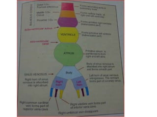
1. The document discusses the embryonic development of the heart, including the septation of the common atrium and ventricle to form the four chambers. 2. Septation of the atria occurs through the septum primum and septum secundum, which divide the primitive atrium into the right and left atria. 3. Septation of the ventricles is accomplished by formation of the muscular interventricular septum, membranous interventricular septum, and conus septum, which separate the bulboventricular cavity into the right and left ventricles.

Recommended
More Related Content
What's hot
What's hot (20)
Similar to Development of Heart Septa in Embryonic Heart
Similar to Development of Heart Septa in Embryonic Heart (20)
More from Dr Laxman Khanal
More from Dr Laxman Khanal (20)
Recently uploaded
Recently uploaded (20)
Development of Heart Septa in Embryonic Heart
- 1. Development of CVS-II To MBBS first year Dr. Laxman Khanal Assistant professor Department of anatomy, BPKIHS, Dharan 24-04-2017
- 2. Right ventricle (T) left ventricle (T) Left/right atrium (T) Right atrium (S) Rt/Lt ventricle (S) Coronary sinus Oblique V of left atrium B-V sulcus primary interventricular foramen Fate of heart tube • Ascending aorta • Pulmonary trunk ??? ??? ???
- 3. Part of heart Embryonic components Right atrium Primitive atrium (T) and Rt horn of SV(S) Left atrium Primitive atrium (T) and pulmonary veins(S) Right ventricle Proximal 1/3rd of BC(T) and middle 1/3rd of BC(S) Left ventricle Primitive ventricle(T) and middle 1/3rd of BC (S) Pulmonary trunk and aorta distal 1/3rd of BC ( TA)
- 4. How Septation occurs ? 1. With Endocardial cushion 2. Without Endocardial cushion
- 5. Septation of common atrium • Septation of common atrium is completed by 2 septa- 1. Septum primum 2. Septum secundum • Normal path of blood flow in developing heart should be kept in mind . Rt atrium to Lt atrium due to pressure gradient.
- 6. Endocardial cushions of the A-V canal not only divide this canal into a right and left orifice, but also participate in formation of the membranous portion of the interventricular septum and in closure of the ostium primum.
- 7. Division of primitive atrium
- 8. Septum primum Sickle shaped crest arising from roof of common atrium into the lumen. Ostium primum
- 9. Ostium secundum Cell death in upper part of septum primum before its closing produce ostium secundum.
- 10. Here comes the role of endocardial cushion of AV canal to close the ostium primum.
- 11. Incorporation of right horn of sinus venosus in lumen of right atrium.
- 12. Rt Lt Septum primum- flexible Septum secundum- rigid Septum secundum: Crescent shaped fold just right of septum primum.
- 13. Left valve of the sinus venosus and septum spurium fuse with the right side of septum secundum. What is the fate of right valve of SV???? Valve of IVC and coronary sinus
- 14. Upper part of SP disappears and lower part function as a valve for the passage between Rt. and Lt. atrium (foramen ovale)
- 15. In about 20% of cases, fusion of the SP and SC is incomplete, and a narrow oblique cleft remains between the two atria. This condition is called probe patency of the oval foramen. It does not allow shunting of blood. After birth: Due to reversal of pressure gradient foramen ovale closes.
- 17. Atrial Septal Defect Primum type ASD: Acyanotic
- 18. Secundum type ASD: Acyanotic Mutation in Heart specifying gene (NKX2.5) is associated with the secundum type of ASD.
- 19. Ventricular Septation Ventricle in figure = primitive ventricle+ proximal part of BC + conus cordis.
- 20. Conus Muscular IV septum IV foramen Conus septum Membranous IV septum
- 22. Membranous IV septum formed by- Endocardial cushion + a pair of bulbar ridges Conus septum
- 23. Tetralogy Of Fallot The condition was thought untreatable until surgeon Alfred Blalock, cardiologist Helen B. Taussig, and lab assistant Vivien Thomas at Johns Hopkins University developed (Blalock–Thomas–Taussig shunt ), an anastomosis between the subclavian artery and the pulmonary artery (see movie "Something the Lord Made"). (source: Wikipedia) It is named after French physician Arthur Fallot.
- 24. 1. Pulomary stenosis 2. Overriding of aorta 3. VSD 4. Rt ventricular hypertrophy
- 25. TOF: Cyanotic heart disease
- 27. Persistent Truncus Arteriosus: No division of TA VSD Cyanotic disorder
- 28. Septation of TA and conus cordis • Two truncal swelling grow in opposite direction creating spiral septum dividing TA into aortic and pulmonary channel. • Two conal swelling meet each other and form Conus septum which divide Conus cordis into outflow tract for Rt and Lt ventricle. • Conus septum is also attached with spiral septum and membranous part of IV septum. • Neural crest cell also contribute to form these swellings. Conal swelling ≈ bulbar swelling
- 29. P trunk Aorta Spiral septum
- 30. What, if the septum is no more spiral !! Transposition of great vessels
- 31. Conclusion Part of heart to be separated Components of septa Primitive atrium (into Rt and Lt atrium) Septum primum and septum secundum Bulboventricular cavity (into Rt and Lt ventricle) Muscular septum Conal (Bulbar) septum Membranous septum Conus (outflow tract for both ventricles) conal septum (bulbar septum) formed by conal ridges Truncus Arteriosus (into pulmonary trunk and ascending aorta) spiral septum (Aortico- pulmonary septum) by a pair of truncal ridges