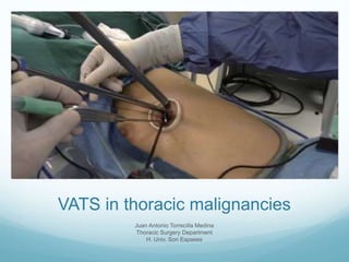
Vats in thoracic malignancies by Juan Antonio Torrecilla
•Download as PPTX, PDF•
7 likes•1,888 views

A very interesting talk about VATS ( Video-assisted Thoracoscopic Surgery) by the Thoracic Surgeon from Son Espases University Hospital in Mallorca

Recommended
More Related Content
What's hot
What's hot (20)
Viewers also liked
Viewers also liked (20)
Similar to Vats in thoracic malignancies by Juan Antonio Torrecilla
Similar to Vats in thoracic malignancies by Juan Antonio Torrecilla (20)
More from Jonathan McFarland
More from Jonathan McFarland (20)
Recently uploaded
Recently uploaded (20)
Vats in thoracic malignancies by Juan Antonio Torrecilla
- 1. VATS in thoracic malignancies Juan Antonio Torrecilla Medina Thoracic Surgery Department H. Univ. Son Espases
- 2. I’d like you to know… What’s VATS? History of video-surgery in Thoracic Surgery Indications of VATS Mediastinum Pleura Lung VATS in major lung resections
- 4. Minimally invasive surgery in thoracic cavity through an utility incision with no rib spreading, looking through an endoscopic camera and a monitor
- 17. Is VATS a new thing?
- 18. 1913
- 19. 1959 - 1973
- 20. 1959
- 21. 1992
- 22. 1992
- 23. 1994
- 24. 1970s
- 25. 1980s
- 26. Stapling devices First stapling device, Hultl (Hungary, 1908) 1950 – 1980s
- 27. Stapling devices First commercially produced re-usable stapling devices (Former USSR, 1950) 1950 – 1980s
- 28. Stapling devices Modern staplers, Hirsch (U.S, 1964) 1950 – 1980s
- 29. Stapling devices Endoscopic staplers (late 1980s) 1950 – 1980s
- 30. Which are the indications?
- 31. Thymoma Enlarged lymph nodes Tumours in posterior mediastinum
- 38. Thoracocentesis: positive in 65%. Close pleural biopsy: sensivity 65% (esp. 99%). Surgery - Biopsy - Poudrage (talc) - Pericardial window Thoracocentesis: positive in 65%. Close pleural biopsy: sensivity 65% (esp. 99%). Surgery - Biopsy - Poudrage (talc) - Pericardial window Pleura Thoracocentesis: positive in 65%. Close pleural biopsy: sensivity 65% (esp. 99%). Surgery - Biopsy - Poudrage (talc) - Pericardial window
- 42. Lung Wedge / anatomic segmentectomy - Pulmonary metastases - NSCLC Lobectomy Pneumonectomy
- 43. Lung metastases
- 44. Lung metastases
- 45. Limited resection in NSCLC
- 48. VATS lobectomy: when? Any resectable NSCLC Same goal as in open surgery (R0, lymph node dissection) contraindications: - Sleeve resections - Endobronchial tumours - Chest wall involvement - Tumours larger than 5 cm - Induction chemotherapy FormerRelative
- 49. VATS lobectomy: pros Less pain, morbidity and early discharge Less postoperative systemic inflammation Less lost of postoperative lung function Best quality of life Best adherence to adjuvant treatment As safe as thoracotomy in long-term survival
- 61. Just to finish… VATS is a minimally invasive procedure using an utility incision with no rib spreading It’s a cutting-edge technique with more than 100 years of history It’s used in the resection of non-advanced thymomas and mediastinal staging in NSCLC It’s a usual technique in the management of malignant effusions
- 62. Just to finish… VATS is an alternative to thoracotomy approach for periferal lung metastases Sublobar resections (by VATS) may be an alternative in early stages of NSLC VATS lobectomy… - Is as safe as thoracotomy, with less perioperative morbidity and it’s related to better quality of life - Is not an easy technique, with a long learning curve - Is cheaper in developed countries, but probably more expensive in the rest of the world
Editor's Notes
- The history of videosurgery will help you to understand how we’ve achieved the actual indications.
- But even the meaning of VATS, I’m going to introduce you the video-assisted surgery in general, including mediastinoscopy.
- I like this diagram because is the original one described by Roviaro.
- Poner una foto del campo desde lejos y con un diagrama que había en uno de los artículos
- Foto de toracotomía antigua
- Foto de toracotomía amiotómica
- Aquí pondré una foto de VATS y una de toracoscopia, para enseñar la diferencia entre ambas técnicas
- Aquí pondré fotos de una monoportal, una biportal.
- Aquí pondré una foto de VATS y una de toracoscopia, para enseñar la diferencia entre ambas técnicas
- Aquí pondré fotos de una triportal y cirugía robótica
- Old instruments just thought to be used in simple procedures as biopsies…
- Material con doble articulación para poder manejarlo a través de la toracotomía de utilidad.
- The development of endosurgery has been possible because: Surgeons have been looking for new indications of this kind of surgery Technological improvements in fiberoptic, mechanical sutures and specific intruments (double-articulated)
- Jacobeus: started the technique in 1910-1913, to divide the pulmonary adhesions in tuberculosis. In 1913 he started the biportal approach. He published the series in 1922. Biopsies were started to be done afterwards, and the indicacion of adhesiolysis stopped in 1945, when the streptomice appeared, which was progressively forgotten (in tuberculosis) because of the high risk of complications.
- In 1973 the first large serie of patients operated of diagnostic thoracoscopy was published. The serie was from 1957 till 1972 (126 patients).
- Carlens y la mediastinoscopia
- It was a RLL lobectomy.
- Walker reported a serie of 6 patients (3 UL, 1 LL, 1 UR and 1 LR).
- And after that: 1999: first robotic surgery 2001: first transcontinental robotic surgery. The first prototype of a surgical robot is dated from 1983. 2000: Da Vinci 2001: first uses in thoracic surgery But this data is not properly for this presentation.
- The technique was pioneered by a Hungarian surgeon, Humor Hultl,[2] known as the "father of surgical stapling".[3] Hultl's prototype stapler of 1908 weighed eight pounds (3.6 kg), and required two hours to assemble and load. Many hours were spent trying to achieve a consistent staple line and reliably patent anastomoses.
- The technology was refined in the 1950s in the former Soviet Union, allowing for the first commercially produced re-usable stapling devices for creation of bowel and vascular anastomoses.
- Mark M. Ravitch, brought a sample of stapling device after attending a surgical conference in USSR, and introduced it to entrepreneur Leon C. Hirsch, who founded the United States Surgical Corporation in 1964 to manufacture surgical staplers under its Auto Suture brand.
- The indication for VATS in thymoma is in Masaoka stages I and IIa (microinvasion of fat tissue). Indications for stages III and IV are controversial.
- Vídeo de timectomia x VATS
- Esquema del mapa ganglionar para explicar la necesidad de estadificación mediastínica y el abordaje de las diferentes estaciones ganglionares. Con cuadros de colores podemos diferenciar cuáles se pueden biopsiar por mediastinoscopia y cuáles por VATS.
- Vídeo de mediastinoscopia
- Vídeo de VATS para ganglios de la ventana aortopulmonar. Poner una imagen del TAC de la paciente (alemana con neo de mama). Explicar la relación con el recurrente.
- Hablar brevemente del VAMLA y el TEMLA como métodos de estadificación. Artículos al respecto (poner las cabeceras). Las recomendaciones de la guía de la ESTS del 2014 son que estas dos técnicas sólo se recomiendan en ensayos clínicos.
- Las recomendaciones de la guía de la ESTS del 2014 son que estas dos técnicas sólo se recomiendan en ensayos clínicos.
- Foto a la izquierda de un derrame pleural. If the first toracocentesis is negative, a second one will be positive en 30% of patients. Poner la VATS como última alternativa para la biopsia y la posibilidad de talcaje. También mencionar la ventana pericárdica cuando hay derrame pleural y pericárdico.
- Video de pleura normal (uno de neumotórax, por ejemplo)
- Vídeo de biopsia pleural y talcaje.
- En las metástasis pulmonares hablar del uso del arpón por la imposibilidad en ocasiones de palpar el nódulo pulmonar. Hablar de la dificultad extrema de encontrar nódulos pulmonares infracentimétricos.
- VATS can be used to resect most metastatic lesions, as the majority are located in the peripheral one-third of the lung [51]. Conversion from a VATS approach to an open thoracotomy is appropriate for lesions located more centrally. As discussed above, there is no consensus regarding a preference for open thoracotomy over a VATS procedure. Some argue that thoracoscopic resection is a valid approach only for patients with a solitary pulmonary nodule on preoperative chest CT, where the risk of additional disease is small [53-56]. On the other hand, proponents of VATS argue that while open surgical exploration identifies radiographically occult nodules, these can be resected with VATS in the future, once they become detectable on surveillance CT scans, without adversely affecting survival.
- VATS can be used to resect most metastatic lesions, as the majority are located in the peripheral one-third of the lung [51]. Conversion from a VATS approach to an open thoracotomy is appropriate for lesions located more centrally. As discussed above, there is no consensus regarding a preference for open thoracotomy over a VATS procedure. Some argue that thoracoscopic resection is a valid approach only for patients with a solitary pulmonary nodule on preoperative chest CT, where the risk of additional disease is small [53-56]. On the other hand, proponents of VATS argue that while open surgical exploration identifies radiographically occult nodules, these can be resected with VATS in the future, once they become detectable on surveillance CT scans, without adversely affecting survival.
- El JCOG 0802 finaliza en agosto de 2018. El CALGB 140503 finaliza en 2016.
- Vídeo de resección atípica simple. Explicar aquí la dificultad para encontrar los nódulos, y la necesidad de guiar por arpón la biopsia en ocasiones. Además, es el momento (si hay tiempo) de explicar que no siempre es fácil realizar una biopsia intraoperatoria (en el caso que se trate de un tumor menos periférico).
- Vídeo de lobectomía (superior derecha).
- Poner foto de artículo de amico (o alguno de estos, de una resección hipermegacompleja).
- Less pain and complications mean less perioperative mortality.
- Less postoperative systemic inflammation
- Less pain and complications (meta-analysis)
- Less pain and complications (randomised study)
- Same long-term survival
- Same long-term survival
- Less lost of postoperative function
- Best quality of life
- Best adherence to adjuvant treatment
- The conclusion is that the technical part is more expensive. It depends on the hospital stay charges that the overall process is more expensive or cheaper.
- The conclusion is that the technical part is more expensive. It depens on the hospital stay charges that the overall process is more expensive or cheaper.