SlideShare a Scribd company logo
1 of 141
1
DENTAL VENEERS
Beauty is in the eye of the beholder
- Margaret Hungerford
Webster’s Third New International Dictionary defines “Aesthetic” as “appreciative
of, responsive to or zealous about the beautiful; having a sense of beauty or fine
culture”. Each of us has a general sense of beauty. However, our own individual
expression, interpretation and experience make it unique.
A desire to look attractive is no longer taken as a sign of vanity. In an economically
and socially competitive world, a pleasing appearance is a necessity. Since the face
is the most exposed part of the body the mouth a prominent feature, teeth are getting
a greater share of attention.
Aesthetic dental treatment can enhance a patient’s own personal image to how he or
she would like to look. As Frush observes ‘A smile can be attractive, a prime asset to
a person’s appearance and it can be a powerful factor in the ego and desirable life
experiences of a human being. He also notes that in any aesthetic treatment, there is
the need for consideration of a patient’s satisfaction with the natural appearance and
function of the result.1
The study of dental aesthetics is a relatively new area of interest in dentistry.
Dentistry has always been concerned with appearance-related treatment, but until
recently, the necessary materials were simply not available easily. The extraor-
dinarily rapid development of adhesive, tooth-colored restorative materials over the
past two decades has established aesthetic dentistry as the major driving force in the
profession. This is not as radical a change as it might seem. After all, aesthetic
dentistry is simply traditional dentistry with an emphasis on appearance and
conservation of healthy tooth structure.8
Aesthetically motivated patients are more likely to undergo routine preventive in the
property of dental care than to wait until pain drives them into the dental office. The
aesthetic practitioner is viewed as a provider of smiles rather than as a purveyor of
pain. Newer techniques and materials make conservative dentistry a reality along
with which the aesthetic results are also readily visible and highly appreciated by the
2
patient. The science that makes all of this possible is the study of adhesion to tooth
structures. Understanding the concepts and materials that are involved in the
property of tooth adhesion is essential to the practice of aesthetic dentistry.
It is very important to integrate aesthetics into the existing restorative and the
speciality areas of dentistry. Comprehensive patient treatment can be contemplated
only if the relationships of various treatment modalities have been established. The
practice of aesthetic dentistry is facilitated by the creation of a suitable and
conducive treatment environment. The rationalization of the traditional dental office
will offer many benefits to both the patient and the dentist.
There are many new treatment modalities, and many more are being developed. It is
essential for the dental practitioner to keep up with these new techniques if the
patient is to be fairly treated. Once the practitioner is more comfortable with
aesthetic dentistry, scientific inquiry requires that the aesthetic envelope be extended.
There is a glimpse of the future as it already exists today, within dentistry.
The development of new materials and techniques in dentistry has required
practitioner to develop new artistic skills. One of the most successful developments
in dentistry has been the meteoric rise of the bonded veneer. Their use has become so
ubiquitous that it is almost hard to imagine that before 1982, when no such
reconstructive modality existed. Much has changed since those first veneers. The
preferred tooth preparation has become more clearly defined, and the number of
clinical applications for this modality has exploded. Materials have improved both
clinically and in the laboratory.
Never before have continuing education and reviewing the literature been more
important to the dentist than they are today. While these efforts may occasionally
seem tedious, it is truly an exciting time for the dentist restoring localized or
generalized defects or intrinsic discoloration.2
3
Definition –
A veneer is a layer of tooth coloured material that is applied to a tooth for
aesthetically restoring localized or generalized defects or intrinsic discolouraton.
RATIONALE FOR VENEERS –
In this ever changing world the appearance or packing of everything is important. A
pleasing appearance is important not only socially and romantically, but also
economically, for it has been found that attractive people tend to get better jobs. The
dominance of a dental composition may be amplified by rendering it more visible.
Increasing the crown size or/and using lighter teeth, placing them more anterior or
increasing the exposed gingival length, may produce this effect. It is for that reason
that the teeth and smile play a major role in whether we perceive the face of an
individual as attractive or not. It is now possible for the aesthetic dentist to
“beautify” the patient’s smile while creating a more “youthful” appearance. The
desire to veneer teeth easily without resorting to full crowns has long been a goal of
dentists. Clinical situations are well known in which strong, sound teeth are present
but appear discoloured because of the administration of tetracycline, excessive
fluoride use, smoking, age, or other reasons. Minimal tooth reduction or none at all
is highly desirable in such situations.3 Various types of veneers requiring minimal
tooth reduction have been used for over 15 years. They have had various
characteristics of longevity ranging from a few months to several years and have
been variable in their aesthetic characteristics. At the present time, many dentists are
accomplishing veneers as a routine procedure. They have become a major part of the
so-called “aesthetic dentistry” movement.4
Pilkington (1936), Defined dental esthetics as “the science of copying or
harmonizing our work with that of nature, making our art inconspicuous”
The term “esthetics” is borrowed from the Greek world “aesthesia” Which means
sensation or sensibility. It can be defined as “belonging to the appreciation of the
beautiful esthete” the noun form of the same word may be used to describe a person
who enjoys or perceives a pleasant sensation similarly the meaning of the term in
4
adjective form indicates ability to respond to beauty in art and nature the relation of
this term to dentistry has been differentiated from the word “Cosmetic” which is
derived from the Greek word “Kosmos” or adornment. It is further stated that
aesthetic dentistry enhances the natural beauty of the mouth and face and that the
term is used specifically to imply and improved relationship rather than a superficial
one.5
HISTORY
As early as 1937, Pincus developed thin facings made of air-fired porcelain.
He attached these thin labial porcelain veneers temporarily with denture adhesive
powder to enhance the appearance of the Hollywood actors for their close up
photographs. Pincus was fully aware of the importance of the “Hollywood smile” as
an integral part of the image and public opinion. These facings presented a viable
option to the full crown for the actors who needed to temporarily change their smile,
yet they possessed very little strength, and the technology necessary to provide a
permanent means of attaching the veneers to tooth structure was lacking. This
reversible technique provided an alternate for those who wanted their smiles to
improve without the need of more aggressive crown preparations. 6, 7
The art of veneering teeth has progressed over 30 years to the current generation of
concepts and materials, which can be divided into two categories:
1. Directly fabricated composite resin veneers.
2. Indirectly fabricated veneers such as preformed laminates or laboratory
fabricated acrylic resin, micro-fill resin, or porcelain veneers. 4
Direct Veneers
Not until the 1970s did the practice of bonding composite resin directly to
teeth for aesthetic improvement grew in popularity.
5
The introduction of light-cured resins in the early to mid 1970s allowed the
dentist greater flexibility. The advantages of visible light- cured composite resins
such as working time and improved chemistry versus the self cured composite resins
marked the entry into the next generation of aesthetic materials. Visible light-cured
composite resins were replacing self-cured composite resins by the late 1970s and
were preferred for aesthetic anterior restoration.
Direct acid-etched bonding proved to be advantageous, yet susceptibility to
stain, poor wear resistance, and lack of natural fluorescence spurred the continued
search for improved materials.
Indirect Veneers –
Faunce and Mayers (1970) described a one piece acrylic resin prefabricated
veneer as an improved alternative to direct acid etched bonding by using a chemical
primer applied to the veneer and a composite resin to lute the veneer onto an etched
tooth; both chemical and mechanical bond attributed to the ethyl acetate. These
hollowed-out structures were treated with ethylacetate, methylene chloride, methyl
methacrylate, and then filled resin cement was used to lute the veneers to etched
teeth. It was more stain resistant than composite resin veneers, but numerous
preformed acrylic resin laminates suffered from delamination at the laminate /
composite interface usually due to the wear of the chemical bond. Like composite
resins they also exhibited poor resistance to abrasion 9
The inherent advantage of laboratory fabricated veneers is the anatomical
accuracy created by the technician, thus alleviating the chair side activity required
with directly applied veneers. 6
PORCELAIN VENEERS –
Porcelain has a long history of use in dentistry as one of the most aesthetic
and biocompatible materials available surpassed only by enamel itself. The
6
utilization of porcelain as a restorative material began a new era in esthetic
dentistry.5
 Porcelain’s abrasion and stain resistance are excellent and it is well tolerated
by gingival tissues.
 The advent of porcelain labial veneers as permanent aesthetic restoration
marked the progression of more than 30 years of dental research in acid-etch,
bonding, and aesthetic restorative techniques.
 Vines et al in 1958. The first major improvement in the esthetic material,
especially in the translucency of all porcelain crowns.
 Weinstein et al first discovered the bonding of porcelain to gold alloys by
means of vacuum firing in the early 1960’s.
 In 1965 Maclean and Hughes described sintered alumina that was originally used
for the fabrication of crowns small fixed partial dentures and individual and custom
built pontics for use in restorative dentistry in the form of prefabricated profiles. 5
 1968, MacCulloch, who first described the methods used for making artificial
teeth, veneers and crowns in glass ceramic, utilized this approach.
 In 1975, Rochette described the innovative restoration of a fractured incisor
with an “etched selected porcelain block”. In early 1980’s the key pioneers in
American laminate dentistry were instrumental in the development of porcelain
veneers and the associated techniques for their fabrication and placement.
 In 1976, McLean and Sced developed the first commercially possible foil-
reinforced crown system. The dental porcelain could be strengthened by the
dispersion of ceramic crystals of high strength and elastic modulus within the glassy
matrix. McLean and Hughes used this method to develop the first aluminous
porcelains for the fabrication of crowns. These reinforced porcelains had strengths of
up to 180 MPa - approximately double that of the more conventional feldspathic
materials.
 Electroforming and the use of tin oxide coatings for the attachment of
conventional metal-bonding porcelain reported by Rogers in 1979, and a number of
foil systems were marketed in the 1980s.
 Although technique sensitive the surface texture, color, florescence, and the
overall aesthetics of porcelain laminate veneers have been regarded as exceptional.
7
In addition, the ability to adjust final color during placement allows maximum
flexibility in final shade adjustment.
 Sadoun refined Count Von Schwerin's slip casting technique in a paper given
at the International Conference on Dental Ceramics held at Leeds Castle, England in
1989. These changes produced high-strength coping, and was marketed under the
name of In-Ceram.
 In-Ceram was not a pure ceramic and represented a step forward in achieving
strengths of upto 630 MPa. In-Ceram made it possible for the laboratory technicians
to make advancements in the esthetics of anterior crowns, without losing strength.
Undoubtedly, this work deserves a notable place in the history of dental ceramics.
 Horn (1983) introduced the platinum foil technique and Calamia reported the
refractory die technique for porcelain laminate veneer fabrication in the laboratory.10
 Simonsen and Calamia (1984) demonstrated good bond strengths for resin
composite to hydrofluoric acid etched porcelain and that the use of silane coupling
agent could further increase the bond strength of resin composite to etched porcelain.
 Other methods such as pressed ceramics (IPS Empress), castable ceramics
(Dicor), and CAD/CAM (Cerec) or copy milling techniques (Celay) have also been
used for veneer fabrication. 11
 1993, Anderson and Oden described a technique for manufacturing
individual all-ceramic crowns made up of densely sintered high-purity alumina.
However, the color of sintered alumina may vary according to the firing conditions,
presenting a serious disadvantage. Similarly, when compared to regular or aluminous
dental porcelains, the sintered alumina is a more difficult material to control.
 Recent studies have shown very good long-term results following placement
of anterior veneers. In one study done by H.D.H. Shaffer (2000), “Porcelain
laminate veneers-A retrospective evaluation after 1 to 10 years of service” had
probability of survival as 97% at 5 years and 91% at 10.5 years, over the entire
observation period, only seven restorations (4%) actually failed.12
 With the recent increase in patient’s demands for esthetic restorations in the
anterior as well as the posterior regions, it has become necessary to develop new
materials that combine the strength and the resistance that are essential in the
posterior region, along with the esthetic qualities desired in the anterior region.
Porcelain laminate veneers (PLVs) have become the esthetic alternative to ceramic
crowns and the traditional porcelain-fused-to-metal. Smiles can be transformed
8
painlessly, conservatively and quickly with dramatic, long-lasting results with the
successful use of the porcelain laminate veneer. Porcelain veneers are now the
restorative choice for esthetics in numerous clinical circumstances that would have
resulted in the use of full crowns in the past. Tissue response is excellent, and the
finished surface is very similar to the natural tooth. Veneers exhibit natural
fluorescence and absorb, reflect, and transmit light exactly as does the natural tooth
structure. Patients are highly enthusiastic about these restorations that represent a
conservative treatment that enhances patient self-image. The subsequent introduction
of special acid etching techniques improved the long-term retention of veneers. Horn
and Simonsen and Calamia, for example, increased interest in porcelain veneers.
They were influential in demonstrating that the bond strength of hydrofluoric acid-
etched and silanated veneers the luting resin composite is generally greater than the
bond strength of the same luting resin to the etched enamel surface. 5
INDICATIONS & CONTRAINDICATIONS
Indications for Veneers-
1. Tetracycline Discoloration - This, unfortunate discoloration of teeth is still very
common and can be a significant aesthetic and emotional detriment to patients.
Slight tetracycline staining can be removed by bleaching. However, moderate to
severe staining is treated best with conservative veneers instead of crowns.13
9
CLASSIFICATION OF TETRACYCLINE STAINS 14
DRUG COLOR OF STAIN ON TEETH
Chlortetracycline (Aureomycin) Grey , Brown
Demethylchlortetracycline
(Ledermycin)
Yellow
Oxy tetracycline (Terramycin) Yellow
Tetracycline (Anchromycin) Yellow
Doxycycline (vibramycin) No reported changes
Minocycline Black
10
Tetracycline Discolouration. Labial reductionof 0.5 to0.57mm.
Incisal reductionof 1.5mmwith
lingual chamfer.
ClassV type cavitypreparation
done instaineddentin.
Opacificationof dentin Opacificationof dentin
Immediately after insertion of veneers.
11
2. Fluorosis Discoloration - This staining is difficult to remove with bleaching,
but it can be bleached in cases of slight to moderate staining. Effective management
of multicoloured or darkly fluorosed teeth can be accomplished best with veneers.13
3. Teeth Darkened by Age - A well-known phenomenon is the darkening of
teeth observed as patient’s age. The darkening is caused by pigmented foods, coffee,
smoking, and so forth. Often, teeth are sound and do not need restorations or crowns
but they are esthetically objectionable. Such teeth are ideal candidates for veneering.
The teeth retain their basic integrity and shape and are not altered significantly except
for a colour change. They become smooth, and the discolorations caused by age are
lightened. 13
12
4. Irregular Tooth Positioning in the Arch. Teeth that are slightly rotated or
in lingual or labial version can receive the appearance of straightening easily by
veneering them. Often these teeth also contain numerous discoloured restorations
because of the patient’s inability to clean them well. The unsightly restorations are
also hidden by the veneers. 13
5. Malformed Teeth - Infrequently patients have one or more teeth that were
malformed by trauma during tooth development, metabolic disturbances of tooth
development caused by systemic disease, or other reasons. These teeth are ideal
candidates for veneers. Their colour is usually acceptable, and they require only
removal of deep pigmentation and veneering of the tooth with a resin matching the
tooth colour. 13
13
6. Teeth Discolored by Endodontic procedures. Slightly discolored non-vital
sound teeth, pigmented during or subsequent to endodontic procedures, can be
covered successfully by properly accomplished veneers. This task is a difficult one
from a color standpoint, but with experience it can be done effectively.
7. Teeth with Numerous Visible Cracks - Athletics, accidental trauma, ice
chewing, and other abuse to teeth cause cracks in the tooth enamel that may also
become stained from foods. Veneers can cover these cracks easily and eliminate
future staining in them. 13
8. Teeth with Numerous Unsightly Restorations. Occasionally, bodily
discolored restorations and those with stained margins are restored best by covering
their entire facial surface with veneers.
14
9. Teeth Denuded of Superficial Structure by Acid Demineralization.
Many people who have gastric disorders or those who regurgitate often because of
illness or by choice have lost significant superficial tooth structure. These teeth are
sensitive to cold, hot, and sweet foods. Veneers satisfy both the sensitivity and the
esthetic problems.4
Contraindications for Veneers
1. Heavy Occlusion - Persons having extremely heavy occlusion exert forces
on veneers that can break them from the surfaces of enamel or dentin. Therefore,
persons exhibiting signs and symptoms of clenching or bruxism are poor candidates
for veneers. Bruxers who have worn incisal guidance and canine rise guidance
excessively break off veneers easily. Other treatment should be used including metal
occlusal surfaces or metal crowns with heat or light cured microfill veneers over the
metal. Porcelain fused-to-metal restorations may be contraindicated in patients with
bruxism. 4
2. Teeth in Severe Labial Version - Veneering “buck teeth” has limited esthetic
potential. Removal of sufficient enamel to allow acceptable colour often eliminates
enamel and forces the dentist to make retention in dentin. This is less successful than
enamel retention. Also, the extreme facial placement of the veneers often increases
the “buck tooth” look, Orthodontic therapy and/or crowns are better treatment for this
type of patient. 5
3. Mouth Breathers - Such patients have irritated gingiva most of the time.
Their dry mouth environment encourages thick mucous collection around the anterior
teeth and subsequently greater gingival irritation when veneers are placed. Correction
of mouth-breathing is suggested before any comprehensive restorative therapy is
initiated.
4. Poor Oral Hygiene - Gingival health is deteriorated around some types of
veneers. Placing veneers in persons with poor oral hygiene can create continuous and
increased periodontal breakdown. Patients often have poor oral hygiene because they
15
dislike their teeth and avoid cleaning them in a subconscious rejection of their esthetic
condition. A trial period to demonstrate improvement of oral hygiene is indicated
before placing veneers. Usually hygiene improves after placement of the veneers. 4
5. Denuded Dentin. Dentin adhesion agents are currently available. However,
it is difficult to obtain optimal bonding to large areas of tooth structure that have only
dentin present. Crowns are indicated in these situations.
6. Highly Fluoridated Teeth. Although fluoridated teeth can be veneered
successfully, longer etching and meticulous cleanliness are required to afford optimal
retention. Etching for 2 minutes is suggested for highly fluorosed teeth. The patient
should be advised of potential veneer retention problems with highly fluoridated
teeth.4
Economic Considerations
An advantage of veneers is a lower cost to the patient than crowns. At this
time, veneers are becoming a routine part of dental practice, and fees for this service
are being stabilized.
This economic advantage allows patients to have one or two replacements of
the veneers before the total cost equals that of a single crown. Of course, the patient
must endure the trauma of additional treatment when the veneers are replaced.
Assuming that the current generation of porcelain fused-to-metal crowns will
serve esthetically for approximately 10 years and that the current generation of
microfill resin veneers will serve for approximately five years, properly placed
veneers are economically feasible for both the patient and the dentist.4
16
CLASSIFICATION
A. Depending on coverage:
Partial veneers
Full Veneers
Full veneer with “window” preparation design that extends to the gingival crest and
terminate, the facio-incisal angle.
17
Full veneer with incisal lapping preparation design extending sub-gingivally that
includes the entire incisal surface.
B. Depending on technique of fabrication
1) Directly fabricated (a) Partial veneers
(b) Full veneers.
2) Indirectly fabricated (a) Partial veneers
(b) Full veneers.
C. Depending on cutting of enamel
1) Intra-enamel preparation
2) Extra-enamel preparation
Intra-enamel preparation-
To achieve ideal aesthetic and physiologically sound results, this
preparation is almost always indicated. Exception to this is when labial surface is
18
under contoured due to severe aberration or erosion. In such cases only the margins
are defined.
The objective of roughening and defining the margin are to-
- provide space for opaquer, without over contouring.
- remove the outer fluoride rich layer which is resistant to acid etching.
- rough surface increases surface area, increasing bond surface.
- is of importance in case of indirectly placed veneers.
Extra-enamel preparation-
Veneer is placed on the entire existing tooth structure without any tooth preparation.
The advantage is that there is no tissue destruction and the complete procedure is
reversible in case of failure. Disadvantage of this technique is over contouring,
decreased bond strength. 4
D. Depending on material
1) Acrylic resin
2) Composite resin
3) Porcelain
4) Castable ceramics (Dicor)
BENEFITS AND SHORT COMINGS
Benefits –
This treatment consists of replacing the visible portion of the dental enamel with an
artificial substitute. This ‘substitute enamel’ helps to replace defective human enamel
19
with bonded artificial enamel.15
Minimally invasive treatment –
This treatment method utilizes minimal tooth preparation, mainly confined within the
dental enamel. It preserves tissue integrity. We all agree that the natural human
enamel would be the best restorative material to use, if that were possible - but
porcelain laminate veneers, being the closest material to natural human enamel, are
the least destructive to the adjoining tissues. These characteristics have established the
porcelain veneers as the preferred restorative material preserving the enamel, which
is-ultimately the most valuable part of the tooth. Numerous authors have suggested
that the minimal thickness for tooth preparation for ceramic veneers should be no
more than 0.5 mm. In practice: however, the actual approximate thickness of a
porcelain laminate veneer is 0.4 to 0.7 mm, which closely resembles that of the
natural tooth enamel. 5
Shape, Position and Surface Appearance-
The shape or position of natural teeth can be affected with functional or
esthetic problems. The major advantages of veneers is that the surface texture can be
transformed permanently and elegantly, eliminating any dysplasia or dystrophy of the
enamel.16
20
Colour –
The smooth surface texture and natural colour of porcelain are exceptional, and the
crystalline structure of porcelain gives it optical refractive properties similar to those
of translucent enamel. When bleaching techniques become ineffective, veneers may
the treatment of choice for improving or changing natural tooth colour. 5
Durability-
Veneers stand up extremely well to biological, chemical and mechanical action.4
Light Transmission-
Using dentin porcelain of varying chromas, as well as transparent, translucent or
opalescent (but not opaque) porcelain, it is possible to achieve moderately thick build
-ups by a layering technique or lateral segmentation that reproduce all the
characteristics of natural enamel such as cracks, fissures and opalescence.13
Illumination of natural tooth. Illumination of all ceramic
crown.
21
Tissue Response –
Extremely good biocompatibility with gingival tissues. The highly glazed porcelain
surface is less of a depository area for plaque accumulation as compared to any other
veneer system. Perhaps one of the most important advantages of porcelain laminate
veneer is that the periodontal reaction is minimal. The smoothly finished margins
help to maintain good periodontal health and oral hygiene. Various studies have been
performed. One of them established that the porcelain-fused-to-metal tissue response
is similar to that of the veneers; yet, another found that there was considerably less
inflammation with porcelain veneers than with porcelain-fused to-metal (PFM)
restorations. In another particular study, Kourkouata et al. found that after the
porcelain veneer restoration was completed, there was an increase in crevicular fluid;
similarly, there was a significant decrease in plaque index, and plaque bacteria
vitality. When selecting a treatment, longevity is a major concern. A more invasive
procedure will result in a higher risk to the tooth and ultimately a more expensive
treatment. 17
Speed and Simplicity-
Laminate veneer treatment procedures are usually carried out under light anesthetic,
with minimal tooth reduction, and at greater speed than other techniques.
Impressions are easier to make, because of the accessible position of the margins.
- Patients appreciate being able to remodel their smile very rapidly.
- Highly acceptable tensile bond strength. The resin to silane treated etched porcelain
veneer has bond strengths ranging from 2600 to 3200 pounds per square inch (psi)
and composite resin veneer to enamel bond strengths of 900 to 1400 psi.
- Porcelain can be used to increase the length of given tooth by extending it over the
incisal edge.
- Long lasting. Once bonded, porcelain veneers develop high tensile and shear
strengths.
- Exceptional resistance to wear and abrasion.
22
- Resistance to stains.
- Surface lustre retention. 15
State of the Art –
The dental profession has always been in search of restorations that have a "natural"
appearance but are, at the same time, long lasting. In the past, the lack of appropriate
dental materials rendered this impossible. However, advances in porcelain veneers
and bonding materials now offer a variety of selections in treatment, with long-lasting
restorations and improved quality in aesthetic results. Porcelain veneers are to be
considered the "state of the art" in aesthetic dentistry, providing abundant advantages
over any previous form of veneer system. They are excellent in terms of their
appearance and durability, and the actual procedure does not disturb the soft tissue or
the adjoining periodontium. Since their introduction almost 20 years ago, porcelain
veneers have become the flagship of most aesthetics-based practices. 5
Strength-
Owing to their ceramic thinness (0.3-0.5mm), the porcelain veneers can be easily
fractured before they are even bonded, similar to kitchen or bathroom tiles. However,
once bonded on a solid foundation, they integrate with the tooth structure and become
extremely durable, in vitro and in vivo. If all conditions have been adhered to and the
appropriate bonding composite has been selected, the dentist will achieve successful
results. The patient's satisfaction is bound to be high as this medium ensures long-
term maintenance, good gingival health and excellent oral hygiene. When it was
proven that function and esthetics could be achieved with porcelain laminate veneers
and without the removal of large amounts of intact tooth structure, porcelain veneers
became the restorative material of choice in addressing the most challenging aspects
of aesthetic dentistry by integrating the individual restorations into the adjacent
natural dentition. 5
23
Longevity –
The union of etched enamel and porcelain, combined with the bonding composite
resin-luting agent with a silane coupling agent, enables the dentist to perform
restorations that are solid as well as long lasting. The long-term data is encouraging
and reports a high success rate for this form of treatment. Materials used in the 1980s
are now considered primitive, whereas the restorations made with laminate veneers
even 15 years ago have shown a high rate of success, increasing interest and
widespread acceptance. Porcelain laminate veneers are continually gaining respect as
the best and most durable restorative treatment, while patients and dentists alike are
delighted with the esthetic results and the minimal tooth preparation required. As
aesthetic dentists are becoming increasingly confident of its application to a variety of
clinical cases, the survival rate of laminate veneers reported in literature ranges any-
where from 18 months to 15 years. Friedman reports in another study that of the
3,500 porcelain veneers that were placed over a 15-year period, 93% of the cases were
successful. 5
Shortcomings –
Porcelain veneers can be easily repaired once bonded to the enamel, but the repairs
are not long lasting due to staining which tends to occur at the margin of composite
resin and porcelain.
-The colour cannot be easily modified once bonded in position.
-Irreversibility of preparation versus little or no preparation for composite resin
bonding.
-Level of difficulty of fabrication and placement, time involved, and expense.
-The extremely fragile veneers are difficult for the dental laboratory to make and
manipulate, and the process requires two appointments, and laboratory fees.
-Technical difficulties in avoiding over contours and obtaining closely-fitted porcelain
enamel margins. The margins can be especially brittle and difficult to finish.
-Lower reparability compared to composite veneers.
24
-Susceptibility to pitting by certain topical fluoride treatments. stannous fluoride
should not be used with porcelain restorations.
At the bonding stage, the slightest error can mean failure- either immediately or later
on. This is a major disadvantage of laminate veneers.
-Handling of such fine ceramics requires very special precautions. Prior to bonding,
they are extremely fragile, and the slightest irregularity could produce fracturing.
-Techniques are becoming simpler, especially with the progress made in phosphate
investment, but the creation of fine natural ceramic veneer restorations remains a
difficult and highly skilled task, the two main drawbacks being:
- Stability during different types of handling.
- Difficulty of getting right balance of powders for layering or segmental build up
with a thickness ranging from 0.3 to 0.7 mm.
-There is practically no possibility of touching up feldspatic porcelain veneers
processed over investment or even using platinum foil, because the ceramic cannot be
re-fired once removed from its support.
-Tooth Sensitivity.
- Marginal gingivitis. 15
25
CASE SELECTION
Patients should be advised of the various treatment options available when
they have malformed, discolored, or otherwise esthetically objectionable teeth. These
options are; (1) to bleach; (2) to veneer with one of the available materials; (3) to
place crowns or, (4) to leave the teeth as they are. 18 Veneering is a good alternative in
most cases. Also, patients should be instructed that they are expected to maintain their
veneers properly for optimal aesthetics and longevity.
Color Selection –
Most shade guides are not as useful as the manufacturers intended. Many
variables influence the color of the underlying tooth and the resin veneers including
thickness of the resin to be placed; moisture in the tooth structure; translucence of the
underlying tooth structure; and location of the resin on the tooth in an incisal-gingival
direction.
One of the adequate ways to determine the best color of resin from the available
materials follows: Place a small amount of the potential resin colours on the un-
etched, damp tooth structure and polymerize them. Selection of the ideal colour is
then simple. Many resins darken when they are polymerized, whereas some lighten in
hue during polymerization. Only a few brands have the same color before and after
polymerization. It is suggested that the color selected be written in the patient’s
record and also on an instruction sheet given to the patient for future reference and
repair.
When veneering medium dark hues, it is often desirable to use one of the medium
opaque materials rather than a translucent hue. The medium opaque hue (for
example, Durafill GO, or Silux GO) will allow some of the original darkened hue to
show through the veneer.
A slight degree of this “show through” allows the veneers to assume a much lightened
hue but gives them some of the original color to blend with the remaining un-
veneered teeth.
26
A technique promoted by some manufacturers suggests placement of a small amount
of stains giving the veneer a different shade and improved smoothness. Such thin
materials are adequate for a short time but soon wear and leave the original shade and
surface characteristics of the underlying resin. It is suggested that the proper shade be
selected for the entire veneer and that this material be finished to optimal smoothness,
thereby eliminating the need for a superficial layer of material. 19
Procedures –
The control, as well as the periodontal, condition of the teeth to be overlaid must be
assessed. A healthy periodontium is imperative for successful results. 20
Examining occlusion
The relative fragility of ceramic veneer restorations require an accurate
analysis of the patient’s occlusion, to ensure that the restorations are not extended into
areas of occlusal stress. The result of this analysis may limit the opportunities for
remodeling. There is no point in aiming to increase the incisor grouping height, for
example, without dealing with canine malocclusion when present. Any attempt to
establish a different height clinically, in cases of attrition or bruxism, carries its share
of risks and should be approached with caution.
Occlusal factors should be considered even with veneer restorations that do not
involve the lingual surfaces (i.e. the incisal edge is preserved). In fact, it is in this
situation that the risks are greater, especially when canines and posterior teeth are
involved. 13
Examining a single tooth –
Once again, shape, position, available enamel and occlusion are all factors to be
considered. Tooth that is outstandingly triangular or every slender raises difficulty,
which will all have to be appraised and dealt with accordingly. 15
27
Normal:
- Line angles
- Cervical convexity
- Vertical lines or ridges
- Horizontal lines or ridges
Widening:
- Displace the line angles laterally
- Flatten facial outline
- Highlight texture and gloss with horizontal lines and ridges
-Decrease facial embrasures
Narrowing:
- Displace line angles medially
- Increase the convexity of the facial outline
Shortening:
- Emphasize the prominence of the cervical convexity
-Displace the cervical convexity coronally
- Highlight texture and gloss with horizontal lines and ridges
-Emphasize the cement –enamel junction
Lengthening:
- Flatten and displace the cervical convexity apically
- Highlight texture and gloss of the line angles and create
vertical ridges
- Lighten the cervical aspect55
28
29
Examining the gingival tissue –
Although the margins of laminate veneers are often located away from the gingival,
the condition of the latter must always be appraised before deciding on any treatment.
Poor dental hygiene, gingival inflammation as well as one or more gingival recession
sites should all be treated before applying ceramic laminate veneers.
Appraising the smile –
Clinical examination should focus not just on the teeth to be restored (their colour,
shape, etc.) but also on the shape of the face, size of the lip, and the lip-to-tooth
relationship during various movements. All these inspections should be carried out
both frontally and from the side. The practitioner must also use other methods of
visualization, such as the following,
1. Diagnostic wax-up
2. Cast models
3. Computer imaging analysis.
A complete upper and lower impression is made before preparing the teeth, followed
by mounting the models on an articulator and making any corrections desired, such as
lengthening or closing up diastema using either light-cured resin or wax. 15
30
The smile must be viewed from the front and sides (both left and right)
 Shape of the face
 Size of the lips
 Visible coronal and gingival levels - At rest
When talking
Broadest smile
 Harmony and proportion - Of the cervical line
Of the line of the incisal edges
Of the lip line
 Tooth colour - Hue
Value
Chroma
Translucency
Texture and luster
 Tooth shape - Size of tooth (height : width ratio)
Incisal edge
Contour
Assessing triangular tooth shape
Analysis of statistic and dynamic occlusion
Spatial arrangement of teeth
31
BONDED RESTORATION IN OCCLUSION AND FUNCTION
Occlusion –
Occlusion refers to the act of closure and the state of being closed. It involves at the
same time dynamic and static components. Static components will be first considered.
22
Static Occlusion –
In a good occlusion, all the teeth in the mouth make simultaneous contacts. It is,
however, acknowledged that in the anterior segment teeth should have lighter
contacts, which clinically may mean: "anterior teeth do just not contact". Indeed, the
perception of a fremitus at interocclusal contact can be detrimental to tooth stability
and lead to interproximal separation.23
The total tooth contact area, which includes the anterior and posterior teeth, averages
4 mm. Considering the number of interocclusal contacts (64 to 100 for the whole
mouth), taking into account varying opinion and the type of occlusion, each single
contact area is estimated to be extremely small. The multiplication of posterior
interocclusal contacts is mandatory for improving stress distribution, as studies have
shown that in the posterior segment, muscle contraction develops whether you have
single or multiple contacts. In nature, cusp to ridge contacts are the rule and
characterize class I occlusions. Cusp to fossa contacts are usually found in class II
occlusions, while the tripodization of interocclusal contacts remains a dream, born
from the imagination of dentists anxious to provide tooth stability.
A coincidental location of tooth interocclusal contact and porcelain-bonded
restoration margin should be avoided, as alterations of the marginal seal of porcelain-
bonded restoration can be more often stated than when the opposing contact is
located elsewhere. 24
Whenever this recommendation finds easy application in the anterior segment,
clinical practice shows that the posterior segment appears as a privileged area where
interocclusal contacts and porcelain-bonded restoration margins are coincidental by
reason of posterior tooth morphology and the nature of interocclusal relationships.
32
Observation shows also that it is in this area that the morphological design of the
restoration is distorted to adapt to antagonist deformities, or inserted in a dental
segment presenting various manifestations of abrasion. As a result, the possibilities
of coincidental location of margin placement and enlarged interocclusal contacts
increase. And with them the chances of altering the marginal seal resulting from
porcelain microfractures under the load of occlusion. 25 It can be assumed that further
deteriorations of the marginal seal of porcelain-bonded restoration can take place
under the impact of parafunctional habits such as clenching, when margin placement
and interocclusal contacts are coincidental. This stresses the importance of tooth
preparation for porcelain-bonded restorations that has not only to foresee
interocclusal contact location but provide sufficient porcelain thickness to allow
porcelain-bonded restoration to offer adequate resistance to constraining parafunc-
tional conditions.
Functional Occlusion –
Function takes place at the very moment teeth enter into action. This dynamic part of
occlusion refers to the engagement of mandibular teeth towards centric, and to their
disengagement. The spatial development of functional movements is dictated by
condylar pathway, antagonist tooth morphology and tooth position. It displays at the
registration a three-dimensional tracing delimiting an area defined as "the envelope
of mandibular function" characterized by a droplet shape displaying, as a result of
antagonist tooth morphology and position, infinite individual variations. 26
This makes teeth the primary factors in tooth-closing patterns. It has been established
that tooth-closing patterns remain under neuro-muscular control - even if the exact
nature of this phenomenon has always escaped scientific explanation.
If we know most of the parameters that participate in function, the observation of
unworn long lasting dentitions clearly indicates the type of oral architecture that best
integrates these parameters. These occlusions are characterized by similar parameters
in the vertical and horizontal planes: that is to say in the overbite and overjet of
incisors and canines. Frontal chewing patterns have an angle of about 70° from the
horizontal for the canines and in eccentric occlusion we find a tooth-to-tooth contact
with a large posterior clearance. This tooth-to-tooth contact in eccentric occlusions
33
can be considered as ideal, as it generates, according to research, a reduced muscular
activity in comparison to group function and bi-balanced occlusion.27
In these dentitions, morphologic integrity is the rule. The teeth can receive porcelain-
bonded restorations without any specific restriction, but in respect of the
environmental morphologic design: function follows forms. Functional integration
will then be achieved by the control of simultaneous interocclusal contacts and the
absence of interferences during tooth closure patterns.
In nature, however, oral architectural ideals are not the rule. Research has shown that
in a selected population the occurrence rate of malocclusion, which includes open-
bite, over-bite, class II and class III occlusions, averages 20%. As a result, one in
every five patients does not present an incisal pathway reflecting usual standards,
which leads them to consider correction by way of orthodontics or orthopedic
surgery. It also appears that out of the remaining 80% of class I occlusions; most
occlusal schemes do not show anterior relationships, duplicating those displayed in
long-lasting dentitions. Variations in the depth of the overbite, width of the overjet,
and related incisal and canine guidance are unlimited, which justifies or imposes
corrective measures necessary to prevent posterior interferences during function. 28
By deduction, it can be advanced that one of the challenges faced by restorative
dentists in restoring a physiological occlusion is the changing of poorly related
anterior relationships. The restoration of an anterior segment should never be
envisaged from the viewpoint of minimal tooth preparation for veneer seating but
from that of occlusal maintenance and functional excellence. 29
Occlusal maintenance involves the evaluation and correction of localized
manifestations of wear affecting one anterior tooth or the whole anterior segment,
concomitant with an appreciation of the degree of incisal or canine guidance
necessary to prevent posterior interferences in eccentric occlusions. The achievement
of these objectives requires an increase of the steepness of the disclusive angle which,
depending on oral conditions, can be achieved either with an increase of vertical
interocclusal relationships by way of a tooth elongation or with a reduction of
horizontal relationships. A combination of these therapeutic measures is not unusual.
Clinically, a modification of the palatal morphology of maxillary incisors and canines
34
appears best suited to improve tooth guidance without compromising aesthetics .The
nature of this modification remains to be defined as long as it leaves open the question
on of the ideal steepness of the interocclusal angle. Mechanically, it appears that the
deeper the angle the greater the likelihood of achieving tooth guidance and the greater
the impossibility for posterior teeth contacting in eccentric occlusions. From a
muscular point of view, the increase of the steepness of the disclusive angle may also
produce a favourable result as it generates a proprioceptive inhibition that will shut
down muscular activity thus reducing the load level in eccentric occlusions. This
muscle inhibition has been explained by an increase of the torsional load affecting the
teeth in their housing. This leads to a triggering effect on proprioceptive receptors
preventing muscles contacting harder through a feedback effect. However, careful
attention must be paid, so that the increase in the disclusive angle does not cause the
teeth, through their morphology or position, to interfere with neurological
programmed tooth closure patterns. This will invariably lead to the abrasion of
antagonist anterior tooth surfaces, mandibular buccal or maxillary lingual surfaces.
Damage of this type affecting antagonist surfaces is mainly generated by
overcontoured porcelain bonded restorations but may also be found in orthodontic and
orthognathic cases.
In the absence-of available clinical possibilities of tracing three-dimensional,
programmed tooth closure patterns for want of an articulator programmed to register
this information, it is left to the dentist's sensibility to prevent restorations impinging
on the area of function. Despite these restrictions, there is no better restorative
material today than porcelain-bonded restoration to achieve functional and aesthetic
excellence or to duplicate the conditions found in long-lasting dentitions. All that
remains is to apply the appropriate treatment at an appropriate time.
Incising –
Mastication starts with the incision of food, which is followed by a lateral chewing
phase. This lateral chewing phase can be divided in an early tooth closing phase and a
late tooth-closing phase. Incising requires an initial mouth-opening necessary to
introduce food followed by a closing movement of the mandible, which develops in
the sagittal plane and places upper and lower incisors in a tip-to-tip position. This
35
movement seems to load incisors along their long axis and bring both condyles in a
forward and downward translation. With unworn incisors there is no more than 1 mm
of shearing load confined in the incisal edge of teeth. This shearing load will increase
dramatically in the presence of worn-out incisal edges. As a result, the tip-to-tip
incisal biting will gradually become a surface-to-surface biting, overloading incisors
away from their long axis, allowing for the posterior confinement of condyles leading
in turn to adverse neuro-muscular response. Finally, the incisal edges of the lower
incisors will glide back along the lingual wall of maxillary incisors, a movement
which develops atraumatically under neuromuscular control. Yet the presence of an
increased incisal tip width affecting lower incisors will defefinitely overload the
anterior teeth forcing lingually mandibular incisors.17
It is therefore essential that porcelain-bonded restorations duplicate in their incisal
design sharp unworn natural teeth and present in their whole configuration the most
adequate compromise in terms of internal stress distribution and stress transfer
following axial or horizontal loading. These mechanical requirements have to be
considered when the presence of interproximal carious lesions, composite
restorations, or tooth fractures impose a lingual extension of anterior maxillary
porcelain-bonded restorations. In the same way, the palatal extension of porcelain -
bonded restorations in the design of maxillary incisors and canines becomes
unavoidable when functional finality demands:
 An improvement of anterior guidance to correct insufficient horizontal or
vertical interocclusal relationships.
The design of cingulum and lateral palatal ridges to create punctiform centric
interocclusal contacts and to prevent the eruption of lingually positioned antagonist
lower incisors.
To optimize the configuration of porcelain bonded restorations one has to know that
the area of the cingulum presents the lowest tensile strength of the whole palatal side
at a tangential blow of 50 N directed against the incisal edge.32 It can therefore be
considered as an ideal location of porcelain-bonded restoration margin. Above all, a
marginal location of porcelain-bonded restoration in this area of low tensile strength
reduces the importance of the design of the finish line: a butt margin cutting the
enamel rods at an angle superior to 50° is perfectly acceptable while a long chamfer
36
will reveal particularly resistant in as much it will cut the rods at 90°, allowing for an
improved adhesion to enamel. Even if studies have shown that this type of porcelain-
bonded restoration configuration presents the lowest relative compliance, knowing
that resilience is a key factor in fracture resistance, fracture rate remains extremely
low in clinical practice, maybe thanks to dentin bonds that have been proven to be a
main determinant in the fracture mechanic. As previously mentioned, the palatal
extension of porcelain-bonded restoration is the rule each time functional conditions
advocate an increase of anterior guidance to allow for sufficient posterior
interocclusal clearance in eccentric occlusions.30
The functional indication for palatal extension varnishes in functionally good
occlusions where tissue preservation predominates. A limited incisal palatal
overlap remains the most valuable configuration as long as it is located in an area
displaying low tensile strength, allowing for a finish line design that can be either a
butt joint or chamfer.
These porcelain-bonded restoration configurations, palatal overlap or palatal coverage
present the qualities to perfectly resist the incising function or the tearing apart of
food. Only a palatal extension of the porcelain-bonded restoration with a finish line
ending up in the palatal concavity has to be avoided, as this thin extension of ceramic
is precisely located in an area of high tensile strength. 27
Normal function not only implies the incision of food but the chewing of food.
Lateral Chewing –
Lateral chewing starts with mouth opening, followed by a lateral movement of the
mandible to the working or chewing side. The working condyle is brought in a
retruded lateral position while the non working condyles move medially. The amount
of this lateral shift varies, depending upon bone configuration of condyle and fossa,
shape and conditions of disc and ligaments. 19
The shifting of the working condyle or Bennet shift affects the mediolateral incline of
the posterior cusps and conditions the direction of the buccal groove of lower molars
whenever the occlusal scheme presents in eccentric occlusions a group function or a
balanced occlusion.
37
In functionally good occlusal schemes, however, the importance of the Bennet shift is
irrelevant. In the early closing phase of mastication, which sees both working canines
starting to proprioceptively engage the closing movement, the importance of the
mediolateral inclination of posterior cusps is irrelevant. 31
In the final closing phase, however, the approach to centric of opposing molar cusps
can lead to interferences because the vertical guidance of the canine does not totally
negate the path of the shifting condyle. As a result, interferences can occur in the
mediolateral inclines of the second and first molars, especially when the condyle-
fossa complex generates in the horizontal plane a shifting in lateroprotrusion or a
shifting in laterosurtrusion in the frontal plane.
The wear resulting from these interferences affects the mediolateral inclines of
mandibular or maxillary molars predominantly in areas characterized by a cusp to
fossa relationship such as the central groove of maxillary and mandibular molars. This
is best visualized in natural unworn dentitions. In good occlusal schemes this occlusal
wear is irrelevant but it may reach a pathological level with a decrease of the anterior
interocclusal angle. Under these conditions the introduction of a restoration in these
areas may reveal itself detrimental to marginal material integrity as interocclusal
contacts and margin location are often coincidental. The integrity of the porcelain-
bonded restoration can even be compromised when mesial or distal transitional zones
of tooth preparation present a too-abrupt angulation. 27
Clinical observation shows that porcelain bonded restoration fracture rate increases
precisely in the mesial part of maxillary molars and in the distal part of mandibular
molars in occlusal schemes showing poor canine guidance. These elements need a
configuration of posterior porcelain-bonded restoration with sufficient material
thickness and suggest coverage of mediolateral cusps designed with a reduction of the
inclination of the mediolateral slopes and a reduction of the MO or DO transitional
angle.
Dysfunction –
The observation of unworn long-lasting dentitions has put in evidence harmonious
relationships between skeletal and dental morphology and integrated function. It has
long been speculated that abnormal function is mainly caused by abnormal tooth
38
guidance. In turn, abnormal tooth guidance results from abnormal occlusal contact
relationships. 32
We know that one out of five patients do not have an incisal pathway reflecting an
appropriate standard. Yet we can observe daily that out of the remaining patients
reflecting appropriate standards in anterior pathways, few show or will show over the
years an occlusal scheme free of wearing manifestations.
It can therefore be deduced from these observations that even appropriate standards in
anterior pathways can display abnormal occlusal guidance. 19
Submitted to registration these elements of abnormal guidance have been classified in
a recent study in lack of guidance, poor guidance, over guidance, abnormal guidance,
and interference.
The presence of interferences in the development of tooth closure patterns has long
been identified as the prime element of dysfunction. It has been even assumed that it
could be at the origin of bruxism. There are two basic types of interferences:
 Centric relation: anterior slide, lateral slide, protrusive-retrusive
 Lateral border: non working and working side.
The elimination of centric interferences in the treatment of bruxism has always been
controversial. In 1928, Tischter recommended occlusal adjustment in order to
eliminate the so called "trigger contacts". A large number of authors stress also the
importance of eliminating these interferences in the treatment of bruxism, while
others deny any relationship between interferences and bruxism. 33
It appears, however, that clinically experienced practitioners never hesitate to
proceed to occlusal correction before restorative procedure by reason of the good
results they obtained.
Dawson (1998) has written: "It is my experience that, when all occlusal interferences
in centric relation and excursions have been eliminated on the posterior teeth,
parafunctional movements resulting in anterior wear are eliminated or reduced to a
point where they are of no clinical concern." His statement gives the restorative den-
tist the best reasons to proceed to occlusal controls, not only before restorative
procedures but also following porcelain-bonded restoration placement, to eliminate
39
the interferences that were either present or could have been introduced with new
restorations. 34
This post bonding control is made necessary because of the great difficulty in
appreciating before bonding porcelain-bonded restorations, the occlusal parameters
because of the risks of fracture. Aside from interocclusal overcontacts in centric
occlusion, centric interferences - lateral or anterior - can be found on posterior
porcelain bonded restorations, while protrusive retrusive interferences are
systematically present on anterior porcelain-bonded restorations, especially those
showing a configuration with a palatal extension.
Clinical observation shows, however, that contrary to Dawson's statement,
parafunctional habits can then be reduced to a point where they are of no clinical
concern when both centric interferences have been eliminated and anterior guidance
steepness has been concomitantly increased. 27
Indeed, nothing else but this increase of anterior guidance steepness can prevent
possibilities of posterior eccentric interferences that may take place in the area of
mandibular motion, clinically undetectable but made possible by increased muscle
and ligament laxness, pillow or hang pressure. These interferences also can be the
initiator of parafunctional movements.28
Occlusal schemes showing moderate but apparently sufficient posterior clearance in
eccentric occlusions must be looked at with suspicious eyes, especially those
presenting deficiencies in tooth alignment. They open the possibility of unexpected
eccentric contacts generator of avoidance patterns leading in an initial phase to the
erosion of the elements of the anterior segment. 19
The rotation of the mandible around a posterior obstacle followed by a mandibular
projection against the maxillary incisors has been observed in clinical tests, and the
follow-up of muscles participation clearly described. It has even been demonstrated
that parafunctional avoidance patterns could not only project the mandible against
maxillary incisors but be moved outside the confine of the maxilla that is to say in a
cross-over position. 35
40
This uncontrolled back and forth movement will progressively erase antagonist
anterior teeth whose progressive morphological reduction will finally permit
posterior contact, and posterior wear faceting.
As a consequence, the set-up of veneers, that is to say porcelain-bonded restoration,
covering the facial part of the tooth and provided with a reduced palatal overlap, is
restricted to occlusal schemes showing a total absence of faceting. The presence of
wear faceting in the occlusal scheme (it could be a simple chipping, a localized or
generalized chevron-shape anterior wear), requires a modification of veneer con-
figuration with the design of either an elongated porcelain-bonded restoration or a
porcelain bonded restoration with a palatal extension, provided an increase of anterior
guidance can be achieved to prevent any possibility of posterior interference in
eccentric occlusions. 36
The predictability of any restorative process rests on the precise evaluation of oral and
occlusal conditions. Porcelain veneer restorations can be considered as ideal
restorations for the corrections of poor anterior relationships and anterior-guidance.
Conversely, they can also be destructive for the dentition when occlusal conditions
have been neglected or wrongly appraised. 5
PERIODONTALCONSIDERATIONSIN AESTHETICTREATMENTPLANNING
Aesthetic dental treatment is not limited to the technical skills and aesthetic
perspectives of the dentist and the dental technician; the patient's perceptions and his
or her understanding of the problems and solutions are also important. 37 Patients
should be instructed that aesthetic dental rehabilitation is a complex and complicated
form of treatment and that she or he is a-party to that treatment not merely a recipient
of it. The patient needs to see treatment as a collaborative effort in particular because
oral hygiene habits and periodontal status dictate the outcome and longevity of the
aesthetic result. The rehabilitation of a smile relies not only on restorative dental
procedures, but also on a healthy and aesthetic gingival appearance. 38
41
Aesthetic periodontal problems can only be tackled after efficient plaque control and
proper initial periodontal, therapy have been addressed. Soft tissue defects may
become more apparent as gingival shrinkage occurs after the resolution of the
inflammation.
The aesthetic outcome of a case is determined by the relationship of the crown with
the soft tissues, whether it is a natural tooth, a pontic, or an implant. A sound
knowledge of the normal anatomy of the dento-gingival unit may initially prevent
violation of the basic and then one can understand the point where physiological
tolerance may shift to inflammatory reaction. 13
Periodontal Tissues in Health –
The Dentogingival Unit represents a unique anatomical feature concerned with
attachment of gingiva to the tooth. It is composed of epithelial and connective tissue
compartments. Three types of anatomically distinct epithelia are associated with the
dentogingival unit. The first, gingival epithelium, consisting of stratified squamous
keratinized epithelium which is continuous at the gingival crest with the second type,
stratified non keratinized or parakeratinized epithelium. 20 Sulcular epithelium relines
the soft tissue wall of the gingival sulcus facing the tooth but not attaching to it and is
continuous at the base of the sulcus with the junctional epithelium, or the epithelial
attachment. The lack of keratinization of the sulcular epithelium is the result of
inflammation in its supporting connective tissue. Junctional epithelium forms a collar
around the tooth and the surface cells provide the actual attachment of gingiva to the
tooth surface. In a study of autopsy material by Gargiulo et al. it was determined that
the average sulcus depth was 0.69 mm, and the average length of the junctional
epithelium was 0.97 mm. A significant finding in that study was that the average
depth of gingival sulcus was highly variable from tooth to tooth and around the same
tooth.39
Connective tissue compartment of the dentogingival unit presents fibrous attachment
of the gingiva to the hard tissue wall and support to the epithelium of the
dentogingival junction. Fibrous connective tissue attachment to the root surface is
provided by a gingival collagen fiber system that also maintains the functional
42
integrity of the teeth and its gingival collar. Gargiulo et al. reported that the average
length of the supracrestal periodontal fiber group was 1.07 mm and was constant at
all locations around the teeth. The most important groups of fiber bundles that
compose this gingival unit are dentogingival, alveologingival, dentoperiosteal,
transseptal, circular/semicircular, and interpapillary fiber bundles. 40
The autopsy material reported by Gargiulo et al. showed that the average length of
the junctional epithelium was 0.97 mm, supracrestal collagen fiber group was 1.07
mm, and the average sulcus depth was 0.69 mm. Of these three tissue components
the supracrestal connective fibrous attachment seems to exhibit the least variability,
followed by the junctional epithelium and sulcus depth. In 1962, Cohen defined the
"biologic width" of supracrestal gingival tissue as those junctional epithelium and
connective tissue elements of the dentogingival unit that occupy the space between
the base of the gingival crevice and the alveolar crest, that the total dimension would
be in the vicinity of 2.04 mm. 40
For practical reasons, as suggested by Nevins and Skurow, the "biologic width" can
be expressed as the sum of the supracrestal fibers, the junctional epithelium and the
gingival sulcus. A better name that would prevent confusion could be "biologic
zone" as used by Kois. 21
The minimum dimension of this space between the most coronal level of the alveolar
bone to the most coronal level of gingival margin would be in the vicinity of 3 mm in
a healthy and normal gingiva. 41
This is the minimum space required for the tissues to maintain their continuity in a
healthy status.
Aesthetic Periodontal Problems –
Gingival Inflammation and tissue loss owing to periodontal disease -
Periodontal disease is the result of the accumulation of dental plaque at the gingival
margin leading to inflammation of the periodontal tissues. 10 Traditionally,
periodontal disease has been divided into gingivitis and periodontitis, depending on
whether destruction of the tooth-supporting tissues has occurred. Diagnosis and
43
treatment of periodontal problems are based on clinical findings such as probing
pocket depths, attachment loss, gingival bleeding, and radiographic evaluation of the
alveolar crest. The objective of periodontal treatment can be summarized as the
prevention of tooth loss, maintenance of periodontal support, repair, and regeneration
of damaged tissues and their function. But when the etiology of the periodontal
disease is considered, the main task seems to be the modification of patient habits in
order to constitute an oral hygiene level that would prevent inflammation of
periodontal tissues owing to plaque accumulation. 42
Gingival inflammation and associated periodontal destruction are leading factors that
can reverse the aesthetic outcome in the long term. 43 Establishing definitive
restorations when inflammatory periodontal problems are present not only restrains
aesthetic success, but also accelerates the rate of periodontal destruction. Elimination
of gingival inflammation is mandatory before initiation of any restorative treatment.
Initial periodontal therapy that consists of oral hygiene instruction, elimination of
retentive factors, scaling and root planning will result in the resolution of inflam-
mation. Patients with moderate to severe periodontitis may require advanced
periodontal treatment that could consist of surgical procedures to eliminate residual
periodontal pockets and regeneration of the lost periodontal tissues. Pocket
elimination surgery usually results in long clinical crowns that may be aesthetically
unacceptable. In deep isolated angular periodontal defects, guided tissue regeneration
offers good results to maintain the tooth and avoid an aesthetic disaster. Alveolar
ridge deformities are inevitable after the extraction of hopeless teeth when special
attention was not paid to preserving the height and width of the ridge at the time of
extraction. 36
Restorative Margins and Violation of Biological Principles –
Preservation of the healthy status of periodontal tissues is the most significant factor
in the long term prognosis of a restored tooth. 5
There are five major causes of plaque-related inflammatory periodontal disease
associated with restorative procedures:
1. Severe damage to the periodontal tissues during tooth preparation and impression
making.
44
2. Failure to maintain emergence profile.
3. Inability to adequately finish and/or seal sub-gingival margins.
4. Placement of sub-gingival margins at sites with minimum to no attached gingival.
5. Violation of the biologic width.
Teeth prepared without retraction cord which subsequently receive retraction cord,
electrosurgery, or rotary gingival curettage sustain various degrees of soft tissue
damage, as reported by Dragoo and Williams. Tooth preparations for restorative
procedures should be accomplished atraumatically to avoid irreversible damage to
the supracrestal connective tissue fibers and tissue loss. 40
The cervical one-third of the clinical crown is also referred to as the- emergence
profile of the tooth. This is the region where the crown emerges from the
periodontium and its supra-subgingival contours should have a flat emergence
profile, with the guidance of anatomic information in order to maintain an area that is
cleanable with oral hygiene instruments and which has a good marginal seal of the
restoration .There is a very strict relationship between marginal seal and emergence
profile. Poor adaptation and roughness of the restoration margin results in
mechanical irritation of the sulcular epithelium and can harbour microbial flora.
Overhanging margins not only accumulate more plaque than properly finished
margins, but the plaque undergoes a shift in composition to that seen in association
with destructive periodontitis. An adequate band of keratinized tissue is fundamental
to successful restorative dentistry if the margins of the restorations are extended
under free gingival margin. Approximately 5 mm of keratinized tissue, composed of
2 mm of free gingiva and 3 mm of attached gingiva, is considered necessary to meet
restorative objectives even in cases with ideal finishing and margin placement at the
subgingival area, Although no solid investigation is present to confirm this observa-
tion, augmentation of keratinized gingiva provides stability of the free gingival
margin and surrounding gingival tissues so that the gingival health around the
restoration can be maintained. Besides vertical dimension of the keratinized gingiva,
the thickness of the tissue should be enough to tolerate intracrevicular restorations.
The gingival sulcus extends from the free gingival margin to the epithelial attachment.
In health, its depth is in the vicinity of 0-3 mm, and it is lined with thin sulcular
45
epithelium. The depth of this sulcus may vary from tooth to tooth and from surface to
surface on the same tooth. Compromising the apico-coronal width of the connective
tissue attachment by placement of crown margins deep in to the sulcus initiates per-
sistent and irreversible gingival inflammation owing to violation of the biologic
width. Such violations sever the junctional epithelium and the supragingival
connective tissue fibers. Apical migration of the junctional epithelium encourages
development of periodontal pockets and alveolar bone loss.31 The finish line of
restorations should follow the contour of the cemento-enamel junction and be always
kept at least 2.5 mm away from the bone crest . A 2.5-mm difference means that
although the biologic width is being respected, the finish line is placed equigingivally.
A greater distance between the bone crest and finish line of the restoration is needed
to ensure that margin of the restoration could be well reached by the plaque control
instruments. The golden standard today is to prepare the finish lines of the restorations
0.5 mm below the marginal gingiva if the sulcus depth is 1 mm, 0.5 to1 mm if the
sulcus depth is exceeding 1.5 mm. This leaves enough distance for the junctional
epithelium and supracrestal fibers, and is also close enough to the gingival margin to
be reached by oral hygiene procedures. When the healthy sulcus depth is less than 1
mm, placement of the margin 0.5 mm subgingivaly could result in impinging upon the
junctional epithelium. The margin in such cases should be terminated just at or above
the gingival margin. 15
Gingival asymmetries and discrepancies –
The aesthetic appearance is considerably affected by the symmetry created by dental
and facial midline. In the etiology of gingival asymmetries, altered passive eruption,
different patterns of tooth wear, traumas that modify tooth eruption, tooth positioning
in the dental arch, parafunctional habits and overzealous tooth brushing should be
considered. The selection of an adequate method of treatment depends on adequacy of
attached gingiva, tooth structure, vestibular depth, the distance from the gingival
margin to the crest of the bone, root angulation and tooth positioning and
interproximal level of the alveolar bone when recession is present. Correction of this
disharmony requires surgical elongation or root coverage, orthodontic treatment and
restorative procedures. 8
46
Excessive Gingival Display –
"Gummy smile" can be considered as a significant aesthetic problem by many
patients. Ideally, the smile should expose minimal gingiva around the lateral incisors.
The gingival margins of maxillary central incisors and cuspids should coincide with
the vermillion border of the upper lip. The lip line, assessed when the patient is in full
smile, is classified as high (gummy smile), medium (ideal), and low (maxillary lip
covering a portion of maxillary teeth). Aging leads to a decrease in the amount of
anterior tooth display when smiling. Maxillary overgrowth, insufficient upper lip,
tooth mal-positioning, and delayed apical migration of the gingival margin or a
combination of more than one are among the most common reasons of "gummy
smile". 5
Skeletal deformities that involve maxillary overgrowth can be diagnosed by
evaluation of the proportions of the face and cephalometric analysis. In such cases the
teeth's length-to-width ratio usually remains constant - the ideal being approximately
10:8. The ideally proportioned face is divided into three equal parts from the hairline
to the eyebrow, from the eyebrow to the base of nasal process, and from the base of
the nose to the chin. When the middle one-third of the face is longer than other two,
"gummy smile" is diagnosed owing to maxillary overgrowth. In combined cases that
maxillary overgrowth is superimposed with delayed passive eruption and the latter
should be treated first. Treatment limited to periodontal surgery will be insufficient,
since the level of the incisal edge to lip at rest position cannot be rebuilt. Minor
maxillary overgrowth cases can be successfully treated with periodontic-orthodontics
or periodontic-restorative combinations. Increase in the gingival and mucosal display
necessitates complex treatment options that combine orthognathic-orthodontic proce-
dures and perio-prostho treatment. 13
The normal length of the upper lip is between 18 and 21 mm, vertically. A shorter lip
over a moderate dental arch could result in excessive gingival display. When the
"gummy smile" is solely dependent on insufficient lip dimensions, a perio-restorative
solution composed of surgical crown lengthening and restoring the teeth with
laminates could be the choice of treatment. 44
A less frequent reason of excessive gingival display is the tooth malpositioning.
Orthodontic repositioning and surgical correction are the treatment options in those
cases. 45
47
The last but not least reason for the excessive gingival display is the delayed passive
eruption, or what some authors call "altered passive eruption", Passive eruption is the
exposure of teeth by apical migration of the gingiva, whereas active eruption is the
movement of teeth in the occlusal direction. When the teeth reach their functional
antagonists, the gingival sulcus and junctional epithelium are still on the enamel, and
the clinical crown is approximately two-thirds of the anatomic crown. Delay in the
apical migration of the marginal gingiva and the junctional epithelium results in a
condition in adulthood in which the gingival margin is positioned incisally or
occlusally on the anatomic crown and does not approximate the CEJ. 5
When the gingiva is not inflamed but thick and fibrotic, and displays a nominal
degree of scallop to the free gingival margin resulting in a square tooth form, delayed
passive eruption should be suspected. Diagnosis is made by probing the sulcus and
sounding the alveolar bone margin to determine their relationship with CEJ. Clinical
and anatomic crown length, width of the attached gingiva tooth positioning and
involvement of frenum are additional factors in diagnosis and treatment planning.15
There are two types of delayed passive eruption, as proposed by Coslet, et al. (Table-
8.1). In this classification, gingival/anatomic crown relationship is divided into two
major groups. In type 1 cases, the gingival margin is located incisal or occlusal to the
CEJ, with a wide zone of attached gingiva, and the mucogingival junction is usually
apical to the alveolar crest. There is typically an excessive amount of gingiva as
measured from the free gingival margin to the mucogingival junction. In type 2 cases
the mucogingival junction is located at the level or near CEJ where the band of
attached gingiva is in the normal range as measured from the free gingival margin to
the mucogingival junction. The major distinction between two types is the location of
the mucogingival junction, which also serves as a useful guideline in the treatment
planning. A further subdivision evaluates the alveolar crest and CEJ relationships. 46
48
CLASSIFICATION OF DELAYED PASSIVE MIGRATION OF THE
GINGIVAL MARGIN
Type A B
1. Short clinical crowns with Alveolar crest to Alveolar crest to
muco-gingival junction being CEJ ≥2 mm CEJ <2 mm
apical to the alveolar crest.
Wide band of attached gingiva.
2. Short clinical crowns with Alveolar crest to Alveolar crest to
muco-gingival junction being CEJ ≥2 mm CEJ <2 mm
at the level or near CEJ.
Normal Band Of Attached Gingiva.
Treatment options of delayed passive eruption should be based on diagnosis of the
type. A typical case of type- IA delayed passive eruption exhibits short, square-
looking teeth with a wide zone of attached gingiva and confirms the presence of
sufficient space (biologic width) to maintain supracrestal fiber attachment on trans-
gingival probing. 47 A gingivectomy procedure performed by a scalpel,
electrosurgery or CO2 laser can remove the excess tissue. Tissue removal should be
performed very carefully in order not to compromise interdental papilla and end up
with some loss at the gingival tissue filling the embrasures. Removal of the tissue will
result in more elliptical tooth form that restores the 10:8 length to width ratio. Initial
wound healing takes place in the first couple of weeks where remodeling of the soft
49
tissue continues for as long as 4 to 6 months.
Treatment options of type-1B Delayed passive eruption cases should create sufficient
space for supracrestal fiber attachment to maintain the proper biologic width while
eliminating the excess gingiva. In type -1B cases there is an added dimension
buccolingually to the osseous form. This extra thickness of osseous structure allows
for an apical angulation of the bone crest from the gingival aspect of the periodontal
ligament side allowing for insertion of the connective tissue fibers just apical to the
CEJ. Surgical correction of this reverse anatomy requires relocation of the alveolar
crest, thinning of the alveolar margin, and securing enough space for development of
proper biologic width. Since the level of gingival tissue normally follows the
architecture of the underlying osseous crest, alveolar reduction should meticulously
be performed in order to dictate the final gingival level and contour at the time of
bony recontouring. The height of the interproximal bone should always be 3 to 4 mm
coronal to the height of the newly created alveolar crest on the facial aspect in order to
support the interproximal papilla. Surgical crown lengthening requires reflection of
full thickness flaps and recontouring of the alveolar bone with chisels and burs.
Surgical trauma owing to full thickness reflection of the access flap and bone
recontouring results in resorption of the crestal alveolar bone approximately 0.63 mm.
This inevitable bone loss should be calculated at the time of surgical planning to
prevent unexpected recessions, especially in cases with thin gingiva and facial
alveolar plate where more bone loss is expected.
On the other hand, in a clinical study it was reported that a 3 mm biological zone was
not routinely achieved after surgical crown lengthening procedure, and the amount of
root surface was still short of the initially planned biologic width after healing. A
histometric study in monkeys demonstrated that healing following osseous crown
lengthening procedure resulted in a junctional epithelium that extended to the apical
level of root planing, reestablishing a reduced biologic width. Scaling and root
planing during crown lengthening was recommended to enable the surgeon accurately
to determine the superior termination of the supracrestal connective tissue, since space
for the supracrestal connective tissue fiber groups is created by crestal resorption of
alveolar bone. Initial wound healing takes around 10 days after surgery, and final
remodeling usually continues for as long as 6 months, but the tissue reaches a stable
position after 6 weeks. Gentle handling of the tissues and strict adherence to plastic
50
surgery principles shortens healing and remodeling periods.
Treatment of type II cases requires relocation of the entire dentogingival complex to a
more apical position without rendering loss of attached gingiva with or without
removal of the bony margin. Loss of attached gingiva results in alveolar mucosa
located at the gingival margin that jeopardizes maintenance of the marginal health,
and unacceptable esthetics. Attached gingiva must be preserved at crown-lengthening
procedures in such cases by reflection of full to split thickness flap and apical
positioning of the mucogingival complex. The healing pattern is similar to other
crown-lengthening procedures.
Long-term stability of the gingival margin created by crown-lengthening procedures
relies particularly on the respect paid for the biologic width. As previously mentioned,
approximately 2 mm is needed between the base of the sulcus and the crest of the
bone to provide space for the connective tissue attachment and junctional epithelium.
Adding the depth of the sulcus to biologic width results in a minimum of 3 mm from
the newly created gingival margin to the crest of the bone. Violations of this basic
principle result in poor cosmetic results that require retreatment. 48
Gingival recession –
Chronic trauma caused by vigorous tooth brushing and plaque-induced periodontal
inflammation is the major causative factor in the development of soft tissue
recessions. Beyond those two major causes, malpositioning of teeth, dehiscence in
vestibular alveolar plate, high frenum, and iatrogenic factors contribute significantly
in the etiology of recessions. Elimination of those factors is crucial in both prevention
and treatment of recessions in a predictable long-term outcome. Sullivan and Atkins
made initial classification of the marginal tissue loss in two major vertical categories
of shallow and deep recessions with horizontal subgroups as "narrow" and "wide".
This classification was used until 1985 when Miller presented an expanded classifica-
tion. Based on this classification, the amount of root coverage can be determined
presurgically. Total root coverage is expected in class I and II cases. In class III cases,
the amount of root coverage can be estimated presurgically by placing a periodontal
probe horizontally on an imaginary line connecting the tissue level on the mid-facial
of the two teeth on either side of the tooth exhibiting recession. This is a very
51
practical and important classification for the clinical decision-making process,
because it allows the practitioner and the patient to have realistic expectations
regarding the outcome of the case. 38
Besides aesthetic concerns owing to gingival recessions, increased root sensitivity and
sensitivity related gingival inflammation and/or decay are major problems associated
with exposed root surfaces. With the growing concern for cosmetic dentistry, and the
subsequent need for periodontal procedures that would meet patient expectations,
numerous techniques have been developed to cover the exposed root surfaces. Those
techniques fall into the following four major categories.
Pedicle Flaps –
Coronally and laterally positioned contiguous gingival grafts. The technique is used to
treat shallow areas of recession (Miller class I) where 3 mm of keratinized tissue is
present. A modification of coronally positioned flaps by Tarnow under the term
"semilunar coronally repositioned flap" offers high predictability and superb colour
match to treat class I recessions. This procedure is limited with the height and
thickness of the gingiva apical to the recession. Laterally positioned pedicle flap
(called by some clinicians "rotational flap") received several modifications over the
years but the fundamental principle is covering the exposed root surfaces by the
gingival tissue lateral to the recession. This technique offers the advantages of good
blood supply, excellent match of colour, and a single surgical site. On the other hand,
limitations like the necessity for adequate dimensions of gingiva lateral to the site of
recession, as well as its inability to treat multiple recessions and its requirement of
deep vestibule, confines its application to cases of single recession with an adequate
amount of neighbouring attached gingiva.
Free Autogenous Soft Tissue Grafts –
Free gingiva and subepithelial connective tissue grafts have been reported to be
predictable and successful ways of root coverage. Free gingival grafts are utilized to
increase the zone of attached gingiva around teeth and implants, and to cover the
exposed root surfaces. Having the palatal mucosa on the surface 1.5-2 mm thick, free
52
gingival grafts heal in a lighter and more opaque colour than the gingiva at the
recipient site, and it increases the gingival thickness. Meta-analytic review of success
rate of the free gingival grafts -revealed a high predictability in classes I and II
recessions with mean root coverage around 75%, and complete root coverage
incidence 50%. Subepithelial connective tissue graft is also used in root coverage
surgery. The procedure takes advantage of a double blood supply from the recipient
bed and the gingival flap that covers the graft. It generally results in a better colour
match, but increased thickness can compromise aesthetics in the healing periodontal
the remodeling process is completed within a year. The major contraindication to this
approach is the inability to harvest adequate thickness of donor material from the
palate. 5
Combined Techniques –
These are surgical procedures that combine more than one technique either in the
same stage or at a second stage of surgery. Free gingival graft with a coronally
positioned flap is a common example of this approach. Either a gingival graft is
placed simultaneously with the coronally positioned flap, or following placement of a
gingival graft the entire complex is moved coronally at a second surgical procedure.
Another common combination is the root surface coverage with a subepithelial
connective tissue graft and sliding the lateral two papillae over the grafted tissue.
Other clinicians have suggested several more combinations to furnish the
requirements of specific cases, but none has become popular and widely used. 5
Guided Tissue Regeneration –
The technique is based on the principle to provide and maintain an adequate space to
allow formation of a blood clot that is expected to transform into new cementum, new
connective tissue attachment, and new bone. Results obtained by resorbable and non-
resorbable membranes to cover exposed root surfaces are comparable with
subepithelial connective tissue grafts, and can be used in the treatment of deep and
wide (larger than 5 mm) facial recessions. No requirement for a donor site reduces
patient discomfort during healing. Highly esthetic results can be provided since the
color of the new tissue blends evenly with the adjacent sites. The main disadvantage
is that the treatment is reserved for single tooth application. The specific use is limited
53
to the cases that lacks donor tissue and attached gingiva until improved GTR devices
and surgical methods are developed. 13
Open Interproximal Spaces –
Reconstruction of the lost interdental papilla is one of the most challenging and least
predictable problems in periodontal plastic surgery. Besides creating a black triangle
at the maxillary anterior region, missing interdental papilla results in food impaction
and phonetic problems. The presence and morphology of the interdental papilla is
strictly dependent on the size and shape of interproximal contact, crest of alveolar
bone, and shape of lateral tooth surfaces, as stated earlier. Reconstruction of the lost
interproximal tissues therefore requires techniques that re-establish the prerequisites
of the presence of the papilla. Non-surgical and prosthetic approaches may alter the
contact point, lateral tooth structure and tooth angulation resulting in restoration of the
lost interdental tissue. Surgical techniques that aim to increase the gingival soft tissue
and bone content of the interproximal spaces are complicated with the anatomic
limitations and the blood supply problem. Several surgical techniques have been
suggested in case reports for reconstruction, but none of them seems to be sufficient
and predictable to restore the lost tissue. Limited success has been attained by soft
tissue grafts. Regeneration of the interproximal alveolar bone seems to be the ultimate
treatment method to achieve papilla reconstruction keeping in mind that the gingival
contour follows the crestal alveolar bone and that the distance from the bone crest to
the contact area dictates the interdental soft tissue level.68
TOOTH PREPARATION FOR VENEERS
 Goal is to remove a uniform layer of the tooth structure
 Using burs with calibrated diamond rings
 Reduced diagnostic steps
 Intrinsic principle is the reproduction of the initial situation( form and
function)
 Can lead to dentin exposure
 Cases typically involve patients with discolored teeth
54
PRINCIPLES –
The preparations must fulfill the following four basic principles if perfect, functional,
biological and aesthetic integration is to be achieved: stabilization, reinforcement,
retention and adhesion. Relying on adhesion without taking the other three factors
into account generally leads to immediate or ultimate failure.11
Preserving as much of the natural enamel as possible, although desirable, must never
jeopardize the planned restoration by minimizing the preparation. Enamel reduction
requires special instrumentation that involves the four surfaces of the teeth.
Instrumentation –
The concept of ceramic laminate veneers has become inseparable from that of
controlled enamel preparation, finally resulting in prosthetic substitute enamel with
optical, mechanical and biological properties closely resembling those of natural
enamel.17
It is the shape of the instrument that determines the profile of the preparation. Many
authors, including Garber (1991) and Lustig (1976), have concentrated on the
rationalization of instrumentation in fixed prosthetics. The present authors, too,
contributed to this field, having suggested TPS (Touati) Brasseler kits in 1985. This
instrument kit consists of eight burs, enabling veneer preparations to be accomplished
in total safety (Figure - ). The laminate veneer preparation kit comprises:
 Two instruments (gauges TFC1 and TFC2) to monitor labial reduction;
 Two instruments (TFC3 and TFC4) for reduction of enamel and margins;
 Two instruments (TFC5 and TFC6) for occlusal reduction;
 Two finishing instruments (TFC7 and TFC8).
Brasseler TPS tool kit instruments for preparation and finish of ceramic veneers.
The advantage of this kit lies in simplifying preparation by plain codifying and
offering a limited number of instruments.
The two 'depth-cutter' instruments (TFC1, TFC2) serve to guide, visualize and, in
particular, quantify enamel reduction. In addition, margins can be plotted, owing to
55
the rounded tip. The authors consider it dangerous to gauge reduction in enamel depth
of between. 0.1 and 0.5 mm without some type of depth guide. Goldstein (1984), who
also recommends two instruments based on the same principle in his laminate veneer
preparation kit (Brasseler, LVS), shares the same concern.38
Other techniques exist for monitoring depth, such as those advocated before the
introduction of penetration gauges. A spherical diamond bur (Komet H01 314 009)
traces out a 0.4-0.5mm groove as a guide in the same way as Lusco's enamel depth
cutter' (a small diamond disc with a smooth stop). Uniform enamel preparation must
result in an average tissue reduction of 0.5 mm. One may, in cases of extreme
discoloration, be inclined to increase the depth to 0.7-0.8 mm. A depth below 0.3 mm
is not recommended.
A chart devised by Crispin (1993) that indicates depths of enamel, broken down by
dental aspect and tooth type, can be used to stay within the rule of thumb of
conserving at least 50% of the enamel.
Aesthetic Pre-recontouring (APR) –
The final aesthetic and functional form determines the actual tooth preparation and
entire porcelain Veneer buildup we seek to achieve. In the restoration process, factors
like occlusion, function, interproximal position of the adjacent teeth and their contact
zone, the size of the pulp and the hard and soft tissues, along with the age of the
patient, are all very important. Smile design and the artistic arrangement of the teeth
can only be dealt with in relation to these various factors.73 When necessary, these
parameters can be manipulated into the desired position in order to achieve the finest
aesthetic results. Tooth preparation that has been done without prior evaluation and
planning is doomed negatively to affect the final result.
Chair-side Evaluation –
It is important to identify the primary anatomic structures before the preparation phase
begins. There are numerous components that make up the linguoincisal anatomic form
and incisal edge survey form that interact with each other to create physiologic and
56
bioesthetic harmony in the natural dentition. These components are: the cemen-
toenamel junction, the cervical ridge contour, points of concavity, longitudinal
developmental lobes, proximal concave lines and the mesial and distal line angles.
The varying components of the normal human body are proportionally related to each
other, each contributing towards the body aesthetics as a whole. The proportions of
the teeth are an important factor in the beauty of a smile. The beauty of any smile
depends on the proportion between the length and width of the teeth, their distribution
in the arch, the shape of the arch and the smile configuration. When two teeth are the
same width but different lengths, the longer tooth seems narrower. Consequently, the
width length relationship of each tooth to the adjacent teeth has a significant effect on
the visual appearance of the ensemble.49
Teeth are single objects that are part of a whole - the dental arch. The visibility of the
teeth decreases from the dental mid-line to the posterior area of the arch. The frontal
view only allows a certain amount of visibility, but the important issue here is the
interchange between the visible and invisible aspects which determines the invisible
aspect of morphology. When a lateral incisor is viewed from the front, the tooth can
be seen near the mesial margin. However, the mesial line angle is in the middle of the
line in view.
Position of Teeth –
The position and alignment of teeth in the arch can significantly affect the appearance
and balance of a smile. Rotated lingually or facially positioned or aligned tooth or
teeth will disrupt the total harmony and balance. A smile is more pleasant when the
teeth are adequately positioned and aligned. Poorly positioned or rotated teeth not
only distort the shape of the arch but they also interfere with their apparent relative
proportion. Anterior aesthetics necessitates the contouring and shaping of the teeth in
order to create an illusion that gives the perception of pretty teeth and an attractive
smile. The ability of the dentist to reshape the tooth to conform to or enhance nature's
given contours is truly an art form that is essential to aesthetics. One of the most
important avenues that require this artistic procedure is aesthetic pre-recontouring
(APR). In order to dictate the final outcome of a pleasant smile and to obtain an
Dental veneers / cosmetic dentistry courses

More Related Content

What's hot

implant supported fixed restorations
implant supported fixed restorationsimplant supported fixed restorations
implant supported fixed restorations
Taban Ameen
 

What's hot (20)

Laminate veneers by student at faculty of oral and dental medcine Ahram canad...
Laminate veneers by student at faculty of oral and dental medcine Ahram canad...Laminate veneers by student at faculty of oral and dental medcine Ahram canad...
Laminate veneers by student at faculty of oral and dental medcine Ahram canad...
 
implant supported fixed restorations
implant supported fixed restorationsimplant supported fixed restorations
implant supported fixed restorations
 
post and core techniques, materials and trouble shooting
post and core techniques, materials  and trouble shootingpost and core techniques, materials  and trouble shooting
post and core techniques, materials and trouble shooting
 
Digital Dentistry
Digital DentistryDigital Dentistry
Digital Dentistry
 
Ceramic inlays and onlays
Ceramic inlays and onlaysCeramic inlays and onlays
Ceramic inlays and onlays
 
Overdenture (1) (DR NOOR ADDEEN ABO ARSHEED)
Overdenture (1) (DR NOOR ADDEEN ABO ARSHEED)Overdenture (1) (DR NOOR ADDEEN ABO ARSHEED)
Overdenture (1) (DR NOOR ADDEEN ABO ARSHEED)
 
Impression techniques in implants
Impression techniques in implantsImpression techniques in implants
Impression techniques in implants
 
Cracked tooth syndrom
Cracked tooth syndromCracked tooth syndrom
Cracked tooth syndrom
 
Porcelain Layering On Zirconia Coping
Porcelain Layering On Zirconia CopingPorcelain Layering On Zirconia Coping
Porcelain Layering On Zirconia Coping
 
3 a. management of maxillary and mandibular single complete dentures
3  a. management of maxillary and mandibular single complete dentures3  a. management of maxillary and mandibular single complete dentures
3 a. management of maxillary and mandibular single complete dentures
 
dental implant impression technique
dental implant impression technique dental implant impression technique
dental implant impression technique
 
Porcelain Laminate Veneer
Porcelain Laminate VeneerPorcelain Laminate Veneer
Porcelain Laminate Veneer
 
CAD/CAM in prosthetic dentistry
CAD/CAM in prosthetic dentistryCAD/CAM in prosthetic dentistry
CAD/CAM in prosthetic dentistry
 
Implant failure & its management.pptx
Implant failure & its management.pptxImplant failure & its management.pptx
Implant failure & its management.pptx
 
An introduction to dentistry in the digital age
An introduction to dentistry in the digital ageAn introduction to dentistry in the digital age
An introduction to dentistry in the digital age
 
dental veneers
dental veneersdental veneers
dental veneers
 
Failures in Fixed Partial Denture
Failures in Fixed Partial DentureFailures in Fixed Partial Denture
Failures in Fixed Partial Denture
 
Biofunctional prosthesis system complete denture
Biofunctional prosthesis system complete dentureBiofunctional prosthesis system complete denture
Biofunctional prosthesis system complete denture
 
Repair of esthetic restorations [autosaved]
Repair of esthetic restorations [autosaved]Repair of esthetic restorations [autosaved]
Repair of esthetic restorations [autosaved]
 
DSD - The World of Digital Smile Design
DSD - The World of Digital Smile DesignDSD - The World of Digital Smile Design
DSD - The World of Digital Smile Design
 

Viewers also liked

At chair ross_nash
At chair ross_nashAt chair ross_nash
At chair ross_nash
aleenborder
 
tooth preparation-partial veneers
tooth preparation-partial veneerstooth preparation-partial veneers
tooth preparation-partial veneers
shabeel pn
 
Preparation of partial veneer crown
Preparation of partial veneer crownPreparation of partial veneer crown
Preparation of partial veneer crown
Himanshu Khatri
 
Principles of crown preparation/ orthodontic seminars
Principles of crown preparation/ orthodontic seminarsPrinciples of crown preparation/ orthodontic seminars
Principles of crown preparation/ orthodontic seminars
Indian dental academy
 

Viewers also liked (20)

the Veneer step by step
 the Veneer step by step the Veneer step by step
the Veneer step by step
 
Dental Veneers & Laminates
Dental Veneers & Laminates Dental Veneers & Laminates
Dental Veneers & Laminates
 
Soft And Hard Tissue Management Using Lasers
Soft And Hard Tissue Management Using LasersSoft And Hard Tissue Management Using Lasers
Soft And Hard Tissue Management Using Lasers
 
At chair ross_nash
At chair ross_nashAt chair ross_nash
At chair ross_nash
 
Veneers and dental implants
Veneers and dental implantsVeneers and dental implants
Veneers and dental implants
 
Veneers/ fixed orthodontics courses
Veneers/ fixed orthodontics coursesVeneers/ fixed orthodontics courses
Veneers/ fixed orthodontics courses
 
Dental Veneers
Dental VeneersDental Veneers
Dental Veneers
 
Dental perspective/cosmetic dentistry courses
Dental perspective/cosmetic dentistry coursesDental perspective/cosmetic dentistry courses
Dental perspective/cosmetic dentistry courses
 
tooth preparation-partial veneers
tooth preparation-partial veneerstooth preparation-partial veneers
tooth preparation-partial veneers
 
Dentalcasting alloys/certified fixed orthodontic courses by Indian dental aca...
Dentalcasting alloys/certified fixed orthodontic courses by Indian dental aca...Dentalcasting alloys/certified fixed orthodontic courses by Indian dental aca...
Dentalcasting alloys/certified fixed orthodontic courses by Indian dental aca...
 
Dr Saransh Malot Partial veneer presentation preparation and classification
Dr Saransh Malot Partial veneer presentation preparation and classification Dr Saransh Malot Partial veneer presentation preparation and classification
Dr Saransh Malot Partial veneer presentation preparation and classification
 
porcelain laminate veneers/ dentistry course in india
porcelain laminate veneers/ dentistry course in indiaporcelain laminate veneers/ dentistry course in india
porcelain laminate veneers/ dentistry course in india
 
Dental implant/ oral surgery courses  
Dental implant/ oral surgery courses  Dental implant/ oral surgery courses  
Dental implant/ oral surgery courses  
 
Properties of Denture base materials /rotary endodontic courses
Properties of Denture base materials /rotary endodontic coursesProperties of Denture base materials /rotary endodontic courses
Properties of Denture base materials /rotary endodontic courses
 
Preparation of partial veneer crown
Preparation of partial veneer crownPreparation of partial veneer crown
Preparation of partial veneer crown
 
Dental tissues and their replacements/ oral surgery courses  
Dental tissues and their replacements/ oral surgery courses  Dental tissues and their replacements/ oral surgery courses  
Dental tissues and their replacements/ oral surgery courses  
 
Dentin bonding agents final/cosmetic dentistry courses
Dentin bonding agents final/cosmetic dentistry coursesDentin bonding agents final/cosmetic dentistry courses
Dentin bonding agents final/cosmetic dentistry courses
 
Principles of crown preparation/ orthodontic seminars
Principles of crown preparation/ orthodontic seminarsPrinciples of crown preparation/ orthodontic seminars
Principles of crown preparation/ orthodontic seminars
 
Dental casting alloys/ oral surgery courses  
Dental casting alloys/ oral surgery courses  Dental casting alloys/ oral surgery courses  
Dental casting alloys/ oral surgery courses  
 
Dental casting investment materials/endodontic courses
Dental casting investment materials/endodontic coursesDental casting investment materials/endodontic courses
Dental casting investment materials/endodontic courses
 

Similar to Dental veneers / cosmetic dentistry courses

Anterior dental esthetics in primary teeth
Anterior dental esthetics in primary teethAnterior dental esthetics in primary teeth
Anterior dental esthetics in primary teeth
Abu-Hussein Muhamad
 
cosmetic dentistry article by Dr. Borshch
cosmetic dentistry article by Dr. Borshchcosmetic dentistry article by Dr. Borshch
cosmetic dentistry article by Dr. Borshch
doctoroleg
 
Introduction to operative dentistry
Introduction to operative dentistryIntroduction to operative dentistry
Introduction to operative dentistry
Anciya Nazar
 
Lambeth college removable partial dentures
Lambeth college removable partial denturesLambeth college removable partial dentures
Lambeth college removable partial dentures
cheesesaladbaguette
 
PD article Cosmetic, aesthetic or restorative_ (1)
PD article Cosmetic, aesthetic or restorative_ (1)PD article Cosmetic, aesthetic or restorative_ (1)
PD article Cosmetic, aesthetic or restorative_ (1)
Dominic Hassall
 

Similar to Dental veneers / cosmetic dentistry courses (20)

Anterior dental esthetics in primary teeth
Anterior dental esthetics in primary teethAnterior dental esthetics in primary teeth
Anterior dental esthetics in primary teeth
 
cosmetic dentistry article by Dr. Borshch
cosmetic dentistry article by Dr. Borshchcosmetic dentistry article by Dr. Borshch
cosmetic dentistry article by Dr. Borshch
 
published article by Dr. Borshch
published article by Dr. Borshchpublished article by Dr. Borshch
published article by Dr. Borshch
 
Denture esthetics
Denture estheticsDenture esthetics
Denture esthetics
 
Copy of dentin bonding agents1/ rotary endodontic courses by indian dental ac...
Copy of dentin bonding agents1/ rotary endodontic courses by indian dental ac...Copy of dentin bonding agents1/ rotary endodontic courses by indian dental ac...
Copy of dentin bonding agents1/ rotary endodontic courses by indian dental ac...
 
Smile esthetics in othodontics. /certified fixed orthodontic courses by India...
Smile esthetics in othodontics. /certified fixed orthodontic courses by India...Smile esthetics in othodontics. /certified fixed orthodontic courses by India...
Smile esthetics in othodontics. /certified fixed orthodontic courses by India...
 
Introduction to operative dentistry
Introduction to operative dentistryIntroduction to operative dentistry
Introduction to operative dentistry
 
Dental bleaching ppt
Dental bleaching pptDental bleaching ppt
Dental bleaching ppt
 
Lambeth college removable partial dentures
Lambeth college removable partial denturesLambeth college removable partial dentures
Lambeth college removable partial dentures
 
PD article Cosmetic, aesthetic or restorative_ (1)
PD article Cosmetic, aesthetic or restorative_ (1)PD article Cosmetic, aesthetic or restorative_ (1)
PD article Cosmetic, aesthetic or restorative_ (1)
 
Cosmetic dentistry to get beautiful smile
Cosmetic dentistry to get beautiful smileCosmetic dentistry to get beautiful smile
Cosmetic dentistry to get beautiful smile
 
Aesthetic Dentistry, Everything you need to know
Aesthetic Dentistry, Everything you need to knowAesthetic Dentistry, Everything you need to know
Aesthetic Dentistry, Everything you need to know
 
Dentistry
DentistryDentistry
Dentistry
 
Characterization in complete dentures
Characterization in complete denturesCharacterization in complete dentures
Characterization in complete dentures
 
January 10, 2010 Adi
January 10, 2010 AdiJanuary 10, 2010 Adi
January 10, 2010 Adi
 
Loose dentures - Dr Muzzafar Zaman
Loose dentures - Dr Muzzafar ZamanLoose dentures - Dr Muzzafar Zaman
Loose dentures - Dr Muzzafar Zaman
 
Esthetics Congenitally Missing Lateral Incisors: Single-Tooth Implants
 Esthetics Congenitally Missing Lateral Incisors: Single-Tooth Implants    Esthetics Congenitally Missing Lateral Incisors: Single-Tooth Implants
Esthetics Congenitally Missing Lateral Incisors: Single-Tooth Implants
 
Introduction to Dentistry 7
Introduction to Dentistry 7Introduction to Dentistry 7
Introduction to Dentistry 7
 
The endo restorative interface, dcna 2010, ree & schwartz
The endo restorative interface, dcna 2010, ree & schwartzThe endo restorative interface, dcna 2010, ree & schwartz
The endo restorative interface, dcna 2010, ree & schwartz
 
151st publication jamdsr- 3rd name
151st publication  jamdsr- 3rd name151st publication  jamdsr- 3rd name
151st publication jamdsr- 3rd name
 

More from Indian dental academy

More from Indian dental academy (20)

Indian Dentist - relocate to united kingdom
Indian Dentist - relocate to united kingdomIndian Dentist - relocate to united kingdom
Indian Dentist - relocate to united kingdom
 
1ST, 2ND AND 3RD ORDER BENDS IN STANDARD EDGEWISE APPLIANCE SYSTEM /Fixed ort...
1ST, 2ND AND 3RD ORDER BENDS IN STANDARD EDGEWISE APPLIANCE SYSTEM /Fixed ort...1ST, 2ND AND 3RD ORDER BENDS IN STANDARD EDGEWISE APPLIANCE SYSTEM /Fixed ort...
1ST, 2ND AND 3RD ORDER BENDS IN STANDARD EDGEWISE APPLIANCE SYSTEM /Fixed ort...
 
Invisalign -invisible aligners course in india
Invisalign -invisible aligners course in india Invisalign -invisible aligners course in india
Invisalign -invisible aligners course in india
 
Invisible aligners for your orthodontics pratice
Invisible aligners for your orthodontics praticeInvisible aligners for your orthodontics pratice
Invisible aligners for your orthodontics pratice
 
online fixed orthodontics course
online fixed orthodontics courseonline fixed orthodontics course
online fixed orthodontics course
 
online orthodontics course
online orthodontics courseonline orthodontics course
online orthodontics course
 
Development of muscles of mastication / dental implant courses
Development of muscles of mastication / dental implant coursesDevelopment of muscles of mastication / dental implant courses
Development of muscles of mastication / dental implant courses
 
Corticosteriods uses in dentistry/ oral surgery courses  
Corticosteriods uses in dentistry/ oral surgery courses  Corticosteriods uses in dentistry/ oral surgery courses  
Corticosteriods uses in dentistry/ oral surgery courses  
 
Cytotoxicity of silicone materials used in maxillofacial prosthesis / dental ...
Cytotoxicity of silicone materials used in maxillofacial prosthesis / dental ...Cytotoxicity of silicone materials used in maxillofacial prosthesis / dental ...
Cytotoxicity of silicone materials used in maxillofacial prosthesis / dental ...
 
Diagnosis and treatment planning in completely endntulous arches/dental courses
Diagnosis and treatment planning in completely endntulous arches/dental coursesDiagnosis and treatment planning in completely endntulous arches/dental courses
Diagnosis and treatment planning in completely endntulous arches/dental courses
 
Use of modified tooth forms in complete denture occlusion / dental implant...
Use of modified  tooth forms  in  complete denture occlusion / dental implant...Use of modified  tooth forms  in  complete denture occlusion / dental implant...
Use of modified tooth forms in complete denture occlusion / dental implant...
 
Dental luting cements / oral surgery courses  
Dental   luting cements / oral surgery courses  Dental   luting cements / oral surgery courses  
Dental luting cements / oral surgery courses  
 
Dental casting waxes/ oral surgery courses  
Dental casting waxes/ oral surgery courses  Dental casting waxes/ oral surgery courses  
Dental casting waxes/ oral surgery courses  
 
Dental ceramics/prosthodontic courses
Dental ceramics/prosthodontic coursesDental ceramics/prosthodontic courses
Dental ceramics/prosthodontic courses
 
Dentofacial perspective / dental courses
Dentofacial perspective  / dental coursesDentofacial perspective  / dental courses
Dentofacial perspective / dental courses
 
Dentofacial perspective /prosthodontic courses
Dentofacial perspective  /prosthodontic coursesDentofacial perspective  /prosthodontic courses
Dentofacial perspective /prosthodontic courses
 
Denture adhesives and cleansers/ oral surgery courses  
Denture adhesives and cleansers/ oral surgery courses  Denture adhesives and cleansers/ oral surgery courses  
Denture adhesives and cleansers/ oral surgery courses  
 
Denture base and teeth /orthodontic courses by Indian dental academy 
Denture base and teeth /orthodontic courses by Indian dental academy Denture base and teeth /orthodontic courses by Indian dental academy 
Denture base and teeth /orthodontic courses by Indian dental academy 
 
Denture base considerations (2)/certified fixed orthodontic courses by Indian...
Denture base considerations (2)/certified fixed orthodontic courses by Indian...Denture base considerations (2)/certified fixed orthodontic courses by Indian...
Denture base considerations (2)/certified fixed orthodontic courses by Indian...
 
Denture base considerations/endodontic courses
Denture base considerations/endodontic coursesDenture base considerations/endodontic courses
Denture base considerations/endodontic courses
 

Recently uploaded

Vishram Singh - Textbook of Anatomy Upper Limb and Thorax.. Volume 1 (1).pdf
Vishram Singh - Textbook of Anatomy  Upper Limb and Thorax.. Volume 1 (1).pdfVishram Singh - Textbook of Anatomy  Upper Limb and Thorax.. Volume 1 (1).pdf
Vishram Singh - Textbook of Anatomy Upper Limb and Thorax.. Volume 1 (1).pdf
ssuserdda66b
 
1029-Danh muc Sach Giao Khoa khoi 6.pdf
1029-Danh muc Sach Giao Khoa khoi  6.pdf1029-Danh muc Sach Giao Khoa khoi  6.pdf
1029-Danh muc Sach Giao Khoa khoi 6.pdf
QucHHunhnh
 
Salient Features of India constitution especially power and functions
Salient Features of India constitution especially power and functionsSalient Features of India constitution especially power and functions
Salient Features of India constitution especially power and functions
KarakKing
 

Recently uploaded (20)

Understanding Accommodations and Modifications
Understanding  Accommodations and ModificationsUnderstanding  Accommodations and Modifications
Understanding Accommodations and Modifications
 
Accessible Digital Futures project (20/03/2024)
Accessible Digital Futures project (20/03/2024)Accessible Digital Futures project (20/03/2024)
Accessible Digital Futures project (20/03/2024)
 
Unit-IV- Pharma. Marketing Channels.pptx
Unit-IV- Pharma. Marketing Channels.pptxUnit-IV- Pharma. Marketing Channels.pptx
Unit-IV- Pharma. Marketing Channels.pptx
 
Sociology 101 Demonstration of Learning Exhibit
Sociology 101 Demonstration of Learning ExhibitSociology 101 Demonstration of Learning Exhibit
Sociology 101 Demonstration of Learning Exhibit
 
ComPTIA Overview | Comptia Security+ Book SY0-701
ComPTIA Overview | Comptia Security+ Book SY0-701ComPTIA Overview | Comptia Security+ Book SY0-701
ComPTIA Overview | Comptia Security+ Book SY0-701
 
Unit-V; Pricing (Pharma Marketing Management).pptx
Unit-V; Pricing (Pharma Marketing Management).pptxUnit-V; Pricing (Pharma Marketing Management).pptx
Unit-V; Pricing (Pharma Marketing Management).pptx
 
Vishram Singh - Textbook of Anatomy Upper Limb and Thorax.. Volume 1 (1).pdf
Vishram Singh - Textbook of Anatomy  Upper Limb and Thorax.. Volume 1 (1).pdfVishram Singh - Textbook of Anatomy  Upper Limb and Thorax.. Volume 1 (1).pdf
Vishram Singh - Textbook of Anatomy Upper Limb and Thorax.. Volume 1 (1).pdf
 
Graduate Outcomes Presentation Slides - English
Graduate Outcomes Presentation Slides - EnglishGraduate Outcomes Presentation Slides - English
Graduate Outcomes Presentation Slides - English
 
Basic Civil Engineering first year Notes- Chapter 4 Building.pptx
Basic Civil Engineering first year Notes- Chapter 4 Building.pptxBasic Civil Engineering first year Notes- Chapter 4 Building.pptx
Basic Civil Engineering first year Notes- Chapter 4 Building.pptx
 
Introduction to Nonprofit Accounting: The Basics
Introduction to Nonprofit Accounting: The BasicsIntroduction to Nonprofit Accounting: The Basics
Introduction to Nonprofit Accounting: The Basics
 
1029-Danh muc Sach Giao Khoa khoi 6.pdf
1029-Danh muc Sach Giao Khoa khoi  6.pdf1029-Danh muc Sach Giao Khoa khoi  6.pdf
1029-Danh muc Sach Giao Khoa khoi 6.pdf
 
This PowerPoint helps students to consider the concept of infinity.
This PowerPoint helps students to consider the concept of infinity.This PowerPoint helps students to consider the concept of infinity.
This PowerPoint helps students to consider the concept of infinity.
 
Holdier Curriculum Vitae (April 2024).pdf
Holdier Curriculum Vitae (April 2024).pdfHoldier Curriculum Vitae (April 2024).pdf
Holdier Curriculum Vitae (April 2024).pdf
 
Mehran University Newsletter Vol-X, Issue-I, 2024
Mehran University Newsletter Vol-X, Issue-I, 2024Mehran University Newsletter Vol-X, Issue-I, 2024
Mehran University Newsletter Vol-X, Issue-I, 2024
 
Salient Features of India constitution especially power and functions
Salient Features of India constitution especially power and functionsSalient Features of India constitution especially power and functions
Salient Features of India constitution especially power and functions
 
Mixin Classes in Odoo 17 How to Extend Models Using Mixin Classes
Mixin Classes in Odoo 17  How to Extend Models Using Mixin ClassesMixin Classes in Odoo 17  How to Extend Models Using Mixin Classes
Mixin Classes in Odoo 17 How to Extend Models Using Mixin Classes
 
Application orientated numerical on hev.ppt
Application orientated numerical on hev.pptApplication orientated numerical on hev.ppt
Application orientated numerical on hev.ppt
 
Explore beautiful and ugly buildings. Mathematics helps us create beautiful d...
Explore beautiful and ugly buildings. Mathematics helps us create beautiful d...Explore beautiful and ugly buildings. Mathematics helps us create beautiful d...
Explore beautiful and ugly buildings. Mathematics helps us create beautiful d...
 
ICT role in 21st century education and it's challenges.
ICT role in 21st century education and it's challenges.ICT role in 21st century education and it's challenges.
ICT role in 21st century education and it's challenges.
 
Spatium Project Simulation student brief
Spatium Project Simulation student briefSpatium Project Simulation student brief
Spatium Project Simulation student brief
 

Dental veneers / cosmetic dentistry courses

  • 1. 1 DENTAL VENEERS Beauty is in the eye of the beholder - Margaret Hungerford Webster’s Third New International Dictionary defines “Aesthetic” as “appreciative of, responsive to or zealous about the beautiful; having a sense of beauty or fine culture”. Each of us has a general sense of beauty. However, our own individual expression, interpretation and experience make it unique. A desire to look attractive is no longer taken as a sign of vanity. In an economically and socially competitive world, a pleasing appearance is a necessity. Since the face is the most exposed part of the body the mouth a prominent feature, teeth are getting a greater share of attention. Aesthetic dental treatment can enhance a patient’s own personal image to how he or she would like to look. As Frush observes ‘A smile can be attractive, a prime asset to a person’s appearance and it can be a powerful factor in the ego and desirable life experiences of a human being. He also notes that in any aesthetic treatment, there is the need for consideration of a patient’s satisfaction with the natural appearance and function of the result.1 The study of dental aesthetics is a relatively new area of interest in dentistry. Dentistry has always been concerned with appearance-related treatment, but until recently, the necessary materials were simply not available easily. The extraor- dinarily rapid development of adhesive, tooth-colored restorative materials over the past two decades has established aesthetic dentistry as the major driving force in the profession. This is not as radical a change as it might seem. After all, aesthetic dentistry is simply traditional dentistry with an emphasis on appearance and conservation of healthy tooth structure.8 Aesthetically motivated patients are more likely to undergo routine preventive in the property of dental care than to wait until pain drives them into the dental office. The aesthetic practitioner is viewed as a provider of smiles rather than as a purveyor of pain. Newer techniques and materials make conservative dentistry a reality along with which the aesthetic results are also readily visible and highly appreciated by the
  • 2. 2 patient. The science that makes all of this possible is the study of adhesion to tooth structures. Understanding the concepts and materials that are involved in the property of tooth adhesion is essential to the practice of aesthetic dentistry. It is very important to integrate aesthetics into the existing restorative and the speciality areas of dentistry. Comprehensive patient treatment can be contemplated only if the relationships of various treatment modalities have been established. The practice of aesthetic dentistry is facilitated by the creation of a suitable and conducive treatment environment. The rationalization of the traditional dental office will offer many benefits to both the patient and the dentist. There are many new treatment modalities, and many more are being developed. It is essential for the dental practitioner to keep up with these new techniques if the patient is to be fairly treated. Once the practitioner is more comfortable with aesthetic dentistry, scientific inquiry requires that the aesthetic envelope be extended. There is a glimpse of the future as it already exists today, within dentistry. The development of new materials and techniques in dentistry has required practitioner to develop new artistic skills. One of the most successful developments in dentistry has been the meteoric rise of the bonded veneer. Their use has become so ubiquitous that it is almost hard to imagine that before 1982, when no such reconstructive modality existed. Much has changed since those first veneers. The preferred tooth preparation has become more clearly defined, and the number of clinical applications for this modality has exploded. Materials have improved both clinically and in the laboratory. Never before have continuing education and reviewing the literature been more important to the dentist than they are today. While these efforts may occasionally seem tedious, it is truly an exciting time for the dentist restoring localized or generalized defects or intrinsic discoloration.2
  • 3. 3 Definition – A veneer is a layer of tooth coloured material that is applied to a tooth for aesthetically restoring localized or generalized defects or intrinsic discolouraton. RATIONALE FOR VENEERS – In this ever changing world the appearance or packing of everything is important. A pleasing appearance is important not only socially and romantically, but also economically, for it has been found that attractive people tend to get better jobs. The dominance of a dental composition may be amplified by rendering it more visible. Increasing the crown size or/and using lighter teeth, placing them more anterior or increasing the exposed gingival length, may produce this effect. It is for that reason that the teeth and smile play a major role in whether we perceive the face of an individual as attractive or not. It is now possible for the aesthetic dentist to “beautify” the patient’s smile while creating a more “youthful” appearance. The desire to veneer teeth easily without resorting to full crowns has long been a goal of dentists. Clinical situations are well known in which strong, sound teeth are present but appear discoloured because of the administration of tetracycline, excessive fluoride use, smoking, age, or other reasons. Minimal tooth reduction or none at all is highly desirable in such situations.3 Various types of veneers requiring minimal tooth reduction have been used for over 15 years. They have had various characteristics of longevity ranging from a few months to several years and have been variable in their aesthetic characteristics. At the present time, many dentists are accomplishing veneers as a routine procedure. They have become a major part of the so-called “aesthetic dentistry” movement.4 Pilkington (1936), Defined dental esthetics as “the science of copying or harmonizing our work with that of nature, making our art inconspicuous” The term “esthetics” is borrowed from the Greek world “aesthesia” Which means sensation or sensibility. It can be defined as “belonging to the appreciation of the beautiful esthete” the noun form of the same word may be used to describe a person who enjoys or perceives a pleasant sensation similarly the meaning of the term in
  • 4. 4 adjective form indicates ability to respond to beauty in art and nature the relation of this term to dentistry has been differentiated from the word “Cosmetic” which is derived from the Greek word “Kosmos” or adornment. It is further stated that aesthetic dentistry enhances the natural beauty of the mouth and face and that the term is used specifically to imply and improved relationship rather than a superficial one.5 HISTORY As early as 1937, Pincus developed thin facings made of air-fired porcelain. He attached these thin labial porcelain veneers temporarily with denture adhesive powder to enhance the appearance of the Hollywood actors for their close up photographs. Pincus was fully aware of the importance of the “Hollywood smile” as an integral part of the image and public opinion. These facings presented a viable option to the full crown for the actors who needed to temporarily change their smile, yet they possessed very little strength, and the technology necessary to provide a permanent means of attaching the veneers to tooth structure was lacking. This reversible technique provided an alternate for those who wanted their smiles to improve without the need of more aggressive crown preparations. 6, 7 The art of veneering teeth has progressed over 30 years to the current generation of concepts and materials, which can be divided into two categories: 1. Directly fabricated composite resin veneers. 2. Indirectly fabricated veneers such as preformed laminates or laboratory fabricated acrylic resin, micro-fill resin, or porcelain veneers. 4 Direct Veneers Not until the 1970s did the practice of bonding composite resin directly to teeth for aesthetic improvement grew in popularity.
  • 5. 5 The introduction of light-cured resins in the early to mid 1970s allowed the dentist greater flexibility. The advantages of visible light- cured composite resins such as working time and improved chemistry versus the self cured composite resins marked the entry into the next generation of aesthetic materials. Visible light-cured composite resins were replacing self-cured composite resins by the late 1970s and were preferred for aesthetic anterior restoration. Direct acid-etched bonding proved to be advantageous, yet susceptibility to stain, poor wear resistance, and lack of natural fluorescence spurred the continued search for improved materials. Indirect Veneers – Faunce and Mayers (1970) described a one piece acrylic resin prefabricated veneer as an improved alternative to direct acid etched bonding by using a chemical primer applied to the veneer and a composite resin to lute the veneer onto an etched tooth; both chemical and mechanical bond attributed to the ethyl acetate. These hollowed-out structures were treated with ethylacetate, methylene chloride, methyl methacrylate, and then filled resin cement was used to lute the veneers to etched teeth. It was more stain resistant than composite resin veneers, but numerous preformed acrylic resin laminates suffered from delamination at the laminate / composite interface usually due to the wear of the chemical bond. Like composite resins they also exhibited poor resistance to abrasion 9 The inherent advantage of laboratory fabricated veneers is the anatomical accuracy created by the technician, thus alleviating the chair side activity required with directly applied veneers. 6 PORCELAIN VENEERS – Porcelain has a long history of use in dentistry as one of the most aesthetic and biocompatible materials available surpassed only by enamel itself. The
  • 6. 6 utilization of porcelain as a restorative material began a new era in esthetic dentistry.5  Porcelain’s abrasion and stain resistance are excellent and it is well tolerated by gingival tissues.  The advent of porcelain labial veneers as permanent aesthetic restoration marked the progression of more than 30 years of dental research in acid-etch, bonding, and aesthetic restorative techniques.  Vines et al in 1958. The first major improvement in the esthetic material, especially in the translucency of all porcelain crowns.  Weinstein et al first discovered the bonding of porcelain to gold alloys by means of vacuum firing in the early 1960’s.  In 1965 Maclean and Hughes described sintered alumina that was originally used for the fabrication of crowns small fixed partial dentures and individual and custom built pontics for use in restorative dentistry in the form of prefabricated profiles. 5  1968, MacCulloch, who first described the methods used for making artificial teeth, veneers and crowns in glass ceramic, utilized this approach.  In 1975, Rochette described the innovative restoration of a fractured incisor with an “etched selected porcelain block”. In early 1980’s the key pioneers in American laminate dentistry were instrumental in the development of porcelain veneers and the associated techniques for their fabrication and placement.  In 1976, McLean and Sced developed the first commercially possible foil- reinforced crown system. The dental porcelain could be strengthened by the dispersion of ceramic crystals of high strength and elastic modulus within the glassy matrix. McLean and Hughes used this method to develop the first aluminous porcelains for the fabrication of crowns. These reinforced porcelains had strengths of up to 180 MPa - approximately double that of the more conventional feldspathic materials.  Electroforming and the use of tin oxide coatings for the attachment of conventional metal-bonding porcelain reported by Rogers in 1979, and a number of foil systems were marketed in the 1980s.  Although technique sensitive the surface texture, color, florescence, and the overall aesthetics of porcelain laminate veneers have been regarded as exceptional.
  • 7. 7 In addition, the ability to adjust final color during placement allows maximum flexibility in final shade adjustment.  Sadoun refined Count Von Schwerin's slip casting technique in a paper given at the International Conference on Dental Ceramics held at Leeds Castle, England in 1989. These changes produced high-strength coping, and was marketed under the name of In-Ceram.  In-Ceram was not a pure ceramic and represented a step forward in achieving strengths of upto 630 MPa. In-Ceram made it possible for the laboratory technicians to make advancements in the esthetics of anterior crowns, without losing strength. Undoubtedly, this work deserves a notable place in the history of dental ceramics.  Horn (1983) introduced the platinum foil technique and Calamia reported the refractory die technique for porcelain laminate veneer fabrication in the laboratory.10  Simonsen and Calamia (1984) demonstrated good bond strengths for resin composite to hydrofluoric acid etched porcelain and that the use of silane coupling agent could further increase the bond strength of resin composite to etched porcelain.  Other methods such as pressed ceramics (IPS Empress), castable ceramics (Dicor), and CAD/CAM (Cerec) or copy milling techniques (Celay) have also been used for veneer fabrication. 11  1993, Anderson and Oden described a technique for manufacturing individual all-ceramic crowns made up of densely sintered high-purity alumina. However, the color of sintered alumina may vary according to the firing conditions, presenting a serious disadvantage. Similarly, when compared to regular or aluminous dental porcelains, the sintered alumina is a more difficult material to control.  Recent studies have shown very good long-term results following placement of anterior veneers. In one study done by H.D.H. Shaffer (2000), “Porcelain laminate veneers-A retrospective evaluation after 1 to 10 years of service” had probability of survival as 97% at 5 years and 91% at 10.5 years, over the entire observation period, only seven restorations (4%) actually failed.12  With the recent increase in patient’s demands for esthetic restorations in the anterior as well as the posterior regions, it has become necessary to develop new materials that combine the strength and the resistance that are essential in the posterior region, along with the esthetic qualities desired in the anterior region. Porcelain laminate veneers (PLVs) have become the esthetic alternative to ceramic crowns and the traditional porcelain-fused-to-metal. Smiles can be transformed
  • 8. 8 painlessly, conservatively and quickly with dramatic, long-lasting results with the successful use of the porcelain laminate veneer. Porcelain veneers are now the restorative choice for esthetics in numerous clinical circumstances that would have resulted in the use of full crowns in the past. Tissue response is excellent, and the finished surface is very similar to the natural tooth. Veneers exhibit natural fluorescence and absorb, reflect, and transmit light exactly as does the natural tooth structure. Patients are highly enthusiastic about these restorations that represent a conservative treatment that enhances patient self-image. The subsequent introduction of special acid etching techniques improved the long-term retention of veneers. Horn and Simonsen and Calamia, for example, increased interest in porcelain veneers. They were influential in demonstrating that the bond strength of hydrofluoric acid- etched and silanated veneers the luting resin composite is generally greater than the bond strength of the same luting resin to the etched enamel surface. 5 INDICATIONS & CONTRAINDICATIONS Indications for Veneers- 1. Tetracycline Discoloration - This, unfortunate discoloration of teeth is still very common and can be a significant aesthetic and emotional detriment to patients. Slight tetracycline staining can be removed by bleaching. However, moderate to severe staining is treated best with conservative veneers instead of crowns.13
  • 9. 9 CLASSIFICATION OF TETRACYCLINE STAINS 14 DRUG COLOR OF STAIN ON TEETH Chlortetracycline (Aureomycin) Grey , Brown Demethylchlortetracycline (Ledermycin) Yellow Oxy tetracycline (Terramycin) Yellow Tetracycline (Anchromycin) Yellow Doxycycline (vibramycin) No reported changes Minocycline Black
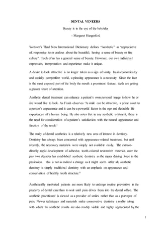
  • 10. 10 Tetracycline Discolouration. Labial reductionof 0.5 to0.57mm. Incisal reductionof 1.5mmwith lingual chamfer. ClassV type cavitypreparation done instaineddentin. Opacificationof dentin Opacificationof dentin Immediately after insertion of veneers.
  • 11. 11 2. Fluorosis Discoloration - This staining is difficult to remove with bleaching, but it can be bleached in cases of slight to moderate staining. Effective management of multicoloured or darkly fluorosed teeth can be accomplished best with veneers.13 3. Teeth Darkened by Age - A well-known phenomenon is the darkening of teeth observed as patient’s age. The darkening is caused by pigmented foods, coffee, smoking, and so forth. Often, teeth are sound and do not need restorations or crowns but they are esthetically objectionable. Such teeth are ideal candidates for veneering. The teeth retain their basic integrity and shape and are not altered significantly except for a colour change. They become smooth, and the discolorations caused by age are lightened. 13
  • 12. 12 4. Irregular Tooth Positioning in the Arch. Teeth that are slightly rotated or in lingual or labial version can receive the appearance of straightening easily by veneering them. Often these teeth also contain numerous discoloured restorations because of the patient’s inability to clean them well. The unsightly restorations are also hidden by the veneers. 13 5. Malformed Teeth - Infrequently patients have one or more teeth that were malformed by trauma during tooth development, metabolic disturbances of tooth development caused by systemic disease, or other reasons. These teeth are ideal candidates for veneers. Their colour is usually acceptable, and they require only removal of deep pigmentation and veneering of the tooth with a resin matching the tooth colour. 13
  • 13. 13 6. Teeth Discolored by Endodontic procedures. Slightly discolored non-vital sound teeth, pigmented during or subsequent to endodontic procedures, can be covered successfully by properly accomplished veneers. This task is a difficult one from a color standpoint, but with experience it can be done effectively. 7. Teeth with Numerous Visible Cracks - Athletics, accidental trauma, ice chewing, and other abuse to teeth cause cracks in the tooth enamel that may also become stained from foods. Veneers can cover these cracks easily and eliminate future staining in them. 13 8. Teeth with Numerous Unsightly Restorations. Occasionally, bodily discolored restorations and those with stained margins are restored best by covering their entire facial surface with veneers.
  • 14. 14 9. Teeth Denuded of Superficial Structure by Acid Demineralization. Many people who have gastric disorders or those who regurgitate often because of illness or by choice have lost significant superficial tooth structure. These teeth are sensitive to cold, hot, and sweet foods. Veneers satisfy both the sensitivity and the esthetic problems.4 Contraindications for Veneers 1. Heavy Occlusion - Persons having extremely heavy occlusion exert forces on veneers that can break them from the surfaces of enamel or dentin. Therefore, persons exhibiting signs and symptoms of clenching or bruxism are poor candidates for veneers. Bruxers who have worn incisal guidance and canine rise guidance excessively break off veneers easily. Other treatment should be used including metal occlusal surfaces or metal crowns with heat or light cured microfill veneers over the metal. Porcelain fused-to-metal restorations may be contraindicated in patients with bruxism. 4 2. Teeth in Severe Labial Version - Veneering “buck teeth” has limited esthetic potential. Removal of sufficient enamel to allow acceptable colour often eliminates enamel and forces the dentist to make retention in dentin. This is less successful than enamel retention. Also, the extreme facial placement of the veneers often increases the “buck tooth” look, Orthodontic therapy and/or crowns are better treatment for this type of patient. 5 3. Mouth Breathers - Such patients have irritated gingiva most of the time. Their dry mouth environment encourages thick mucous collection around the anterior teeth and subsequently greater gingival irritation when veneers are placed. Correction of mouth-breathing is suggested before any comprehensive restorative therapy is initiated. 4. Poor Oral Hygiene - Gingival health is deteriorated around some types of veneers. Placing veneers in persons with poor oral hygiene can create continuous and increased periodontal breakdown. Patients often have poor oral hygiene because they
  • 15. 15 dislike their teeth and avoid cleaning them in a subconscious rejection of their esthetic condition. A trial period to demonstrate improvement of oral hygiene is indicated before placing veneers. Usually hygiene improves after placement of the veneers. 4 5. Denuded Dentin. Dentin adhesion agents are currently available. However, it is difficult to obtain optimal bonding to large areas of tooth structure that have only dentin present. Crowns are indicated in these situations. 6. Highly Fluoridated Teeth. Although fluoridated teeth can be veneered successfully, longer etching and meticulous cleanliness are required to afford optimal retention. Etching for 2 minutes is suggested for highly fluorosed teeth. The patient should be advised of potential veneer retention problems with highly fluoridated teeth.4 Economic Considerations An advantage of veneers is a lower cost to the patient than crowns. At this time, veneers are becoming a routine part of dental practice, and fees for this service are being stabilized. This economic advantage allows patients to have one or two replacements of the veneers before the total cost equals that of a single crown. Of course, the patient must endure the trauma of additional treatment when the veneers are replaced. Assuming that the current generation of porcelain fused-to-metal crowns will serve esthetically for approximately 10 years and that the current generation of microfill resin veneers will serve for approximately five years, properly placed veneers are economically feasible for both the patient and the dentist.4
  • 16. 16 CLASSIFICATION A. Depending on coverage: Partial veneers Full Veneers Full veneer with “window” preparation design that extends to the gingival crest and terminate, the facio-incisal angle.
  • 17. 17 Full veneer with incisal lapping preparation design extending sub-gingivally that includes the entire incisal surface. B. Depending on technique of fabrication 1) Directly fabricated (a) Partial veneers (b) Full veneers. 2) Indirectly fabricated (a) Partial veneers (b) Full veneers. C. Depending on cutting of enamel 1) Intra-enamel preparation 2) Extra-enamel preparation Intra-enamel preparation- To achieve ideal aesthetic and physiologically sound results, this preparation is almost always indicated. Exception to this is when labial surface is
  • 18. 18 under contoured due to severe aberration or erosion. In such cases only the margins are defined. The objective of roughening and defining the margin are to- - provide space for opaquer, without over contouring. - remove the outer fluoride rich layer which is resistant to acid etching. - rough surface increases surface area, increasing bond surface. - is of importance in case of indirectly placed veneers. Extra-enamel preparation- Veneer is placed on the entire existing tooth structure without any tooth preparation. The advantage is that there is no tissue destruction and the complete procedure is reversible in case of failure. Disadvantage of this technique is over contouring, decreased bond strength. 4 D. Depending on material 1) Acrylic resin 2) Composite resin 3) Porcelain 4) Castable ceramics (Dicor) BENEFITS AND SHORT COMINGS Benefits – This treatment consists of replacing the visible portion of the dental enamel with an artificial substitute. This ‘substitute enamel’ helps to replace defective human enamel
  • 19. 19 with bonded artificial enamel.15 Minimally invasive treatment – This treatment method utilizes minimal tooth preparation, mainly confined within the dental enamel. It preserves tissue integrity. We all agree that the natural human enamel would be the best restorative material to use, if that were possible - but porcelain laminate veneers, being the closest material to natural human enamel, are the least destructive to the adjoining tissues. These characteristics have established the porcelain veneers as the preferred restorative material preserving the enamel, which is-ultimately the most valuable part of the tooth. Numerous authors have suggested that the minimal thickness for tooth preparation for ceramic veneers should be no more than 0.5 mm. In practice: however, the actual approximate thickness of a porcelain laminate veneer is 0.4 to 0.7 mm, which closely resembles that of the natural tooth enamel. 5 Shape, Position and Surface Appearance- The shape or position of natural teeth can be affected with functional or esthetic problems. The major advantages of veneers is that the surface texture can be transformed permanently and elegantly, eliminating any dysplasia or dystrophy of the enamel.16
  • 20. 20 Colour – The smooth surface texture and natural colour of porcelain are exceptional, and the crystalline structure of porcelain gives it optical refractive properties similar to those of translucent enamel. When bleaching techniques become ineffective, veneers may the treatment of choice for improving or changing natural tooth colour. 5 Durability- Veneers stand up extremely well to biological, chemical and mechanical action.4 Light Transmission- Using dentin porcelain of varying chromas, as well as transparent, translucent or opalescent (but not opaque) porcelain, it is possible to achieve moderately thick build -ups by a layering technique or lateral segmentation that reproduce all the characteristics of natural enamel such as cracks, fissures and opalescence.13 Illumination of natural tooth. Illumination of all ceramic crown.
  • 21. 21 Tissue Response – Extremely good biocompatibility with gingival tissues. The highly glazed porcelain surface is less of a depository area for plaque accumulation as compared to any other veneer system. Perhaps one of the most important advantages of porcelain laminate veneer is that the periodontal reaction is minimal. The smoothly finished margins help to maintain good periodontal health and oral hygiene. Various studies have been performed. One of them established that the porcelain-fused-to-metal tissue response is similar to that of the veneers; yet, another found that there was considerably less inflammation with porcelain veneers than with porcelain-fused to-metal (PFM) restorations. In another particular study, Kourkouata et al. found that after the porcelain veneer restoration was completed, there was an increase in crevicular fluid; similarly, there was a significant decrease in plaque index, and plaque bacteria vitality. When selecting a treatment, longevity is a major concern. A more invasive procedure will result in a higher risk to the tooth and ultimately a more expensive treatment. 17 Speed and Simplicity- Laminate veneer treatment procedures are usually carried out under light anesthetic, with minimal tooth reduction, and at greater speed than other techniques. Impressions are easier to make, because of the accessible position of the margins. - Patients appreciate being able to remodel their smile very rapidly. - Highly acceptable tensile bond strength. The resin to silane treated etched porcelain veneer has bond strengths ranging from 2600 to 3200 pounds per square inch (psi) and composite resin veneer to enamel bond strengths of 900 to 1400 psi. - Porcelain can be used to increase the length of given tooth by extending it over the incisal edge. - Long lasting. Once bonded, porcelain veneers develop high tensile and shear strengths. - Exceptional resistance to wear and abrasion.
  • 22. 22 - Resistance to stains. - Surface lustre retention. 15 State of the Art – The dental profession has always been in search of restorations that have a "natural" appearance but are, at the same time, long lasting. In the past, the lack of appropriate dental materials rendered this impossible. However, advances in porcelain veneers and bonding materials now offer a variety of selections in treatment, with long-lasting restorations and improved quality in aesthetic results. Porcelain veneers are to be considered the "state of the art" in aesthetic dentistry, providing abundant advantages over any previous form of veneer system. They are excellent in terms of their appearance and durability, and the actual procedure does not disturb the soft tissue or the adjoining periodontium. Since their introduction almost 20 years ago, porcelain veneers have become the flagship of most aesthetics-based practices. 5 Strength- Owing to their ceramic thinness (0.3-0.5mm), the porcelain veneers can be easily fractured before they are even bonded, similar to kitchen or bathroom tiles. However, once bonded on a solid foundation, they integrate with the tooth structure and become extremely durable, in vitro and in vivo. If all conditions have been adhered to and the appropriate bonding composite has been selected, the dentist will achieve successful results. The patient's satisfaction is bound to be high as this medium ensures long- term maintenance, good gingival health and excellent oral hygiene. When it was proven that function and esthetics could be achieved with porcelain laminate veneers and without the removal of large amounts of intact tooth structure, porcelain veneers became the restorative material of choice in addressing the most challenging aspects of aesthetic dentistry by integrating the individual restorations into the adjacent natural dentition. 5
  • 23. 23 Longevity – The union of etched enamel and porcelain, combined with the bonding composite resin-luting agent with a silane coupling agent, enables the dentist to perform restorations that are solid as well as long lasting. The long-term data is encouraging and reports a high success rate for this form of treatment. Materials used in the 1980s are now considered primitive, whereas the restorations made with laminate veneers even 15 years ago have shown a high rate of success, increasing interest and widespread acceptance. Porcelain laminate veneers are continually gaining respect as the best and most durable restorative treatment, while patients and dentists alike are delighted with the esthetic results and the minimal tooth preparation required. As aesthetic dentists are becoming increasingly confident of its application to a variety of clinical cases, the survival rate of laminate veneers reported in literature ranges any- where from 18 months to 15 years. Friedman reports in another study that of the 3,500 porcelain veneers that were placed over a 15-year period, 93% of the cases were successful. 5 Shortcomings – Porcelain veneers can be easily repaired once bonded to the enamel, but the repairs are not long lasting due to staining which tends to occur at the margin of composite resin and porcelain. -The colour cannot be easily modified once bonded in position. -Irreversibility of preparation versus little or no preparation for composite resin bonding. -Level of difficulty of fabrication and placement, time involved, and expense. -The extremely fragile veneers are difficult for the dental laboratory to make and manipulate, and the process requires two appointments, and laboratory fees. -Technical difficulties in avoiding over contours and obtaining closely-fitted porcelain enamel margins. The margins can be especially brittle and difficult to finish. -Lower reparability compared to composite veneers.
  • 24. 24 -Susceptibility to pitting by certain topical fluoride treatments. stannous fluoride should not be used with porcelain restorations. At the bonding stage, the slightest error can mean failure- either immediately or later on. This is a major disadvantage of laminate veneers. -Handling of such fine ceramics requires very special precautions. Prior to bonding, they are extremely fragile, and the slightest irregularity could produce fracturing. -Techniques are becoming simpler, especially with the progress made in phosphate investment, but the creation of fine natural ceramic veneer restorations remains a difficult and highly skilled task, the two main drawbacks being: - Stability during different types of handling. - Difficulty of getting right balance of powders for layering or segmental build up with a thickness ranging from 0.3 to 0.7 mm. -There is practically no possibility of touching up feldspatic porcelain veneers processed over investment or even using platinum foil, because the ceramic cannot be re-fired once removed from its support. -Tooth Sensitivity. - Marginal gingivitis. 15
  • 25. 25 CASE SELECTION Patients should be advised of the various treatment options available when they have malformed, discolored, or otherwise esthetically objectionable teeth. These options are; (1) to bleach; (2) to veneer with one of the available materials; (3) to place crowns or, (4) to leave the teeth as they are. 18 Veneering is a good alternative in most cases. Also, patients should be instructed that they are expected to maintain their veneers properly for optimal aesthetics and longevity. Color Selection – Most shade guides are not as useful as the manufacturers intended. Many variables influence the color of the underlying tooth and the resin veneers including thickness of the resin to be placed; moisture in the tooth structure; translucence of the underlying tooth structure; and location of the resin on the tooth in an incisal-gingival direction. One of the adequate ways to determine the best color of resin from the available materials follows: Place a small amount of the potential resin colours on the un- etched, damp tooth structure and polymerize them. Selection of the ideal colour is then simple. Many resins darken when they are polymerized, whereas some lighten in hue during polymerization. Only a few brands have the same color before and after polymerization. It is suggested that the color selected be written in the patient’s record and also on an instruction sheet given to the patient for future reference and repair. When veneering medium dark hues, it is often desirable to use one of the medium opaque materials rather than a translucent hue. The medium opaque hue (for example, Durafill GO, or Silux GO) will allow some of the original darkened hue to show through the veneer. A slight degree of this “show through” allows the veneers to assume a much lightened hue but gives them some of the original color to blend with the remaining un- veneered teeth.
  • 26. 26 A technique promoted by some manufacturers suggests placement of a small amount of stains giving the veneer a different shade and improved smoothness. Such thin materials are adequate for a short time but soon wear and leave the original shade and surface characteristics of the underlying resin. It is suggested that the proper shade be selected for the entire veneer and that this material be finished to optimal smoothness, thereby eliminating the need for a superficial layer of material. 19 Procedures – The control, as well as the periodontal, condition of the teeth to be overlaid must be assessed. A healthy periodontium is imperative for successful results. 20 Examining occlusion The relative fragility of ceramic veneer restorations require an accurate analysis of the patient’s occlusion, to ensure that the restorations are not extended into areas of occlusal stress. The result of this analysis may limit the opportunities for remodeling. There is no point in aiming to increase the incisor grouping height, for example, without dealing with canine malocclusion when present. Any attempt to establish a different height clinically, in cases of attrition or bruxism, carries its share of risks and should be approached with caution. Occlusal factors should be considered even with veneer restorations that do not involve the lingual surfaces (i.e. the incisal edge is preserved). In fact, it is in this situation that the risks are greater, especially when canines and posterior teeth are involved. 13 Examining a single tooth – Once again, shape, position, available enamel and occlusion are all factors to be considered. Tooth that is outstandingly triangular or every slender raises difficulty, which will all have to be appraised and dealt with accordingly. 15
  • 27. 27 Normal: - Line angles - Cervical convexity - Vertical lines or ridges - Horizontal lines or ridges Widening: - Displace the line angles laterally - Flatten facial outline - Highlight texture and gloss with horizontal lines and ridges -Decrease facial embrasures Narrowing: - Displace line angles medially - Increase the convexity of the facial outline Shortening: - Emphasize the prominence of the cervical convexity -Displace the cervical convexity coronally - Highlight texture and gloss with horizontal lines and ridges -Emphasize the cement –enamel junction Lengthening: - Flatten and displace the cervical convexity apically - Highlight texture and gloss of the line angles and create vertical ridges - Lighten the cervical aspect55
  • 28. 28
  • 29. 29 Examining the gingival tissue – Although the margins of laminate veneers are often located away from the gingival, the condition of the latter must always be appraised before deciding on any treatment. Poor dental hygiene, gingival inflammation as well as one or more gingival recession sites should all be treated before applying ceramic laminate veneers. Appraising the smile – Clinical examination should focus not just on the teeth to be restored (their colour, shape, etc.) but also on the shape of the face, size of the lip, and the lip-to-tooth relationship during various movements. All these inspections should be carried out both frontally and from the side. The practitioner must also use other methods of visualization, such as the following, 1. Diagnostic wax-up 2. Cast models 3. Computer imaging analysis. A complete upper and lower impression is made before preparing the teeth, followed by mounting the models on an articulator and making any corrections desired, such as lengthening or closing up diastema using either light-cured resin or wax. 15
  • 30. 30 The smile must be viewed from the front and sides (both left and right)  Shape of the face  Size of the lips  Visible coronal and gingival levels - At rest When talking Broadest smile  Harmony and proportion - Of the cervical line Of the line of the incisal edges Of the lip line  Tooth colour - Hue Value Chroma Translucency Texture and luster  Tooth shape - Size of tooth (height : width ratio) Incisal edge Contour Assessing triangular tooth shape Analysis of statistic and dynamic occlusion Spatial arrangement of teeth
  • 31. 31 BONDED RESTORATION IN OCCLUSION AND FUNCTION Occlusion – Occlusion refers to the act of closure and the state of being closed. It involves at the same time dynamic and static components. Static components will be first considered. 22 Static Occlusion – In a good occlusion, all the teeth in the mouth make simultaneous contacts. It is, however, acknowledged that in the anterior segment teeth should have lighter contacts, which clinically may mean: "anterior teeth do just not contact". Indeed, the perception of a fremitus at interocclusal contact can be detrimental to tooth stability and lead to interproximal separation.23 The total tooth contact area, which includes the anterior and posterior teeth, averages 4 mm. Considering the number of interocclusal contacts (64 to 100 for the whole mouth), taking into account varying opinion and the type of occlusion, each single contact area is estimated to be extremely small. The multiplication of posterior interocclusal contacts is mandatory for improving stress distribution, as studies have shown that in the posterior segment, muscle contraction develops whether you have single or multiple contacts. In nature, cusp to ridge contacts are the rule and characterize class I occlusions. Cusp to fossa contacts are usually found in class II occlusions, while the tripodization of interocclusal contacts remains a dream, born from the imagination of dentists anxious to provide tooth stability. A coincidental location of tooth interocclusal contact and porcelain-bonded restoration margin should be avoided, as alterations of the marginal seal of porcelain- bonded restoration can be more often stated than when the opposing contact is located elsewhere. 24 Whenever this recommendation finds easy application in the anterior segment, clinical practice shows that the posterior segment appears as a privileged area where interocclusal contacts and porcelain-bonded restoration margins are coincidental by reason of posterior tooth morphology and the nature of interocclusal relationships.
  • 32. 32 Observation shows also that it is in this area that the morphological design of the restoration is distorted to adapt to antagonist deformities, or inserted in a dental segment presenting various manifestations of abrasion. As a result, the possibilities of coincidental location of margin placement and enlarged interocclusal contacts increase. And with them the chances of altering the marginal seal resulting from porcelain microfractures under the load of occlusion. 25 It can be assumed that further deteriorations of the marginal seal of porcelain-bonded restoration can take place under the impact of parafunctional habits such as clenching, when margin placement and interocclusal contacts are coincidental. This stresses the importance of tooth preparation for porcelain-bonded restorations that has not only to foresee interocclusal contact location but provide sufficient porcelain thickness to allow porcelain-bonded restoration to offer adequate resistance to constraining parafunc- tional conditions. Functional Occlusion – Function takes place at the very moment teeth enter into action. This dynamic part of occlusion refers to the engagement of mandibular teeth towards centric, and to their disengagement. The spatial development of functional movements is dictated by condylar pathway, antagonist tooth morphology and tooth position. It displays at the registration a three-dimensional tracing delimiting an area defined as "the envelope of mandibular function" characterized by a droplet shape displaying, as a result of antagonist tooth morphology and position, infinite individual variations. 26 This makes teeth the primary factors in tooth-closing patterns. It has been established that tooth-closing patterns remain under neuro-muscular control - even if the exact nature of this phenomenon has always escaped scientific explanation. If we know most of the parameters that participate in function, the observation of unworn long lasting dentitions clearly indicates the type of oral architecture that best integrates these parameters. These occlusions are characterized by similar parameters in the vertical and horizontal planes: that is to say in the overbite and overjet of incisors and canines. Frontal chewing patterns have an angle of about 70° from the horizontal for the canines and in eccentric occlusion we find a tooth-to-tooth contact with a large posterior clearance. This tooth-to-tooth contact in eccentric occlusions
  • 33. 33 can be considered as ideal, as it generates, according to research, a reduced muscular activity in comparison to group function and bi-balanced occlusion.27 In these dentitions, morphologic integrity is the rule. The teeth can receive porcelain- bonded restorations without any specific restriction, but in respect of the environmental morphologic design: function follows forms. Functional integration will then be achieved by the control of simultaneous interocclusal contacts and the absence of interferences during tooth closure patterns. In nature, however, oral architectural ideals are not the rule. Research has shown that in a selected population the occurrence rate of malocclusion, which includes open- bite, over-bite, class II and class III occlusions, averages 20%. As a result, one in every five patients does not present an incisal pathway reflecting usual standards, which leads them to consider correction by way of orthodontics or orthopedic surgery. It also appears that out of the remaining 80% of class I occlusions; most occlusal schemes do not show anterior relationships, duplicating those displayed in long-lasting dentitions. Variations in the depth of the overbite, width of the overjet, and related incisal and canine guidance are unlimited, which justifies or imposes corrective measures necessary to prevent posterior interferences during function. 28 By deduction, it can be advanced that one of the challenges faced by restorative dentists in restoring a physiological occlusion is the changing of poorly related anterior relationships. The restoration of an anterior segment should never be envisaged from the viewpoint of minimal tooth preparation for veneer seating but from that of occlusal maintenance and functional excellence. 29 Occlusal maintenance involves the evaluation and correction of localized manifestations of wear affecting one anterior tooth or the whole anterior segment, concomitant with an appreciation of the degree of incisal or canine guidance necessary to prevent posterior interferences in eccentric occlusions. The achievement of these objectives requires an increase of the steepness of the disclusive angle which, depending on oral conditions, can be achieved either with an increase of vertical interocclusal relationships by way of a tooth elongation or with a reduction of horizontal relationships. A combination of these therapeutic measures is not unusual. Clinically, a modification of the palatal morphology of maxillary incisors and canines
  • 34. 34 appears best suited to improve tooth guidance without compromising aesthetics .The nature of this modification remains to be defined as long as it leaves open the question on of the ideal steepness of the interocclusal angle. Mechanically, it appears that the deeper the angle the greater the likelihood of achieving tooth guidance and the greater the impossibility for posterior teeth contacting in eccentric occlusions. From a muscular point of view, the increase of the steepness of the disclusive angle may also produce a favourable result as it generates a proprioceptive inhibition that will shut down muscular activity thus reducing the load level in eccentric occlusions. This muscle inhibition has been explained by an increase of the torsional load affecting the teeth in their housing. This leads to a triggering effect on proprioceptive receptors preventing muscles contacting harder through a feedback effect. However, careful attention must be paid, so that the increase in the disclusive angle does not cause the teeth, through their morphology or position, to interfere with neurological programmed tooth closure patterns. This will invariably lead to the abrasion of antagonist anterior tooth surfaces, mandibular buccal or maxillary lingual surfaces. Damage of this type affecting antagonist surfaces is mainly generated by overcontoured porcelain bonded restorations but may also be found in orthodontic and orthognathic cases. In the absence-of available clinical possibilities of tracing three-dimensional, programmed tooth closure patterns for want of an articulator programmed to register this information, it is left to the dentist's sensibility to prevent restorations impinging on the area of function. Despite these restrictions, there is no better restorative material today than porcelain-bonded restoration to achieve functional and aesthetic excellence or to duplicate the conditions found in long-lasting dentitions. All that remains is to apply the appropriate treatment at an appropriate time. Incising – Mastication starts with the incision of food, which is followed by a lateral chewing phase. This lateral chewing phase can be divided in an early tooth closing phase and a late tooth-closing phase. Incising requires an initial mouth-opening necessary to introduce food followed by a closing movement of the mandible, which develops in the sagittal plane and places upper and lower incisors in a tip-to-tip position. This
  • 35. 35 movement seems to load incisors along their long axis and bring both condyles in a forward and downward translation. With unworn incisors there is no more than 1 mm of shearing load confined in the incisal edge of teeth. This shearing load will increase dramatically in the presence of worn-out incisal edges. As a result, the tip-to-tip incisal biting will gradually become a surface-to-surface biting, overloading incisors away from their long axis, allowing for the posterior confinement of condyles leading in turn to adverse neuro-muscular response. Finally, the incisal edges of the lower incisors will glide back along the lingual wall of maxillary incisors, a movement which develops atraumatically under neuromuscular control. Yet the presence of an increased incisal tip width affecting lower incisors will defefinitely overload the anterior teeth forcing lingually mandibular incisors.17 It is therefore essential that porcelain-bonded restorations duplicate in their incisal design sharp unworn natural teeth and present in their whole configuration the most adequate compromise in terms of internal stress distribution and stress transfer following axial or horizontal loading. These mechanical requirements have to be considered when the presence of interproximal carious lesions, composite restorations, or tooth fractures impose a lingual extension of anterior maxillary porcelain-bonded restorations. In the same way, the palatal extension of porcelain - bonded restorations in the design of maxillary incisors and canines becomes unavoidable when functional finality demands:  An improvement of anterior guidance to correct insufficient horizontal or vertical interocclusal relationships. The design of cingulum and lateral palatal ridges to create punctiform centric interocclusal contacts and to prevent the eruption of lingually positioned antagonist lower incisors. To optimize the configuration of porcelain bonded restorations one has to know that the area of the cingulum presents the lowest tensile strength of the whole palatal side at a tangential blow of 50 N directed against the incisal edge.32 It can therefore be considered as an ideal location of porcelain-bonded restoration margin. Above all, a marginal location of porcelain-bonded restoration in this area of low tensile strength reduces the importance of the design of the finish line: a butt margin cutting the enamel rods at an angle superior to 50° is perfectly acceptable while a long chamfer
  • 36. 36 will reveal particularly resistant in as much it will cut the rods at 90°, allowing for an improved adhesion to enamel. Even if studies have shown that this type of porcelain- bonded restoration configuration presents the lowest relative compliance, knowing that resilience is a key factor in fracture resistance, fracture rate remains extremely low in clinical practice, maybe thanks to dentin bonds that have been proven to be a main determinant in the fracture mechanic. As previously mentioned, the palatal extension of porcelain-bonded restoration is the rule each time functional conditions advocate an increase of anterior guidance to allow for sufficient posterior interocclusal clearance in eccentric occlusions.30 The functional indication for palatal extension varnishes in functionally good occlusions where tissue preservation predominates. A limited incisal palatal overlap remains the most valuable configuration as long as it is located in an area displaying low tensile strength, allowing for a finish line design that can be either a butt joint or chamfer. These porcelain-bonded restoration configurations, palatal overlap or palatal coverage present the qualities to perfectly resist the incising function or the tearing apart of food. Only a palatal extension of the porcelain-bonded restoration with a finish line ending up in the palatal concavity has to be avoided, as this thin extension of ceramic is precisely located in an area of high tensile strength. 27 Normal function not only implies the incision of food but the chewing of food. Lateral Chewing – Lateral chewing starts with mouth opening, followed by a lateral movement of the mandible to the working or chewing side. The working condyle is brought in a retruded lateral position while the non working condyles move medially. The amount of this lateral shift varies, depending upon bone configuration of condyle and fossa, shape and conditions of disc and ligaments. 19 The shifting of the working condyle or Bennet shift affects the mediolateral incline of the posterior cusps and conditions the direction of the buccal groove of lower molars whenever the occlusal scheme presents in eccentric occlusions a group function or a balanced occlusion.
  • 37. 37 In functionally good occlusal schemes, however, the importance of the Bennet shift is irrelevant. In the early closing phase of mastication, which sees both working canines starting to proprioceptively engage the closing movement, the importance of the mediolateral inclination of posterior cusps is irrelevant. 31 In the final closing phase, however, the approach to centric of opposing molar cusps can lead to interferences because the vertical guidance of the canine does not totally negate the path of the shifting condyle. As a result, interferences can occur in the mediolateral inclines of the second and first molars, especially when the condyle- fossa complex generates in the horizontal plane a shifting in lateroprotrusion or a shifting in laterosurtrusion in the frontal plane. The wear resulting from these interferences affects the mediolateral inclines of mandibular or maxillary molars predominantly in areas characterized by a cusp to fossa relationship such as the central groove of maxillary and mandibular molars. This is best visualized in natural unworn dentitions. In good occlusal schemes this occlusal wear is irrelevant but it may reach a pathological level with a decrease of the anterior interocclusal angle. Under these conditions the introduction of a restoration in these areas may reveal itself detrimental to marginal material integrity as interocclusal contacts and margin location are often coincidental. The integrity of the porcelain- bonded restoration can even be compromised when mesial or distal transitional zones of tooth preparation present a too-abrupt angulation. 27 Clinical observation shows that porcelain bonded restoration fracture rate increases precisely in the mesial part of maxillary molars and in the distal part of mandibular molars in occlusal schemes showing poor canine guidance. These elements need a configuration of posterior porcelain-bonded restoration with sufficient material thickness and suggest coverage of mediolateral cusps designed with a reduction of the inclination of the mediolateral slopes and a reduction of the MO or DO transitional angle. Dysfunction – The observation of unworn long-lasting dentitions has put in evidence harmonious relationships between skeletal and dental morphology and integrated function. It has long been speculated that abnormal function is mainly caused by abnormal tooth
  • 38. 38 guidance. In turn, abnormal tooth guidance results from abnormal occlusal contact relationships. 32 We know that one out of five patients do not have an incisal pathway reflecting an appropriate standard. Yet we can observe daily that out of the remaining patients reflecting appropriate standards in anterior pathways, few show or will show over the years an occlusal scheme free of wearing manifestations. It can therefore be deduced from these observations that even appropriate standards in anterior pathways can display abnormal occlusal guidance. 19 Submitted to registration these elements of abnormal guidance have been classified in a recent study in lack of guidance, poor guidance, over guidance, abnormal guidance, and interference. The presence of interferences in the development of tooth closure patterns has long been identified as the prime element of dysfunction. It has been even assumed that it could be at the origin of bruxism. There are two basic types of interferences:  Centric relation: anterior slide, lateral slide, protrusive-retrusive  Lateral border: non working and working side. The elimination of centric interferences in the treatment of bruxism has always been controversial. In 1928, Tischter recommended occlusal adjustment in order to eliminate the so called "trigger contacts". A large number of authors stress also the importance of eliminating these interferences in the treatment of bruxism, while others deny any relationship between interferences and bruxism. 33 It appears, however, that clinically experienced practitioners never hesitate to proceed to occlusal correction before restorative procedure by reason of the good results they obtained. Dawson (1998) has written: "It is my experience that, when all occlusal interferences in centric relation and excursions have been eliminated on the posterior teeth, parafunctional movements resulting in anterior wear are eliminated or reduced to a point where they are of no clinical concern." His statement gives the restorative den- tist the best reasons to proceed to occlusal controls, not only before restorative procedures but also following porcelain-bonded restoration placement, to eliminate
  • 39. 39 the interferences that were either present or could have been introduced with new restorations. 34 This post bonding control is made necessary because of the great difficulty in appreciating before bonding porcelain-bonded restorations, the occlusal parameters because of the risks of fracture. Aside from interocclusal overcontacts in centric occlusion, centric interferences - lateral or anterior - can be found on posterior porcelain bonded restorations, while protrusive retrusive interferences are systematically present on anterior porcelain-bonded restorations, especially those showing a configuration with a palatal extension. Clinical observation shows, however, that contrary to Dawson's statement, parafunctional habits can then be reduced to a point where they are of no clinical concern when both centric interferences have been eliminated and anterior guidance steepness has been concomitantly increased. 27 Indeed, nothing else but this increase of anterior guidance steepness can prevent possibilities of posterior eccentric interferences that may take place in the area of mandibular motion, clinically undetectable but made possible by increased muscle and ligament laxness, pillow or hang pressure. These interferences also can be the initiator of parafunctional movements.28 Occlusal schemes showing moderate but apparently sufficient posterior clearance in eccentric occlusions must be looked at with suspicious eyes, especially those presenting deficiencies in tooth alignment. They open the possibility of unexpected eccentric contacts generator of avoidance patterns leading in an initial phase to the erosion of the elements of the anterior segment. 19 The rotation of the mandible around a posterior obstacle followed by a mandibular projection against the maxillary incisors has been observed in clinical tests, and the follow-up of muscles participation clearly described. It has even been demonstrated that parafunctional avoidance patterns could not only project the mandible against maxillary incisors but be moved outside the confine of the maxilla that is to say in a cross-over position. 35
  • 40. 40 This uncontrolled back and forth movement will progressively erase antagonist anterior teeth whose progressive morphological reduction will finally permit posterior contact, and posterior wear faceting. As a consequence, the set-up of veneers, that is to say porcelain-bonded restoration, covering the facial part of the tooth and provided with a reduced palatal overlap, is restricted to occlusal schemes showing a total absence of faceting. The presence of wear faceting in the occlusal scheme (it could be a simple chipping, a localized or generalized chevron-shape anterior wear), requires a modification of veneer con- figuration with the design of either an elongated porcelain-bonded restoration or a porcelain bonded restoration with a palatal extension, provided an increase of anterior guidance can be achieved to prevent any possibility of posterior interference in eccentric occlusions. 36 The predictability of any restorative process rests on the precise evaluation of oral and occlusal conditions. Porcelain veneer restorations can be considered as ideal restorations for the corrections of poor anterior relationships and anterior-guidance. Conversely, they can also be destructive for the dentition when occlusal conditions have been neglected or wrongly appraised. 5 PERIODONTALCONSIDERATIONSIN AESTHETICTREATMENTPLANNING Aesthetic dental treatment is not limited to the technical skills and aesthetic perspectives of the dentist and the dental technician; the patient's perceptions and his or her understanding of the problems and solutions are also important. 37 Patients should be instructed that aesthetic dental rehabilitation is a complex and complicated form of treatment and that she or he is a-party to that treatment not merely a recipient of it. The patient needs to see treatment as a collaborative effort in particular because oral hygiene habits and periodontal status dictate the outcome and longevity of the aesthetic result. The rehabilitation of a smile relies not only on restorative dental procedures, but also on a healthy and aesthetic gingival appearance. 38
  • 41. 41 Aesthetic periodontal problems can only be tackled after efficient plaque control and proper initial periodontal, therapy have been addressed. Soft tissue defects may become more apparent as gingival shrinkage occurs after the resolution of the inflammation. The aesthetic outcome of a case is determined by the relationship of the crown with the soft tissues, whether it is a natural tooth, a pontic, or an implant. A sound knowledge of the normal anatomy of the dento-gingival unit may initially prevent violation of the basic and then one can understand the point where physiological tolerance may shift to inflammatory reaction. 13 Periodontal Tissues in Health – The Dentogingival Unit represents a unique anatomical feature concerned with attachment of gingiva to the tooth. It is composed of epithelial and connective tissue compartments. Three types of anatomically distinct epithelia are associated with the dentogingival unit. The first, gingival epithelium, consisting of stratified squamous keratinized epithelium which is continuous at the gingival crest with the second type, stratified non keratinized or parakeratinized epithelium. 20 Sulcular epithelium relines the soft tissue wall of the gingival sulcus facing the tooth but not attaching to it and is continuous at the base of the sulcus with the junctional epithelium, or the epithelial attachment. The lack of keratinization of the sulcular epithelium is the result of inflammation in its supporting connective tissue. Junctional epithelium forms a collar around the tooth and the surface cells provide the actual attachment of gingiva to the tooth surface. In a study of autopsy material by Gargiulo et al. it was determined that the average sulcus depth was 0.69 mm, and the average length of the junctional epithelium was 0.97 mm. A significant finding in that study was that the average depth of gingival sulcus was highly variable from tooth to tooth and around the same tooth.39 Connective tissue compartment of the dentogingival unit presents fibrous attachment of the gingiva to the hard tissue wall and support to the epithelium of the dentogingival junction. Fibrous connective tissue attachment to the root surface is provided by a gingival collagen fiber system that also maintains the functional
  • 42. 42 integrity of the teeth and its gingival collar. Gargiulo et al. reported that the average length of the supracrestal periodontal fiber group was 1.07 mm and was constant at all locations around the teeth. The most important groups of fiber bundles that compose this gingival unit are dentogingival, alveologingival, dentoperiosteal, transseptal, circular/semicircular, and interpapillary fiber bundles. 40 The autopsy material reported by Gargiulo et al. showed that the average length of the junctional epithelium was 0.97 mm, supracrestal collagen fiber group was 1.07 mm, and the average sulcus depth was 0.69 mm. Of these three tissue components the supracrestal connective fibrous attachment seems to exhibit the least variability, followed by the junctional epithelium and sulcus depth. In 1962, Cohen defined the "biologic width" of supracrestal gingival tissue as those junctional epithelium and connective tissue elements of the dentogingival unit that occupy the space between the base of the gingival crevice and the alveolar crest, that the total dimension would be in the vicinity of 2.04 mm. 40 For practical reasons, as suggested by Nevins and Skurow, the "biologic width" can be expressed as the sum of the supracrestal fibers, the junctional epithelium and the gingival sulcus. A better name that would prevent confusion could be "biologic zone" as used by Kois. 21 The minimum dimension of this space between the most coronal level of the alveolar bone to the most coronal level of gingival margin would be in the vicinity of 3 mm in a healthy and normal gingiva. 41 This is the minimum space required for the tissues to maintain their continuity in a healthy status. Aesthetic Periodontal Problems – Gingival Inflammation and tissue loss owing to periodontal disease - Periodontal disease is the result of the accumulation of dental plaque at the gingival margin leading to inflammation of the periodontal tissues. 10 Traditionally, periodontal disease has been divided into gingivitis and periodontitis, depending on whether destruction of the tooth-supporting tissues has occurred. Diagnosis and
  • 43. 43 treatment of periodontal problems are based on clinical findings such as probing pocket depths, attachment loss, gingival bleeding, and radiographic evaluation of the alveolar crest. The objective of periodontal treatment can be summarized as the prevention of tooth loss, maintenance of periodontal support, repair, and regeneration of damaged tissues and their function. But when the etiology of the periodontal disease is considered, the main task seems to be the modification of patient habits in order to constitute an oral hygiene level that would prevent inflammation of periodontal tissues owing to plaque accumulation. 42 Gingival inflammation and associated periodontal destruction are leading factors that can reverse the aesthetic outcome in the long term. 43 Establishing definitive restorations when inflammatory periodontal problems are present not only restrains aesthetic success, but also accelerates the rate of periodontal destruction. Elimination of gingival inflammation is mandatory before initiation of any restorative treatment. Initial periodontal therapy that consists of oral hygiene instruction, elimination of retentive factors, scaling and root planning will result in the resolution of inflam- mation. Patients with moderate to severe periodontitis may require advanced periodontal treatment that could consist of surgical procedures to eliminate residual periodontal pockets and regeneration of the lost periodontal tissues. Pocket elimination surgery usually results in long clinical crowns that may be aesthetically unacceptable. In deep isolated angular periodontal defects, guided tissue regeneration offers good results to maintain the tooth and avoid an aesthetic disaster. Alveolar ridge deformities are inevitable after the extraction of hopeless teeth when special attention was not paid to preserving the height and width of the ridge at the time of extraction. 36 Restorative Margins and Violation of Biological Principles – Preservation of the healthy status of periodontal tissues is the most significant factor in the long term prognosis of a restored tooth. 5 There are five major causes of plaque-related inflammatory periodontal disease associated with restorative procedures: 1. Severe damage to the periodontal tissues during tooth preparation and impression making.
  • 44. 44 2. Failure to maintain emergence profile. 3. Inability to adequately finish and/or seal sub-gingival margins. 4. Placement of sub-gingival margins at sites with minimum to no attached gingival. 5. Violation of the biologic width. Teeth prepared without retraction cord which subsequently receive retraction cord, electrosurgery, or rotary gingival curettage sustain various degrees of soft tissue damage, as reported by Dragoo and Williams. Tooth preparations for restorative procedures should be accomplished atraumatically to avoid irreversible damage to the supracrestal connective tissue fibers and tissue loss. 40 The cervical one-third of the clinical crown is also referred to as the- emergence profile of the tooth. This is the region where the crown emerges from the periodontium and its supra-subgingival contours should have a flat emergence profile, with the guidance of anatomic information in order to maintain an area that is cleanable with oral hygiene instruments and which has a good marginal seal of the restoration .There is a very strict relationship between marginal seal and emergence profile. Poor adaptation and roughness of the restoration margin results in mechanical irritation of the sulcular epithelium and can harbour microbial flora. Overhanging margins not only accumulate more plaque than properly finished margins, but the plaque undergoes a shift in composition to that seen in association with destructive periodontitis. An adequate band of keratinized tissue is fundamental to successful restorative dentistry if the margins of the restorations are extended under free gingival margin. Approximately 5 mm of keratinized tissue, composed of 2 mm of free gingiva and 3 mm of attached gingiva, is considered necessary to meet restorative objectives even in cases with ideal finishing and margin placement at the subgingival area, Although no solid investigation is present to confirm this observa- tion, augmentation of keratinized gingiva provides stability of the free gingival margin and surrounding gingival tissues so that the gingival health around the restoration can be maintained. Besides vertical dimension of the keratinized gingiva, the thickness of the tissue should be enough to tolerate intracrevicular restorations. The gingival sulcus extends from the free gingival margin to the epithelial attachment. In health, its depth is in the vicinity of 0-3 mm, and it is lined with thin sulcular
  • 45. 45 epithelium. The depth of this sulcus may vary from tooth to tooth and from surface to surface on the same tooth. Compromising the apico-coronal width of the connective tissue attachment by placement of crown margins deep in to the sulcus initiates per- sistent and irreversible gingival inflammation owing to violation of the biologic width. Such violations sever the junctional epithelium and the supragingival connective tissue fibers. Apical migration of the junctional epithelium encourages development of periodontal pockets and alveolar bone loss.31 The finish line of restorations should follow the contour of the cemento-enamel junction and be always kept at least 2.5 mm away from the bone crest . A 2.5-mm difference means that although the biologic width is being respected, the finish line is placed equigingivally. A greater distance between the bone crest and finish line of the restoration is needed to ensure that margin of the restoration could be well reached by the plaque control instruments. The golden standard today is to prepare the finish lines of the restorations 0.5 mm below the marginal gingiva if the sulcus depth is 1 mm, 0.5 to1 mm if the sulcus depth is exceeding 1.5 mm. This leaves enough distance for the junctional epithelium and supracrestal fibers, and is also close enough to the gingival margin to be reached by oral hygiene procedures. When the healthy sulcus depth is less than 1 mm, placement of the margin 0.5 mm subgingivaly could result in impinging upon the junctional epithelium. The margin in such cases should be terminated just at or above the gingival margin. 15 Gingival asymmetries and discrepancies – The aesthetic appearance is considerably affected by the symmetry created by dental and facial midline. In the etiology of gingival asymmetries, altered passive eruption, different patterns of tooth wear, traumas that modify tooth eruption, tooth positioning in the dental arch, parafunctional habits and overzealous tooth brushing should be considered. The selection of an adequate method of treatment depends on adequacy of attached gingiva, tooth structure, vestibular depth, the distance from the gingival margin to the crest of the bone, root angulation and tooth positioning and interproximal level of the alveolar bone when recession is present. Correction of this disharmony requires surgical elongation or root coverage, orthodontic treatment and restorative procedures. 8
  • 46. 46 Excessive Gingival Display – "Gummy smile" can be considered as a significant aesthetic problem by many patients. Ideally, the smile should expose minimal gingiva around the lateral incisors. The gingival margins of maxillary central incisors and cuspids should coincide with the vermillion border of the upper lip. The lip line, assessed when the patient is in full smile, is classified as high (gummy smile), medium (ideal), and low (maxillary lip covering a portion of maxillary teeth). Aging leads to a decrease in the amount of anterior tooth display when smiling. Maxillary overgrowth, insufficient upper lip, tooth mal-positioning, and delayed apical migration of the gingival margin or a combination of more than one are among the most common reasons of "gummy smile". 5 Skeletal deformities that involve maxillary overgrowth can be diagnosed by evaluation of the proportions of the face and cephalometric analysis. In such cases the teeth's length-to-width ratio usually remains constant - the ideal being approximately 10:8. The ideally proportioned face is divided into three equal parts from the hairline to the eyebrow, from the eyebrow to the base of nasal process, and from the base of the nose to the chin. When the middle one-third of the face is longer than other two, "gummy smile" is diagnosed owing to maxillary overgrowth. In combined cases that maxillary overgrowth is superimposed with delayed passive eruption and the latter should be treated first. Treatment limited to periodontal surgery will be insufficient, since the level of the incisal edge to lip at rest position cannot be rebuilt. Minor maxillary overgrowth cases can be successfully treated with periodontic-orthodontics or periodontic-restorative combinations. Increase in the gingival and mucosal display necessitates complex treatment options that combine orthognathic-orthodontic proce- dures and perio-prostho treatment. 13 The normal length of the upper lip is between 18 and 21 mm, vertically. A shorter lip over a moderate dental arch could result in excessive gingival display. When the "gummy smile" is solely dependent on insufficient lip dimensions, a perio-restorative solution composed of surgical crown lengthening and restoring the teeth with laminates could be the choice of treatment. 44 A less frequent reason of excessive gingival display is the tooth malpositioning. Orthodontic repositioning and surgical correction are the treatment options in those cases. 45
  • 47. 47 The last but not least reason for the excessive gingival display is the delayed passive eruption, or what some authors call "altered passive eruption", Passive eruption is the exposure of teeth by apical migration of the gingiva, whereas active eruption is the movement of teeth in the occlusal direction. When the teeth reach their functional antagonists, the gingival sulcus and junctional epithelium are still on the enamel, and the clinical crown is approximately two-thirds of the anatomic crown. Delay in the apical migration of the marginal gingiva and the junctional epithelium results in a condition in adulthood in which the gingival margin is positioned incisally or occlusally on the anatomic crown and does not approximate the CEJ. 5 When the gingiva is not inflamed but thick and fibrotic, and displays a nominal degree of scallop to the free gingival margin resulting in a square tooth form, delayed passive eruption should be suspected. Diagnosis is made by probing the sulcus and sounding the alveolar bone margin to determine their relationship with CEJ. Clinical and anatomic crown length, width of the attached gingiva tooth positioning and involvement of frenum are additional factors in diagnosis and treatment planning.15 There are two types of delayed passive eruption, as proposed by Coslet, et al. (Table- 8.1). In this classification, gingival/anatomic crown relationship is divided into two major groups. In type 1 cases, the gingival margin is located incisal or occlusal to the CEJ, with a wide zone of attached gingiva, and the mucogingival junction is usually apical to the alveolar crest. There is typically an excessive amount of gingiva as measured from the free gingival margin to the mucogingival junction. In type 2 cases the mucogingival junction is located at the level or near CEJ where the band of attached gingiva is in the normal range as measured from the free gingival margin to the mucogingival junction. The major distinction between two types is the location of the mucogingival junction, which also serves as a useful guideline in the treatment planning. A further subdivision evaluates the alveolar crest and CEJ relationships. 46
  • 48. 48 CLASSIFICATION OF DELAYED PASSIVE MIGRATION OF THE GINGIVAL MARGIN Type A B 1. Short clinical crowns with Alveolar crest to Alveolar crest to muco-gingival junction being CEJ ≥2 mm CEJ <2 mm apical to the alveolar crest. Wide band of attached gingiva. 2. Short clinical crowns with Alveolar crest to Alveolar crest to muco-gingival junction being CEJ ≥2 mm CEJ <2 mm at the level or near CEJ. Normal Band Of Attached Gingiva. Treatment options of delayed passive eruption should be based on diagnosis of the type. A typical case of type- IA delayed passive eruption exhibits short, square- looking teeth with a wide zone of attached gingiva and confirms the presence of sufficient space (biologic width) to maintain supracrestal fiber attachment on trans- gingival probing. 47 A gingivectomy procedure performed by a scalpel, electrosurgery or CO2 laser can remove the excess tissue. Tissue removal should be performed very carefully in order not to compromise interdental papilla and end up with some loss at the gingival tissue filling the embrasures. Removal of the tissue will result in more elliptical tooth form that restores the 10:8 length to width ratio. Initial wound healing takes place in the first couple of weeks where remodeling of the soft
  • 49. 49 tissue continues for as long as 4 to 6 months. Treatment options of type-1B Delayed passive eruption cases should create sufficient space for supracrestal fiber attachment to maintain the proper biologic width while eliminating the excess gingiva. In type -1B cases there is an added dimension buccolingually to the osseous form. This extra thickness of osseous structure allows for an apical angulation of the bone crest from the gingival aspect of the periodontal ligament side allowing for insertion of the connective tissue fibers just apical to the CEJ. Surgical correction of this reverse anatomy requires relocation of the alveolar crest, thinning of the alveolar margin, and securing enough space for development of proper biologic width. Since the level of gingival tissue normally follows the architecture of the underlying osseous crest, alveolar reduction should meticulously be performed in order to dictate the final gingival level and contour at the time of bony recontouring. The height of the interproximal bone should always be 3 to 4 mm coronal to the height of the newly created alveolar crest on the facial aspect in order to support the interproximal papilla. Surgical crown lengthening requires reflection of full thickness flaps and recontouring of the alveolar bone with chisels and burs. Surgical trauma owing to full thickness reflection of the access flap and bone recontouring results in resorption of the crestal alveolar bone approximately 0.63 mm. This inevitable bone loss should be calculated at the time of surgical planning to prevent unexpected recessions, especially in cases with thin gingiva and facial alveolar plate where more bone loss is expected. On the other hand, in a clinical study it was reported that a 3 mm biological zone was not routinely achieved after surgical crown lengthening procedure, and the amount of root surface was still short of the initially planned biologic width after healing. A histometric study in monkeys demonstrated that healing following osseous crown lengthening procedure resulted in a junctional epithelium that extended to the apical level of root planing, reestablishing a reduced biologic width. Scaling and root planing during crown lengthening was recommended to enable the surgeon accurately to determine the superior termination of the supracrestal connective tissue, since space for the supracrestal connective tissue fiber groups is created by crestal resorption of alveolar bone. Initial wound healing takes around 10 days after surgery, and final remodeling usually continues for as long as 6 months, but the tissue reaches a stable position after 6 weeks. Gentle handling of the tissues and strict adherence to plastic
  • 50. 50 surgery principles shortens healing and remodeling periods. Treatment of type II cases requires relocation of the entire dentogingival complex to a more apical position without rendering loss of attached gingiva with or without removal of the bony margin. Loss of attached gingiva results in alveolar mucosa located at the gingival margin that jeopardizes maintenance of the marginal health, and unacceptable esthetics. Attached gingiva must be preserved at crown-lengthening procedures in such cases by reflection of full to split thickness flap and apical positioning of the mucogingival complex. The healing pattern is similar to other crown-lengthening procedures. Long-term stability of the gingival margin created by crown-lengthening procedures relies particularly on the respect paid for the biologic width. As previously mentioned, approximately 2 mm is needed between the base of the sulcus and the crest of the bone to provide space for the connective tissue attachment and junctional epithelium. Adding the depth of the sulcus to biologic width results in a minimum of 3 mm from the newly created gingival margin to the crest of the bone. Violations of this basic principle result in poor cosmetic results that require retreatment. 48 Gingival recession – Chronic trauma caused by vigorous tooth brushing and plaque-induced periodontal inflammation is the major causative factor in the development of soft tissue recessions. Beyond those two major causes, malpositioning of teeth, dehiscence in vestibular alveolar plate, high frenum, and iatrogenic factors contribute significantly in the etiology of recessions. Elimination of those factors is crucial in both prevention and treatment of recessions in a predictable long-term outcome. Sullivan and Atkins made initial classification of the marginal tissue loss in two major vertical categories of shallow and deep recessions with horizontal subgroups as "narrow" and "wide". This classification was used until 1985 when Miller presented an expanded classifica- tion. Based on this classification, the amount of root coverage can be determined presurgically. Total root coverage is expected in class I and II cases. In class III cases, the amount of root coverage can be estimated presurgically by placing a periodontal probe horizontally on an imaginary line connecting the tissue level on the mid-facial of the two teeth on either side of the tooth exhibiting recession. This is a very
  • 51. 51 practical and important classification for the clinical decision-making process, because it allows the practitioner and the patient to have realistic expectations regarding the outcome of the case. 38 Besides aesthetic concerns owing to gingival recessions, increased root sensitivity and sensitivity related gingival inflammation and/or decay are major problems associated with exposed root surfaces. With the growing concern for cosmetic dentistry, and the subsequent need for periodontal procedures that would meet patient expectations, numerous techniques have been developed to cover the exposed root surfaces. Those techniques fall into the following four major categories. Pedicle Flaps – Coronally and laterally positioned contiguous gingival grafts. The technique is used to treat shallow areas of recession (Miller class I) where 3 mm of keratinized tissue is present. A modification of coronally positioned flaps by Tarnow under the term "semilunar coronally repositioned flap" offers high predictability and superb colour match to treat class I recessions. This procedure is limited with the height and thickness of the gingiva apical to the recession. Laterally positioned pedicle flap (called by some clinicians "rotational flap") received several modifications over the years but the fundamental principle is covering the exposed root surfaces by the gingival tissue lateral to the recession. This technique offers the advantages of good blood supply, excellent match of colour, and a single surgical site. On the other hand, limitations like the necessity for adequate dimensions of gingiva lateral to the site of recession, as well as its inability to treat multiple recessions and its requirement of deep vestibule, confines its application to cases of single recession with an adequate amount of neighbouring attached gingiva. Free Autogenous Soft Tissue Grafts – Free gingiva and subepithelial connective tissue grafts have been reported to be predictable and successful ways of root coverage. Free gingival grafts are utilized to increase the zone of attached gingiva around teeth and implants, and to cover the exposed root surfaces. Having the palatal mucosa on the surface 1.5-2 mm thick, free
  • 52. 52 gingival grafts heal in a lighter and more opaque colour than the gingiva at the recipient site, and it increases the gingival thickness. Meta-analytic review of success rate of the free gingival grafts -revealed a high predictability in classes I and II recessions with mean root coverage around 75%, and complete root coverage incidence 50%. Subepithelial connective tissue graft is also used in root coverage surgery. The procedure takes advantage of a double blood supply from the recipient bed and the gingival flap that covers the graft. It generally results in a better colour match, but increased thickness can compromise aesthetics in the healing periodontal the remodeling process is completed within a year. The major contraindication to this approach is the inability to harvest adequate thickness of donor material from the palate. 5 Combined Techniques – These are surgical procedures that combine more than one technique either in the same stage or at a second stage of surgery. Free gingival graft with a coronally positioned flap is a common example of this approach. Either a gingival graft is placed simultaneously with the coronally positioned flap, or following placement of a gingival graft the entire complex is moved coronally at a second surgical procedure. Another common combination is the root surface coverage with a subepithelial connective tissue graft and sliding the lateral two papillae over the grafted tissue. Other clinicians have suggested several more combinations to furnish the requirements of specific cases, but none has become popular and widely used. 5 Guided Tissue Regeneration – The technique is based on the principle to provide and maintain an adequate space to allow formation of a blood clot that is expected to transform into new cementum, new connective tissue attachment, and new bone. Results obtained by resorbable and non- resorbable membranes to cover exposed root surfaces are comparable with subepithelial connective tissue grafts, and can be used in the treatment of deep and wide (larger than 5 mm) facial recessions. No requirement for a donor site reduces patient discomfort during healing. Highly esthetic results can be provided since the color of the new tissue blends evenly with the adjacent sites. The main disadvantage is that the treatment is reserved for single tooth application. The specific use is limited
  • 53. 53 to the cases that lacks donor tissue and attached gingiva until improved GTR devices and surgical methods are developed. 13 Open Interproximal Spaces – Reconstruction of the lost interdental papilla is one of the most challenging and least predictable problems in periodontal plastic surgery. Besides creating a black triangle at the maxillary anterior region, missing interdental papilla results in food impaction and phonetic problems. The presence and morphology of the interdental papilla is strictly dependent on the size and shape of interproximal contact, crest of alveolar bone, and shape of lateral tooth surfaces, as stated earlier. Reconstruction of the lost interproximal tissues therefore requires techniques that re-establish the prerequisites of the presence of the papilla. Non-surgical and prosthetic approaches may alter the contact point, lateral tooth structure and tooth angulation resulting in restoration of the lost interdental tissue. Surgical techniques that aim to increase the gingival soft tissue and bone content of the interproximal spaces are complicated with the anatomic limitations and the blood supply problem. Several surgical techniques have been suggested in case reports for reconstruction, but none of them seems to be sufficient and predictable to restore the lost tissue. Limited success has been attained by soft tissue grafts. Regeneration of the interproximal alveolar bone seems to be the ultimate treatment method to achieve papilla reconstruction keeping in mind that the gingival contour follows the crestal alveolar bone and that the distance from the bone crest to the contact area dictates the interdental soft tissue level.68 TOOTH PREPARATION FOR VENEERS  Goal is to remove a uniform layer of the tooth structure  Using burs with calibrated diamond rings  Reduced diagnostic steps  Intrinsic principle is the reproduction of the initial situation( form and function)  Can lead to dentin exposure  Cases typically involve patients with discolored teeth
  • 54. 54 PRINCIPLES – The preparations must fulfill the following four basic principles if perfect, functional, biological and aesthetic integration is to be achieved: stabilization, reinforcement, retention and adhesion. Relying on adhesion without taking the other three factors into account generally leads to immediate or ultimate failure.11 Preserving as much of the natural enamel as possible, although desirable, must never jeopardize the planned restoration by minimizing the preparation. Enamel reduction requires special instrumentation that involves the four surfaces of the teeth. Instrumentation – The concept of ceramic laminate veneers has become inseparable from that of controlled enamel preparation, finally resulting in prosthetic substitute enamel with optical, mechanical and biological properties closely resembling those of natural enamel.17 It is the shape of the instrument that determines the profile of the preparation. Many authors, including Garber (1991) and Lustig (1976), have concentrated on the rationalization of instrumentation in fixed prosthetics. The present authors, too, contributed to this field, having suggested TPS (Touati) Brasseler kits in 1985. This instrument kit consists of eight burs, enabling veneer preparations to be accomplished in total safety (Figure - ). The laminate veneer preparation kit comprises:  Two instruments (gauges TFC1 and TFC2) to monitor labial reduction;  Two instruments (TFC3 and TFC4) for reduction of enamel and margins;  Two instruments (TFC5 and TFC6) for occlusal reduction;  Two finishing instruments (TFC7 and TFC8). Brasseler TPS tool kit instruments for preparation and finish of ceramic veneers. The advantage of this kit lies in simplifying preparation by plain codifying and offering a limited number of instruments. The two 'depth-cutter' instruments (TFC1, TFC2) serve to guide, visualize and, in particular, quantify enamel reduction. In addition, margins can be plotted, owing to
  • 55. 55 the rounded tip. The authors consider it dangerous to gauge reduction in enamel depth of between. 0.1 and 0.5 mm without some type of depth guide. Goldstein (1984), who also recommends two instruments based on the same principle in his laminate veneer preparation kit (Brasseler, LVS), shares the same concern.38 Other techniques exist for monitoring depth, such as those advocated before the introduction of penetration gauges. A spherical diamond bur (Komet H01 314 009) traces out a 0.4-0.5mm groove as a guide in the same way as Lusco's enamel depth cutter' (a small diamond disc with a smooth stop). Uniform enamel preparation must result in an average tissue reduction of 0.5 mm. One may, in cases of extreme discoloration, be inclined to increase the depth to 0.7-0.8 mm. A depth below 0.3 mm is not recommended. A chart devised by Crispin (1993) that indicates depths of enamel, broken down by dental aspect and tooth type, can be used to stay within the rule of thumb of conserving at least 50% of the enamel. Aesthetic Pre-recontouring (APR) – The final aesthetic and functional form determines the actual tooth preparation and entire porcelain Veneer buildup we seek to achieve. In the restoration process, factors like occlusion, function, interproximal position of the adjacent teeth and their contact zone, the size of the pulp and the hard and soft tissues, along with the age of the patient, are all very important. Smile design and the artistic arrangement of the teeth can only be dealt with in relation to these various factors.73 When necessary, these parameters can be manipulated into the desired position in order to achieve the finest aesthetic results. Tooth preparation that has been done without prior evaluation and planning is doomed negatively to affect the final result. Chair-side Evaluation – It is important to identify the primary anatomic structures before the preparation phase begins. There are numerous components that make up the linguoincisal anatomic form and incisal edge survey form that interact with each other to create physiologic and
  • 56. 56 bioesthetic harmony in the natural dentition. These components are: the cemen- toenamel junction, the cervical ridge contour, points of concavity, longitudinal developmental lobes, proximal concave lines and the mesial and distal line angles. The varying components of the normal human body are proportionally related to each other, each contributing towards the body aesthetics as a whole. The proportions of the teeth are an important factor in the beauty of a smile. The beauty of any smile depends on the proportion between the length and width of the teeth, their distribution in the arch, the shape of the arch and the smile configuration. When two teeth are the same width but different lengths, the longer tooth seems narrower. Consequently, the width length relationship of each tooth to the adjacent teeth has a significant effect on the visual appearance of the ensemble.49 Teeth are single objects that are part of a whole - the dental arch. The visibility of the teeth decreases from the dental mid-line to the posterior area of the arch. The frontal view only allows a certain amount of visibility, but the important issue here is the interchange between the visible and invisible aspects which determines the invisible aspect of morphology. When a lateral incisor is viewed from the front, the tooth can be seen near the mesial margin. However, the mesial line angle is in the middle of the line in view. Position of Teeth – The position and alignment of teeth in the arch can significantly affect the appearance and balance of a smile. Rotated lingually or facially positioned or aligned tooth or teeth will disrupt the total harmony and balance. A smile is more pleasant when the teeth are adequately positioned and aligned. Poorly positioned or rotated teeth not only distort the shape of the arch but they also interfere with their apparent relative proportion. Anterior aesthetics necessitates the contouring and shaping of the teeth in order to create an illusion that gives the perception of pretty teeth and an attractive smile. The ability of the dentist to reshape the tooth to conform to or enhance nature's given contours is truly an art form that is essential to aesthetics. One of the most important avenues that require this artistic procedure is aesthetic pre-recontouring (APR). In order to dictate the final outcome of a pleasant smile and to obtain an