Fibroid uterus by Dr waseem sajjad
•Download as PPTX, PDF•
100 likes•14,494 views

obstetrics & gynecology,

Recommended
More Related Content
What's hot
What's hot (20)
Viewers also liked
Viewers also liked (20)
Similar to Fibroid uterus by Dr waseem sajjad
Similar to Fibroid uterus by Dr waseem sajjad (20)
More from Ayub Medical College
More from Ayub Medical College (20)
Recently uploaded
Recently uploaded (20)
Fibroid uterus by Dr waseem sajjad
- 2. Definition • “A benign(non-caseous) tumor arising from the smooth muscles layer and accompanying connective tissue of the uterus” • fibroid is chiefly composed of smooth muscle fibres & a small amount of connective tissue. • The name fibroid is a misnomer, more appropriate term for this tumor of smooth muscle is Myoma or Leiomyoma.
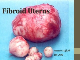
- 3. Pathology • GROSS: • nodular structures • Oval or rounded shaped ,firm in consistency, whorled appearance on cut section • single but mostly multiple (up to 125) • Size typically size of grape fruit but varies Tiny seedling to a huge abdominal mass
- 4. Uterine leiomyoma ,external surface on left, cut surface on right.
- 5. Pathology • Microscopy: • Smooth muscle cell bundles arranged in whorled pattern with variable amount of connective tissue • Predominance of fibrous tissues rarely seen.
- 6. Epidemiology • most common tumor of the female body • Present in 20-30 % of women of reproductive age,only a fraction of these will require treatment. • Age : never occurs before menarche , regresses after menopause ,peak incidence 4th & 5th decades • Parity : higher in Infertile & women of low parity. • Race : twice common in black women, African American women are three times more likely to get fibroids than Caucasian women. • Hereditary factor : women with family history , twice more likely to develop fibroids.
- 7. Etiology • Exact aetiology is unknown, current working hypothesis is that genetic predispositions, prenatal hormone exposure and the effects of hormones ,growth factors and xenoestrogens cause fibroid growth. • 50% cases shows karyotypically detectable chromosomal abnormalities . • 70% cases with fibroids have specific mutations MED12 protein. • Risk factors: African-American descent, nulliparity , obesity (fat aromatase), polycystic ovary syndrome ,diabetes and hypertension. • While pregnancy & smoking decreases risk of fibroids
- 8. • Fibroid growth is strongly dependent on estrogen and progesterone , number of observations support the idea that fibroid is an ovarian hormone dependent tumor. 1. Higher concentration of estrogen and progesterone receptors in fibroid. 2. GnRH analouges reduces size of fibroid by reducing the estogens levels. 3. Oral contraceptive pills when taken by women with fibroids ,increases fibroid size while taken by a female without fibroids pills reduces the incidence of fibroids. 4. Obese women are at high risk , because of high serum estrogen levels. Fat aromatase converting androgen(androstenedione) to estrogen(estradiol)
- 9. Classification of fibroids • Intramural fibroids • Subserosal fibroids • Submucous fibroids Within Body of uterus Cervical Intraligamentary
- 10. • Intramural fibroid : Within uterine wall , surrounded by myometrium , non capsulated but pseudocapsule form with growth , blood supply is through nutrient arteries entering through the pseudocapsule.
- 11. • Subserosal fibroid Originates from outer myometrium & projects outwards from uterus covered with peritoneum, attain large size to lack of surrounding myometrium. large subserosal fibroid
- 12. • Submucous fibroids : Arises from inner myometrium, covered with endometrium . Projects inwards from uterine wall into uterine cavity ,may get pedunculated.
- 13. • Cervical fibroid : Less common(1-2%), arises from cervix , usually single, Confined to the supravaginal portion of cervix, Either intramural or intraluminal. • Intraligamentary fibroids : Arises from smooth muscles fibres with in the broad ligament e.g round ligament & ovarian ligament
- 14. Symptomatology • Fibroids are mostly asymptomatic , particularly when small in size. • Menorrhagia is common intramural & sub mucous fibroids, with increased blood loss but regular cycle. • Due to : Increased endometrial surface area. ulcerated and damaged endometrium over the fibroid. mechanical compression of venous drainage by fibroid . • Intermenstrual bleeding in case of submucous fibroid • Postcoital bleeding caused by pedunculated submucous fibroid
- 15. • Subfertility : 30% of patients with fibroid have problems related to fertility. However Its unclear whether fibroid is a cause or effect of infertility , possible explanations are a) delay in child bearing predispose to development of fibroid b) fibroid causes interference in implantation • Pain : pain usually start when complications occurs e.g torsion red degeneration sarcomatous degeneration
- 16. • Urinary symptoms : cervical fibroid – irritation of bladder – increased frequency large cervical fibroid – impaction of pelvis – urinary retention • Pressure symptoms : Large fibroids causes interference with venous and lymphatic drainage of the lower limb causing edema and varicosities. Pressure on pelvic vein may cause hemorrhoids. • Abdominopelvic mass : A large fibroid may fill the abdominal cavity causing dyspepsia due to stomach irritation & dyspnea due to pressure on lungs.
- 17. Examination General physical examination • No specific findings • Excessive loss of blood may cause anemia ,presenting with pallor and in extreme cases with breathlessness • Edema and varicosities of limbs are rare findings with large fibroids . Abdominal examination : Uterus palpable abdominally after 12weeks size of pregnancy • Single fibroid -- central uterus with smooth surface • Multiple fibroids – irregular mass maybe shifted to a side • Fibroids – firm ,non tender unless undergone degeneration. Pevic examination : • Protuding fibroids easily seen • Speculum examination – patients with mennorrhagia ,intermestrual and postcoital bleeding. • Bimanual examination – • Single fibroid -- central uterus with smooth surface • Multiple fibroids – irregular mass maybe shifted to a side • Fibroids – firm ,non tender unless undergone degeneration
- 18. Complications 1. Related to site of fibroid • Subserous fibroid : if pedunculated ,can undergo torsion or twist in the pedicle occludes blood supply leading to ischemic necrosis presenting with acute pain. • Sessile subserous fibroid may get adherent with the bowel or omentum ,if develop its own blood supply and get separated from the uterus forming Parasitic fibroid.
- 19. • Submucous fibroid : if pedunculated may prolapse through cervix causing Intermenstrual and postcoital bleeding Get infected and ulcerated May cause uterine inversion • Cervical fibroid : may get impacted in pelvis causing ureteric obstruction and urinary retention.
- 20. 2. Degeneration : Blood supply to fibroid is from periphery and the central area is relatively deprived of circulation so a rapidly growing fibroid easily undergo degeneration ,these include • Hyaline degeneration Soft , on cut section whorled pattern lost & become cystic.
- 21. • Fatty degeneration : Fatty change occurs in the fibroid Require differentiation from uterine lipoma A longitudinal (a) and transverse (b) image of a 1.46 x 1.16 x 1.56 anterior leiomyoma that has undergone fatty degeneration.
- 22. Leiomyoma with fatty degeneration
- 23. • Red degeneration : Thrombosis of peripheral blood vessels , while other become distended and engorged with red blood cells. Cut section: appear reddish due to presence of thrombotic and hemolytic changes. • Calcification due to poor blood supply. • Rarely sarcomatous degeneration also occurs (0.1%) Leimyoma with extensive red degenration
- 24. 3. Infections : Subserous fibroid can acquire infection from Appendicitis Diverticullitis Pyosalpinges Submural fibroids – after abortion and during perpurium Submucosal fibroids –after ulceration 4. Hematological complications : menorrhagia-exessive bleeding -anemia fibroids--increased erythropoietin production- polycythemia
- 25. Investigations Investigation of choice • Ultrasonography Under special circumstances • Hysteroscopy and curettage • Laparoscopy Other investigation • Hysterosalpingogram • Modern imaging techniques • Complete blood picture
- 26. Ultrasonography • Investigation of choice • Typical fibroid appearance :: mild to moderate echogenic mass in the uterine wall that causes nodular distortion of uterine outline. • Small intramural or Submucous fibroid :: recognized by distortion of the normally linear central endometrial echoes. • Fibroids with hyaline degeneration :: anechoic area within fibroid • Fibroids with cystic degeneration:: will give Snow storm appearance..
- 28. Hysteroscopy & Curettage • Hysteroscopy provides a direct veiw of uterine cavity, & is indicated during Abnormal uterine bleeding Small submucous fibroids missed during ultrasound Investigation under special circumstances
- 29. Curettage may help to diagnose a co existing endometrial pathology ,which may be the actual cause of menorrhagia.
- 30. • Indication when the mass cannot be differentiated on the ultrasound fibroid associated with infertility or pelvic pain laparoscopy
- 31. Other investigations 1. Hysterosalpingogram : carried out as a part of infertility investigation and can pick small submucous fibroids. Hysterosalpingogram showing two submucous leiomyoma(arrow) Hysterosalpingogram
- 32. 1. Modern Imaging Technique : CT scan and MRI are more accurate in describing pelvic mass but too expensive for routine examination. 1. Complete Blood Picture : In severe menorrhagia hemoglobin will be low and polycythemia can also be diagnosed. A very large (9cm) fibroid of the uterus seen on CT
- 34. Conservative Treatment • Asymptomatic fibroid of size less than 12 weeks pregnancy in a patient of 42 years of age is left alone in a hope that I would regress after menopause. • Even an asymptomatic fibroid of size more that 12 weeks of pregnancy does not justify prophylactic removal as risk of sarcomatous change is less than 0.1%. • Only mangment required is a regular follow up till menopause. • These days, Removal only indicated in case of a very large fibroid or a rapidly increasing in size due to concern about the nature of the mass . •
- 35. Medical Treatment • Ideal drug – complete regression of fibroids • GnRH analogues is the only drugs which has shown promising results . • GnRH analogues : Monthly IM depot injection Daily Nasal spray prescribed for 3 months improved 80% cases of menorrhegia, 50% of the fibroid size is reduced Disadvantages: expensive , effects only last during therapy , cause post menopausal symptoms (hot flushes , night sweats , psychological disturbance)
- 36. • Therefore only given when reduction in size and vascularity is required prior to myomectomy & hysterectomy. • Long term use (6months or more) only allowed when patient is unfit for surgery ( obses ,extensive adhesions) or approaching her menopause. • Other drugs : these shows reduction in size of fibroids up to some extent • Danazol • Gestrinone
- 37. Surgical treatment • Surgical treatment is present in the form of a) Myomectomy b) Hysterectomy Myomectomy term myomectomy is used for an operation where the uterus is conserved and fibroid is removed. Preferred treatment in following conditions, 1. Symptomatic fibroids in young patient, 2. Infertile patients when fibroids are only pathology, 3. Patients wishing to have more childrens, 4. Patients with recurrent abortion ,fibroids likely to be the underlying cause, 5. Patients wishing to conserve her uterus.
- 38. Routes of Myomectomy • Abdominal myomectomy Most common method, perform through abdomen. • Vaginal myomectomy For pedunculated submucosal fibroids protruding through cervix removed vaginally by ligating its pedicle with cautery. • Endoscopic myomectomy
- 39. Disadvantages of Myomectomy • Hemorrhages Patient hemoglobin less than 11 gm/dl , two pints of cross matched blood should be kept for transfusion Uncontrolled hemorrhages may lead to hysterectomy. • Early post operative complications Post operative oozing from the uterine wound causes pyrexia and paralytic ileus thus prolonging post operative recovery • Delayed complications Intraperitoneal adhesions causing infertility and intestinal obstruction. • Recurrence 15% risk
- 40. Hysterectomy • Removal of uterus • Mostly through abdomen although small fibroids can be removed through vaginal hysterectomy • Advantages : a) Low post operative morbidity b) No risk of recurrence Hysterectomy specimen:deformed uterus with one isthmic fibroid
- 41. • Its preferred over myomectomy under theses circumstance 1. Patients above 40 years of age 2. Presence of multiple fibroids 3. Patients with complete family 4. Patients experiencing severe symptoms
- 42. Differential diagnosis • Adenomyosis : Also called adenomyoma Disease of multiparous women Menorrhagia is associated with severe dysmenorrhea Uterus : uniformly enlarged ,tender Ultrasound findings : thickened myometrium with swiss chees appearance Cut surface : lacks whorled appearance and capsule. • Ovarian tumors : Confused with pedunculaed sub serous fluid Menorrhagia often absent Mass feels separate from the uterus while fibroids has limited mobilty. Ultrasound may be helpful but diagnosis is not confirmed until laproscopy or laprotomy is performed..
- 43. thank you