
Recommended
More Related Content
What's hot
What's hot (20)
Similar to Genital tuberculosis
Similar to Genital tuberculosis (20)
Recently uploaded
Recently uploaded (20)
Genital tuberculosis
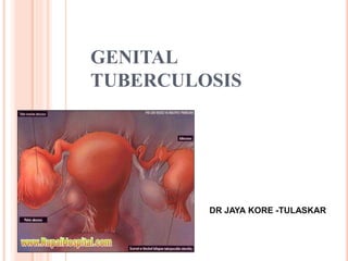
- 1. GENITAL TUBERCULOSIS DR JAYA KORE -TULASKAR
- 2. There is a dread disease ….in which life and death are so strongly blended that death takes the glow and hue of life and life the gaunt and grisly form of death - CHARLE’S DICKENS
- 3. HISTORY In Ancient Vedas tuberculosis was known as Rakshasman-King of diseases Word ‘TUBERCULOSIS’ was coined by Johann Lukas Schönle in 1834. -ROBERT KOCH discovered tubercle bacilli in 1882.
- 4. MILE STONES --- BEING THE FIRSTS First case of genital TB was recognised and described by Morgagni in 1744. First case of tubercular endometritis was reported by kwisch in 1847. Spencer Wales (1862) first operated on clinically diagnosed ovarian cyst – TB loculated cyst. Hegar (1883) removed tubercular tube. Sutherland (1943)-tested endometrial biopsy for TB. Raymond described TB cervicitis.
- 5. INCIDENCE INDIA IS THE HIGHEST TB BURDEN COUNTRY ACCOUNTING FOR MORE THAN ONE-FOURTH OF THE GLOBAL INCIDENCE India 26% China 13% Indonesia 6% Nigeria 5% South Africa 5% Bangladesh 4% Ethiopia 3% Pakistan 3% Phillipines 3% other 13 HBCs 15% other countries 18% Source: WHO Geneva; WHO Report 2014: Global annual incidence = 8.6 million India annual incidence = 2.2 million India is 17th among 22 High Burden Countries (in terms of TB incidence rate)
- 6. In 2013, 8.6 million people fell ill with TB and 1.5 million died from it.(WHO,2014). India alone accounted for 26% of total TB cases globally(WHO,2014). HIV has altered the dynamics of TB worldwide Patients co-infected with HIV contributed about 8.5% of the increase in tuberculosis cases per year.(RCOG,2011).
- 7. Genital tuberculosis is the second most common form of extrapulmonary TB after peripheral lymphadenopathy. Genital TB is found in 5-10% of women with infertility problems, with low rates in Australia (1%) and high rates of up to 19% in India (ICMR,2011) Genital tuberculosis in India has increased to 30 per cent in 2015 from 19 per cent in 2011(ICMR,2015).
- 8. PATHOPHYSIOLOGY Tuberculosis organism is - non-motile obligate aerobe replicating cycle of 17-24 hours (slow growing) -Non capsulated -Acid-fast due to the surface lipids. So resistant to common antibacterial agents and lytic enzymes.
- 9. PATHOPHYSIOLOGY AGE GROUP- -Can occur in any age group, 75% being in the 20–45 yrs . Postmenopausal women ( 7–11%) . o if primary infection occurs close to menarche increased chance of genital T.B Mycobacterium tuberculosis of human type.(90–95%) Rarely M.bovine.(5-10%). Almost always of secondary type.(RCOG,2005) If the bacilli are not eradicated, risk of reactivation, especially in conjunction with diseases (e.g. Hodgkin’s lymphoma, AIDS, steroids, stress, or malnutrition
- 10. ORGAN FREQUENCY Fallopian tubes 90-100% Endometrium 50-60% Ovaries 20-30% Cervix 5-15% Vulva and Vagina 1% Schaefer G: Female genital tuberculosis. Clin Obstet Gynecol 19:23, 1976) Fallopian tubes -primary sites Mode of spread : Hematogenous (90%) Lymphatic/ Direct Ascending Frequency of tuberculosis in genital organs
- 11. TUBERCULOSIS OF THE FALLOPIAN TUBES Both tubes are involved in 100 % of cases.
- 12. Initial: Submucosal layer of Ampulla Spreads medially Spreads Outwards Muscle layer Fibrosis, segmental Perisalpingitis with Thickening, Tobacco Exudation.( dense Pouch appearance adhesions) Pyosapinx Tuboovarian Mass Salpingitis Isthmica Nodosa
- 14. TYPES OF TUBERCULOUS SALPINGITIS Exudative -Acute phase of the process. - large pyosalpinx may form. Frequently, the organs contain a large amount of caseous material plus purulent exudate. -show few adhesions . Productive-Adhesive -Found most frequently at laparoscopy or laparotomy. - Tubes are studded with tubercles and are densely adherent to the surrounding organs. - The tube wall is thickened and nodular. - Eventually, when the process starts healing, it results in calcification and fibrosis.
- 16. TUBERCULOSIS OF ENDOMETRIUM Uterus Tubercles at basal layer of endometrium Shed at each menstruation Reinfection Endometrial ulceration Asherman’s syndrome
- 17. Tubercles at basal layer of endometrium Myometrium(2-3%) Caseation Pyometra(postmenopausal woman)
- 18. TUBERCULOSIS OF THE OVARY The involvement is bilateral. Two forms : PERIOOPHORITIS -Extension of the tuberculosis from the tube - Most common form of tuberculosis. - From the periphery toward the center - Resulting in a tuboovarian mass, which is frequently adherent to omentum and intestines.
- 19. OOPHORITIS -Follows hematogenous spread. -A relatively rare condition - Infection starts in the stroma of the ovary, -Typical tubercles or larger foci with caseous centers may be recognized on cross section in the hilum of the ovary
- 20. TUBERCULOSIS OF CERVIX Descending infection from fallopian tubes and uterus. Sexual parteners may be source of infection. No macroscopic changes in the cervix specific for TB. The cervix may appear normal or inflamed, may resemble invasive carcinoma. Three forms -Ulcerative –most common -Papillomatous -Miliary
- 21. TUBERCULOUS PERITONITIS In combination with female genital tract TB approximately 45% of the time. Often extensive adhesions seen. Two types Serous variety- - more common - Characterized by ascitis, signs of peritoneal inflammation, fever, abdominal pain, weight loss, and anorexia. Plastic variety- - Less common - Characterized by tender abdominal masses and an abdomen “doughy” to palpation.
- 22. TUBERCULOSIS OF THE VULVA AND VAGINA Rarest form of genital TB(1%). Sexual partners may be source of infection. In the vulva begins as a nodule on the labia or in the vestibular region-breaks down - irregular ragged ulcer. -sometimes with sinuses discharging caseous material and pus. - as a hypertrophic, irregular warty growth. In the vagina may simulate carcinoma in its gross appearance.
- 23. TB OF PELVIS Tubercular adenitis of mesenteric or pelvic lymph nodes. Pelvic T.B. is not the same disease as Genital T.B. ‘FROZEN PELVIS’ DDs of frozen pelvis? Florid genital tuberculosis Grade III/IV pelvic endometriosis Advance invasive carcinoma of cervix Following radiotherapy for invasive carcinoma of cervix
- 24. CLINICAL FEATURES 20% have history of T.B. in immediate family. Past history of tuberculosis. Asymptomatic-10-11% Systemic- A history of poor general health persisting over a period of months or years and associated with weight loss, undue fatigue, low-grade fever, or vague lower abdominal pain Infertility Menstrual disturbances Abdominal swelling Postcoital bleeding Vaginal discharge Dyspareunia
- 25. INFERTILITY Most common initial symptom. In most large studies: Infertility presenting c/o in 40% - 50% Past h/o TB /family . LOWER ABDOMINAL PAIN Second common symptom. Pain present for several months which is not usually severe. M/c associated with swelling of abdomen. Episodes of acute lower abdominal pain owing to secondary infection by pyogenic org. In advanced disease pelvic pain becomes severe and gets aggravated by coitus, exercise & mensus.
- 26. MENSTRUAL COMPLAINTS Third common symptom. Menorragia/ Menometrorragia/ Intermenstrual bleeding/ Oligomenorrhoea/ Postmenopausal bleeding. Menstrual cycle may be normal. AMMENORRHOEA Advanced active pulmonary T.B. produce amen. but concomitant genital T.B. is rare. Complete destruction of ovary by genital T.B. seldom occurs so ovarian failure is not the cause. ‘End organ failure’ secondary to endometrial caseation
- 27. H/o primary infertility with no apparent cause on examination & family H/o or personal H/o T.B. H/o vague lower abdominal discomfort with low grade fever/undue fatigue/persistent ill health over months to years associated with weight loss. Adolescent female presenting with ascites pain and low grade fever. Menopausal female enlarged uterus that is tense and tender on examination (pyometra formation) Recurrent Pelvic inflammatory disease not responding to antibiotic therapy.
- 28. SIGNS Normal in 50%.. Abdominal examination-doughy feeling. Abdominal mass Pelvic mass Adnexal mass Abdominal tenderness Pelvic/adnexal tenderness
- 29. Ascites Excessive vaginal discharge Ulcer in the vulva, vagina, and cervix Enlarged uterus with pyometra Fistulal Lesions of the cervix and external genitalia.
- 30. DIAGNOSTIC CRITERIA Clinical suspicion Laboratory evidence Histopathological confirmation Imaging Visual evidence ( laparo / hysteroscopic )
- 31. CLINICAL SUSPICION Unexplained infertility Infertility of any duration with High risk factors - history of previous pulmonary TB infection. - contact with a pulmonary TB sufferer. - recent travel to or migration from high prevalence countries. - residence in high prevalence areas . - low socioeconomic background. - drug abuse. - HIV positive status.
- 32. - Generalised s/s loss of wt / appetite low grade fever / malaise night sweats persistent vaginal discharge non healing of wounds , unexplained ascites chronicity of symptoms Adnexal disease with ascites in virgins Chronic PID refractory to standard antibiotic treatment Postmenopausal women with bleeding, persistent leucorrhoea and pyometra where endometrial neoplasia has been excluded.
- 33. INVESTIGATIONS CBC, ESR CXR Pelvic ultrasound / hystero-salpingography Laparoscopy Histopathology Microbiology:- Mantoux test QTG-T Serology AFB microscopy / culture EA / EB / EC / menstrual blood Urine c/s Molecular tests HIV
- 34. Hemogram is usually normal,may show lymphocytosis ,low Hb ESR is usually normal but may be raised Urine microscopy may show abacteruric pyuria in concomitant GUTB. Raised serum CA125 level
- 35. CHEST X RAY Most chest X-rays are normal Old healed scarred lesions Current or past tuberculous lesions in the lungs o Milliary tubercles if disseminated tuberculosis.
- 36. ENDOMETRIAL BIOPSY Sampled by endometrial aspiration , biopsy Curettage ,premenstrual biopsy/ menstrual blood on1st day within 12 hrs of menses +ive in 50-60 % cases of GTB Reveals granulation , caseation , dilated glands, inflammatory cells ,lymphocytes , plasma cells, destruction of epithelium , fibrosis Microscopic appearance of the granuloma: Multinucleated giant cells, Langhans cells Chr. Inflammatory cell,Epitheloid cells,Central area of caseation necrosis. +ive HPE indicates 100% TB salpingitis -ive HPE does not rule out endometrial / tubal TB
- 37. CULTURE Decisive step for diagnosis, treatment & control of TB Combination of solid and liquid media-Gold Standard For isolation of bacteria – a surest test More sensitive requiring 10 -100 org / ml Sensitivity is 30-40% CONVENTIONAL o Egg based media 3-8 weeks eg Lowenstein Jensen media Agar based < 3 weeks eg- BACTEC medium MGIT: more rapid and sensitive than other methods of culture.
- 38. RADIOMETRIC CULTURE METHOD BACTEC -- is based on measurement of carbon dioxide released by bacteria during growth in liquid medium Radioactive carbon labelled substrate like palmitic acid or formic acid is used as marker for bacterial growth Diagnosis is made in 1-2 weeks Sensitivity is 80-90%
- 39. BACT/ALERT 3D MB Fully automated,Non- invasive,Continuously monitored non- radiometric system. Revised antibiotic supplement kit Medium - modified Middlebrook 7H9 broth with supplements Direct inoculation No processing Decontaminated clinical specimen and sterile body fluid specimen (other than blood) -10 ml Middlebrook 7H9 Broth BSA, Catalase
- 40. For blood and sterile body fluids -30 ml Middlebrook 7H9 Broth SPS, Glycerol. CO2 released by mycobacteria detected by sensor Colour changes - increase in reflectance units Positive broth - 106-107 orgs/ml Mean detection time (days)-11 to15 days.
- 41. SMEAR MICROSCOPY Ziehl-Neelsen Fluorochrome - Auramine-rhodamine (direct fluorescence) Simple & rapid procedure Higher sensitivity; faster screening Requires 10000 mycobacteria/ml for + ive smear Or 1-9 AFB / HPF -- +++ Or 1-9 AFB / 10 HPF ++ Or 1-9 AFB / 100HPF + - ive smear does not rule out GTB Cyto centrifugation enhances sensitivity
- 42. TUBERCULIN (MANTOUX) TESTS 0.1 ML PPD IS INJECTED INTRADERMALLY Delayed hypersensitivity reaction Read between 48 and 72 hours after the injection The tuberculin skin test has a sensitivity of 55% and specificity of 80% in patients with genital tuberculosis.(RCOG ,2005)
- 43. SSSSSS
- 44. QUANTIFERON-GOLD In vitro assay that measure interferon (IFN-γ) released by sensitized T cells after stimulation by M. tuberculosis antigens. FDA-approved in 2001,for detection of latent tuberculosis infection. Indirect test for M. tuberculosis complex Tuberculosis disease OR latent tuberculosis infection (LTBI)- cannot distinguish between them Intended for use in conjunction with risk assessment, radiography, and other medical and diagnostic evaluations
- 45. PRINCIPLE OF QUANTIFERON GOLD Fresh heparinised whole blood from sensitised persons Incubated with mixtures of synthetic peptides (two proteins present in M. tuberculosis) ESAT-6 (early secretory antigenic target-6) CFP-10 (culture filtrate protein-10) Lymphocytes recognize these mycobacterial antigens – Generation and secretion of interferon-γ (IFN-γ) Detection and subsequent quantification of IFN-γ by ELISA
- 46. Result Interpretation Positive (ESAT-6 and/or CFP-10 responsiveness detected) M. tuberculosis infection likely Negative (No ESAT-6 or CFP-10 responsiveness detected) M. tuberculosis infection unlikely, but cannot be excluded in immunocompromised patients, or highly probable cases Indeterminate Test not interpretable
- 47. ADVANTAGES Single patient visit Rapid results (within 24 hours) Higher specificity than Mantoux No booster response (measured by subsequent tests - which can happen with Mantoux) No reader bias (cf Mantoux) Not affected by prior BCG vaccination or Impaired or altered immune function
- 48. T-SPOT®.TB TEST (T-SPOT) T-SPOT®.TB test (T-Spot) - Counts the cells releasing IFN-G visualized as spots with the enzyme-linked immunospot (ELISPOT) technique. Clinical Utility: Can detect both latent and active pulmonary and extra- pulmonary cases. Useful for screening person who has symptoms of TB. Screening suspected Extra-Pulmonary tuberculosis eg: GTB
- 49. ULTRASONOGRAPHY Loculated ascites: The presence of fine lacy strands or particulate matter in fluid is due to thin fibrin strands Bilateral or unilateral tubo-ovarian mass Hydrosalpinx Fluid in cul de sac , peritoneal cavity Mesenteric lymphadenopathy Small uterus with atrophic endometrium Enlarged uterus with caseous pyometra
- 50. HYSTEROSALPINGOGRAPHY (HSG) Hysterosalpingography is contraindicated -in the presence of recent acute pelvic infection -If Tuberculosis is suspected. Vascular or lymphatic extravasation of the dye Rigid (lead-pipe) tubes with nodulations Tobacco-pouch appearance Leopard skin appearance Rosette appearance Moth eaten appearance Sperm head appearance Beaded appearance of the tube Distal tube obstruction Coiling/ calcified shadows Bilateral cornual block Irregular, honey-comb appearance of the uterine cavity
- 51. TOBACCO POUCH •Tube is enlarged and distended, ostium remain patent with recognizable everted fimbriae.
- 52. COTTON WOOL PLUG APPEARANCE.. Focal irregularity and areas of calcification occur within the lumen of the fallopian tubes.
- 53. TUFTED APPEARANCE.. Caseous ulceration of the mucosa of the fallopian tube produces an irregular contour of the lumen of the tubes. Diverticular cavities may surround the ampulla and give a “tuft” like appearance. Thick arrow – hydrosalphinx.
- 54. PIPE STEM APPEARANCE/RIGID PIPE Scarring fallopian tubes. Irregular and rigid. Filling defect in uterine cavity – adhesion.
- 55. BEADED APPEARANCE.. Multiple constrictions along the course of fallopian tube on HSG due to fibrotic strictures.
- 56. T-SHAPED UTERINE CAVITY Scarring results in a “T” shaped uterine cavity with intravasation of contrast.
- 57. FLASK SHAPED FALLOPIAN TUBES.. a flask-shaped dilatation of the fallopian tubes due to obstruction at the fimbria.
- 58. FILLING DEFECT
- 60. SPERM HEAD APPEARANCE/GOLF CLUB
- 61. HYSTEROSCOPY Ostia not visualized Caseous material coming out of ostia Periosteal fibrosis Caseation/ tubercles Endometrial calcification Scanty endometrium Intrauterine adhesions Irregular uterine cavity
- 62. LAPAROSCOPY Blue uterus” when chromopertubation test done with methylene blue. Acute salpingitis: red, swollen edematous tubes. Tiny tubercles 1-4 mm on surface of organs: tubes, uterus. Hydrosalpinx, pyosalpinx. Tobacco-pouch appearance. Unilateral / bilateral TO masses. Straw colored thick jelly like exudate in POD. Fimbrial biopsy, peritoneal fluid may be taken to confirm the diagnosis .
- 63. SEROLOGICAL TESTS Based on recognition of host response Antibodies tested against mycobacterial antigens by ELISA using mono /polyclonal Abs A-60 & 38 kDa –are usual purified Ags tested Useful in chronic & inaccessible disease Active infection IgM develops before ESR is raised or Montoux is +ive Sensitivity is 80-90% in extra pulmonary TB
- 64. PCR MOLECULAR DIAGNOSTIC TESTS PCR based sequencing - Amplification of DNA using specific primers - Highly sensitive ( > 90% ) & specific ( 95% ) - Few bacterial cells used for amplification - Detectable with even 1-10 organisms / ml - Results available in 2-3 days - Cumbersome and expensive - False –ive if contaminated with heparin or high salt concentration - False +ive as can not distinguish between live or dead bacilli
- 65. MOLECULAR DIAGNOSTIC TESTS DNA probes - - Species specific probes that hybridise with RNA - Combined with short term culture - Used for rapid detection of myco TB - Highly sensitive & specific DNA microarrays - Identify mutation rapidly & accurately - By Ligase chain reaction where fragmented primers are amplified
- 66. PCR GENOTYPIC MOLECULAR TESTS PCR assays targetting various gene segments are -- a 65 kDa protein encoding gene -- DNA IS 6110 element -myco TB has 10 -20 copies of this sequence.PCR detect less than 10 pg of DNA =3 genomes -- mpb 64 gene –a specific Ag -- as patch test . Becomes +ive in 3-4 days & lasts for 1 week (100% specific & 98% sensitive)
- 67. Advantages: High specificity and sensitivity, requires only < 10 bacteria/mL Rapid method, results are available within a day of the DNA being extracted from the specimen. Can be applied to sterile fluids like peritoneal fluid where the culture is difficult due to a low bacterial load. Disadvantages: False Negative - absence of even a single AFB in the sample collected, and high salt concentration of a specimen which interferes with the PCR results. False Positive- PCR cannot distinguish between live and killed bacilli
- 68. BIOCHEMICAL MARKERS Adenosine Deaminase activity - Tested in associated peritoneal TB - Levels of > 30 u/L in ascitic fluid significant - Rapid , highly sensitive & specific - Pending confirmatory tests - therapy can be initiated Tuberculstearic acid test - Fatty acid present in mycobacteria only - Gas chromatography /mass spectrometry done with samples containing small number of bacteria
- 69. COMPLICATIONS Subfertility or Sterility Even in patients considered to be “cured,” extensive damage to the fallopian tubes and the endometrium is often irreversible, and chances of successful intrauterine pregnancy drop significantly. Ectopic Pregnancy Congenital Tuberculosis A rare but potentially serious complication . transmission from maternal tuberculous endometrium to the fetus .
- 70. ANTI TUBERCULAR TREATMENT 3 basic principles for chemotherapy for T.B. Regimen must contain multiple drugs to which organism is susceptible. Drugs are to be taken regularly. Drugs should continue for a sufficient period of time.
- 71. RECENT RNTCP GUIDELINES 2016.
- 74. ` Drug Bactericidal activity Isoniazide Bactericidal in-vivo and in-vitro Rifampicin Pyrazinamide Selectively bactericidal Streptomycin
- 75. RESERVE LINE/SECOND LINE DRUGS USED IN TREATMENT OF TB Drug <50 Kg >50 Kg Thiacetazone 150 mg 150 mg Ethionamide 500 mg 750 mg Cyclocerine 750 mg 1000 mg Inj. Kanamycin 750 mg 1000 mg Inj. Capreomycin 750 mg 1000 mg Inj. Viomycin 750 mg 1000 mg
- 76. NEWER DRUGS USED IN THE TREATMENT OF DRUG RESISTANT TB Fluoroquinolones: Ciprofloxacin (1500 mg/day), and Ofloxacin (400-600 mg/day) for 6 months Anti-leprosy drugs: Clofazimine (100-200 mg/day) Beta-lactam antibiotics: amoxicillin+ clavulanic acid (Augmentin) Macrolides: azithromycin, roxithromycin, and clarithromycin
- 77. SURGICAL MANAGEMENT Tubal reconstructive surgery is contraindicated because there is usually irreparable damage of tubes (cilia are destroyed) May result in reactivation and dissemination Those with infertility must be treated with medical treatment; and if this does not help artificial reproductive techniques may be considered.
- 78. SURGICAL MANAGEMENT Indications:- Persistent & recurrent disease/pelvic masses/pelvic pain/abnormal bleeding despite adequate treatment Persistent non healing fistula Multi drug resistant disease Concomitant neoplasia of genital tract Chemotherapy should precede surgery by 1-2 weeks. Surgery should be done at mid cycle in premenopausal. Chemotherapy should be continued for 6-12 months post op. Premenopausal-save ovaries if normal, otherwise TAH with BSO followed by HRT. (Glob. libr. women's med., The International Federation of Gynecology and Obstetrics,2008)
- 79. GTB & ENDOMETRIAL RECEPTIVITY Implantation failure reasons are Inability to express Integrin molecules in the endometrium during implantation window Inhibition by Myco. TB. Of basal production of progesterone & stimulatory effect of HCG leading to ovulation failure Fibrosis , endometrial atrophy , adhesion formation .
- 82. UTERINE BIOPHYSICAL PROFILE Applebaum uterine scoring system for reproduction (USSR on USG ) Endometrial thickness > 7mm in A.P. diameter 5 lined appearance of endometrium -from D1 to midcycle – progressive increase in thickness Blood flow within zone 3 on color doppler Myometrial contractions giving wave like motion of endometrium Uterine arterial blood flow as Pulsatility Index <3.0 Homogenous myometrial echogenicity Myometrial blood flow seen on grey scale exam. (internal to arcuate vessel)
- 83. POOR PREDICTORS FOR CONCEPTION Poor endometrial receptivity Blocked tubes Elderly age Infertility of longer duration Sec. amenorrhea / oligomenorrhea Endometrial atrophy , caseation Endometrial scarring , synechiae
- 84. PREDICTORS OF GOOD RECEPTIVITY Endometrial thickness Endometrial layering Myometrial echogenicity
- 85. Each parameter is scored as follows: 1. endometrial thickness a. < 7 mm = 0 b. 7 - 9 mm = 2 c. 10 - 14 mm = 3 d. > 14 mm = 1 2. endometrial layering a. no layering = 0 b. hazy 5-line appearance = 1 c. distinct 5-line appearance = 3 3. myometrial contractions (seen as wave-like endometrial motion high-speed playback from videotape) a. < 3 contractions in 2 minutes (real-time) = 0 b. > 3 contractions in 2 minutes (real-time) = 3
- 86. 4. myometrial echogenicity a. coarse/inhomogeneous echogenicity = 1 b. relatively homogeneous echogenicity = 2 5. uterine artery Doppler flow evaluation a. PI > 3.0 = 0 b. PI< 2.5 - 2.99 = 0 c. PI < 2.2 - 2.49 = 1 d. PI < 2.19 = 2 6. endometrial blood flow within Zone 3 a. absent = 0 b. present, but sparse = 2 c. present multifocally = 5 7. myometrial blood flow internal to the arcuate vessels seen on gray-scale examination a. absent = 0 b. present = 2