SlideShare a Scribd company logo
1 of 12
Download to read offline
1
Anatomy and Physiology of the Skin
Paul A.J. Kolarsick, BS, Maria Ann Kolarsick, MSN, ARNP-C,
and Carolyn Goodwin, APRN-BC, FNP
C H A P T E R 1
Introduction
The skin is the largest organ of the body, accounting for
about 15% of the total adult body weight. It performs many
vital functions, including protection against external physical,
chemical, and biologic assailants, as well as prevention of ex-
cess water loss from the body and a role in thermoregulation.
The skin is continuous, with the mucous membranes lining
the body’s surface (Kanitakis, 2002).
The integumentary system is formed by the skin and
its derivative structures (see Figure 1-1). The skin is
composed of three layers: the epidermis, the dermis, and
subcutaneous tissue (Kanitakis, 2002). The outermost
level, the epidermis, consists of a specific constellation of
cells known as keratinocytes, which function to synthesize
keratin, a long, threadlike protein with a protective role.
The middle layer, the dermis, is fundamentally made up of
the fibrillar structural protein known as collagen. The der-
mis lies on the subcutaneous tissue, or panniculus, which
contains small lobes of fat cells known as lipocytes. The
thickness of these layers varies considerably, depending on
the geographic location on the anatomy of the body. The
eyelid, for example, has the thinnest layer of the epidermis,
measuring less than 0.1 mm, whereas the palms and soles
of the feet have the thickest epidermal layer, measuring
approximately 1.5 mm. The dermis is thickest on the back,
where it is 30–40 times as thick as the overlying epidermis
(James, Berger, & Elston, 2006).
Figure 1-1. Cross-Section of Skin and Panniculus
Note. From Andrews’ Diseases of the Skin: Clinical Dermatology (10th ed., p. 1), by W.D. James, T.G. Berger, and D.M. Elston, 2006, Phila-
delphia: Elsevier Saunders. Copyright 2006 by Elsevier Saunders. Reprinted with permission.
Epidermis
Meissner nerve
ending
Dermis
papillary
reticular
Sebaceous gland
Arrector pili muscle
Hair shaft
Pacini nerve ending
Subcutaneous tissue
Apocrine
unit
Straight duct
Coiled gland
Eccrine
sweat unit
Straight duct
Straight duct
Coiled duct
Eccrine gland
Dermal
vasculature
Superficial plexus
Deep plexus
SKIN CANCER
2
Epidermis
The epidermis is a stratified, squamous epithelium layer
that is composed primarily of two types of cells: keratinocytes
and dendritic cells. The keratinocytes differ from the “clear”
dendritic cells by possessing intercellular bridges and ample
amounts of stainable cytoplasm (Murphy, 1997).The epidermis
harborsanumberofothercellpopulations,suchasmelanocytes,
Langerhans cells, and Merkel cells, but the keratinocyte cell
type comprises the majority of the cells by far. The epidermis
commonly is divided into four layers according to keratino-
cyte morphology and position as they differentiate into horny
cells, including the basal cell layer (stratum germinativum),
the squamous cell layer (stratum spinosum), the granular cell
layer (stratum granulosum), and the cornified or horny cell layer
(stratum corneum) (James et al., 2006; Murphy) (see Figure
1-2).The lower three layers that constitute the living, nucleated
cells of the epidermis are sometimes referred to as the stratum
malpighii and rete malpighii (Murphy).
The epidermis is a continually renewing layer and gives rise
to derivative structures, such as pilosebaceous apparatuses,
nails, and sweat glands. The basal cells of the epidermis un-
dergo proliferation cycles that provide for the renewal of the
outer epidermis. The epidermis is a dynamic tissue in which
cells are constantly in unsynchronized motion, as differing
individual cell populations pass not only one another but also
melanocytes and Langerhans cells as they move toward the
surface of the skin (Chu, 2008).
Keratinocytes
At least 80% of cells in the epidermis are the ectodermally
derived keratinocytes.The differentiation process that occurs
as the cells migrate from the basal layer to the surface of the
skin results in keratinization, a process in which the kerati-
nocyte first passes through a synthetic and then a degradative
phase (Chu, 2008). In the synthetic phase, the cell builds up a
cytoplasmic supply of keratin, a fibrous intermediate filament
arranged in an alpha-helical coil pattern that serves as part
of the cell’s cytoskeleton. Bundles of these keratin filaments
converge on and terminate at the plasma membrane forming
the intercellular attachment plates known as desmosomes.
During the degradative phase of keratinization, cellular
organelles are lost, the contents of the cell are consolidated
into a mixture of filaments and amorphous cell envelopes,
and the cell finally is known as a horny cell or corneocyte.
The process of maturation resulting in cell death is known as
terminal differentiation (James et al., 2006).
Basal Layer
The basal layer, also known as the stratum germinativum,
contains column-shaped keratinocytes that attach to the base-
ment membrane zone with their long axis perpendicular to
the dermis. These basal cells form a single layer and adhere
to one another as well as to more superficial squamous cells
through desmosomal junctions (Murphy, 1997). Other dis-
tinguishing features of the basal cells are their dark-staining
oval or elongated nuclei and the presence of melanin pigment
transferred from adjoining melanocytes (Murphy).
The basal layer is the primary location of mitotically active
cellsintheepidermisthatgiverisetocellsoftheouterepidermal
layers. However, not all basal cells have the potential to divide
(Jones, 1996; Lavker & Sun, 1982). Epidermal stem cells in the
basallayerareclonogeniccellswithalonglifespanthatprogress
through the cell cycle very slowly under normal conditions. Hy-
perplasiogenic conditions, such as wounding, can increase the
number of cycling cells in the epidermis by stimulating division
of stem cells. DNA damage caused by carcinogenic agents may
mutate cell proliferation machinery and can also affect the rate
of cellular division. Migration of a basal cell from the basal layer
to the cornified layer in humans takes at least 14 days, and the
transit through the cornified layer to the outermost epidermis
requires another 14 days (Chu, 2008).
Squamous Cell Layer
Overlying the basal cell layer is a layer of the epidermis
that is 5–10 cells thick and known as the squamous cell layer
Figure 1-2.Three Basic Cell Types in the Epidermis
The three basic cell types in the epidermis include keratinocytes
(some labeled K) and Langerhans cells (L) in the Malpighian
layer and melanocytes (M) in the basal layer. Arrows point to
the basement membrane zone, which separates the basal layer
of the epidermis from the underlying dermis (D).
Note. From Andrews’ Diseases of the Skin: Clinical Dermatology
(10th ed., p. 4), by W.D. James, T.G. Berger, and D.M. Elston,
2006, Philadelphia: Elsevier Saunders. Copyright 2006 by El-
sevier Saunders. Reprinted with permission.
M
M
L
LK
K
D
CHAPTER 1. ANATOMY AND PHYSIOLOGY OF THE SKIN
3
or stratum spinosum (Murphy, 1997). The squamous layer is
composed of a variety of cells that differ in shape, structure,
and subcellular properties depending on their location. Supra-
basal spinous cells, for example, are polyhedral in shape and
have a rounded nucleus, whereas cells of the upper spinous
layers are generally larger in size, become flatter as they are
pushed toward the surface of the skin, and contain lamellar
granules (Chu, 2008). Lamellar granules are membrane-bound
organelles containing glycoproteins, glycolipids, phospholip-
ids, free sterols, and a number of acid hydrolases, including
lipases, proteases, acid phosphatases, and glycosidases. The
abundance of hydrolytic enzymes indicates that the lamel-
lar granules are a type of lysosome. Although the lamellar
granules primarily are active in cells at the interface between
the granular and cornified layers, they also function in cells
of the upper spinous layer to deliver precursors of stratum
corneum lipids into the intercellular space (Haake & Hol-
lbrook, 1999).
Intercellular spaces between spinous cells are bridged by
abundant desmosomes that promote mechanical coupling be-
tween cells of the epidermis and provide resistance to physical
stresses. Organized concentrically around the nucleus, keratin
filaments in the cytoplasm are bound to desmosomal plaques
at one end and remain free at the end closer to the nucleus
(Murphy, 1997). The desmosomal plaques are composed of
six polypeptides found on the cytoplasmic side of the cell
membrane that are important in the regulation of the calcium
required for desmosomal assembly and maintenance (Fairley,
Scott, Jensen, Goldsmith, & Diaz, 1991; Hennings & Holbrook,
1983; Lin, Mascaro, Liu, Espana, & Diaz, 1997).The spine-like
appearance of the numerous desmosomes along cell margins is
where the stratum spinosum derives its name (Chu, 2008).
Gap junctions are another type of connection between epi-
dermal cells. Essentially forming an intercellular pore, these
junctions allow for physiologic communication via chemi-
cal signals that is vital in the regulation of cell metabolism,
growth, and differentiation (Caputo & Peluchetti, 1977).
Granular Layer
The most superficial layer of the epidermis containing
living cells, the granular layer or stratum granulosum, is
composed of flattened cells holding abundant keratohyaline
granules in their cytoplasm. These cells are responsible for
further synthesis and modification of proteins involved in
keratinization (Chu, 2008).The granular layer varies in thick-
ness in proportion to that of the overlying horny cell layer. For
example, under thin cornified layer areas, the granular layer
may be only 1–3 cell layers in thickness, whereas under the
palms of the hands and soles of the feet the granular layer
may be 10 times this thickness.A very thin or absent granular
layer can lead to extensive parakeratosis in which the nuclei
of keratinocytes persist as the cells move into the stratum
corneum, resulting in psoriasis (Murphy, 1997).
The keratohyaline granules are deeply basophilic and
irregular in shape and size, and they are necessary in the
formation of both the interfibrillary matrix that holds keratin
filaments together and the inner lining of the horny cells.
Enzymatic action of the keratohyaline granules results in the
production of “soft” keratin in the epidermis by providing
periodic cutting of keratin filaments. In contrast, the hair and
nails do not contain keratohyaline granules, and the tonofibril
filaments traversing the cell cytoplasm will harden because of
the incorporation of disulfide bonds, producing “hard” keratin
in those structures (Matoltsy, 1976; Schwarz, 1979).
Lysosomal enzymes present only in small amounts in the
stratum basalis and stratum spinosum are found at high lev-
els in the stratum granulosum because the granular layer is
a keratogenous zone of the epidermis. Here, the dissolution
of cellular organelles is prepared as the cells of the granular
layer undergo the abrupt terminal differentiation process to a
horny cell of the cornified layer (Chu, 2008).
Cornified Layer
Horny cells (corneocytes) of the cornified layer provide
mechanical protection to the underlying epidermis and a bar-
rier to prevent water loss and invasion by foreign substances
(Jackson, Williams, Feingold, & Elias, 1993). The corneo-
cytes, which are rich in protein and low in lipid content, are
surrounded by a continuous extracellular lipid matrix (Chu,
2008).The large, flat, polyhedral-shaped horny cells have lost
their nuclei during terminal differentiation and technically
are considered to be dead (Chu; Murphy, 1997).The physical
and biochemical properties of cells in the cornified layer vary
in accordance with position in order to promote desquama-
tion moving outward. For instance, cells in the middle have
a much higher capacity for water-binding than the deeper
layers because of the high concentration of free amino acids
found in the cytoplasm of middle layer cells. The deep cells
also are more densely compact and display a greater array
of intercellular attachments than the more superficial layers.
Desmosomes undergo proteolytic degradation as the cells
progress outward, contributing to the shedding of corneocytes
during desquamation (Haake & Hollbrook, 1999).
The Regulation of Epidermal Proliferation
and Differentiation
As a perpetually regenerating tissue, the epidermis must
maintain a relatively constant number of cells as well as regu-
late the interactions and junctions between epidermal cells.
Adhesions between keratinocytes, the interactions of kerati-
nocytes and immigrant cells, the adhesion between the basal
lamina and the underlying dermis, and the process of terminal
differentiation to produce corneocytes must be regulated as
cells relocate during development as well as throughout life
SKIN CANCER
4
(Haake & Hollbrook, 1999). Epidermal morphogenesis and
differentiation is regulated in part by the underlying dermis,
which also plays a critical role in the maintenance of postnatal
structure and function.The epidermal-dermal interface is also
a key site in the development of epidermal appendages.
The maintenance of a constant epidermal thickness de-
pends also on intrinsic properties of epidermal cells, such
as the ability to undergo apoptosis, programmed cell death.
Apoptosis follows an orderly pattern of morphologic and
biochemical changes resulting in cell death without injury to
neighboring cells, as is often the case in necrosis. This major
homeostatic mechanism is regulated by a number of cellular
signaling molecules including hormones, growth factors, and
cytokines. In the skin, apoptosis is important in developmental
remodeling, regulation of cell numbers, and defense against
mutated, virus-infected, or otherwise damaged cells. Terminal
differentiation is a type of apoptosis evolved to convert the ke-
ratinocyte into the protective corneocyte (Haake & Hollbrook,
1999). The disruption of dynamic equilibrium maintaining
constant epidermal thickness can result in conditions such as
psoriasis, whereas the dysregulation of apoptosis is often seen
in tumors of the skin (Kerr, Wyllie, & Currie, 1972).
Nonkeratinocyte Cells of the Epidermis
Melanocytes
The melanocyte is a dendritic, pigment-synthesizing cell
derived from the neural crest and confined in the skin pre-
dominantly to the basal layer (Chu, 2008). Branching into
more superficial layers, extensions of the melanocyte come
into contact with keratinocytes but do not form cellular junc-
tions. Melanocytes are responsible for the production of the
pigment melanin and its transfer to keratinocytes. Melanin is
produced in a rounded, membrane-bound organelle known as
the melanosome via a series of receptor-mediated, hormone-
stimulated, enzyme-catalyzed reactions (Haake & Hollbrook,
1999).
Melanosomes are moved to the end of the melanocyte pro-
cesses that lie closest to the skin surface and are transferred
to keratinocytes (see Figure 1-3). In white skin, these mel-
anosomes are aggregated into membrane-bound melanosome
complexes containing two or three melanosomes, whereas
melanosomes tend to be removed from these complexes more
rapidly in keratinocytes of individuals with dark skin. Heavily
pigmented skin can be attributed to the greater production of
melanosomes in melanocytes, the higher degree of melaniza-
tion in each melanosome, the larger size of melanosomes, the
greater amount of dispersion of melanosomes in keratinocytes,
and the slower rate of melanosome degradation in comparison
to fair skin (Flaxman, Sosis, &Van Scott, 1973; Murphy, 1997;
Olson, Nordquist, & Everett, 1970).
Increased ultraviolet light exposure stimulates an increase
in melanogenesis and a corresponding increase in melanosome
transfer to keratinocytes where the melanosomes will aggre-
gate toward the superficial side of the nucleus.This response,
which results in tanning of the skin, increases the cell’s ability
to absorb light and thus protect genetic information in the
nucleus from damaging radiation.
Merkel Cells
Merkel cells are oval-shaped, slow-adapting, type I
mechanoreceptors located in sites of high tactile sensitiv-
ity that are attached to basal keratinocytes by desmosomal
junctions. Merkel cells are found in the digits, lips, regions
of the oral cavity, and outer root sheath of the hair follicle
and are sometimes assembled into specialized structures
known as tactile discs or touch domes (Moll, 1994). Rela-
tively small deformations of adjoining keratinocytes are
stimulus enough to cause Merkel cells to secrete a chemi-
cal signal that generates an action potential in the adjoin-
ing afferent neuron, which relays the signal to the brain.
The high concentration of Merkel cells in certain regions
such as the fingertips results in smaller and more densely
Figure 1-3. Portion of a Melanocyte From Dark Skin
Melanosomes are indicated by broad arrows. Thin arrows point
to the basement membrane zone between the epidermis and
the underlying dermis (D).
Note. From Andrews’ Diseases of the Skin: Clinical Dermatology
(10th ed., p. 4), by W.D. James, T.G. Berger, and D.M. Elston,
2006, Philadelphia: Elsevier Saunders. Copyright 2006 by El-
sevier Saunders. Reprinted with permission.
D
CHAPTER 1. ANATOMY AND PHYSIOLOGY OF THE SKIN
5
packed receptive fields and thus higher tactile resolution
and sensitivity.
Langerhans Cells
Langerhanscells areinvolvedinavarietyofT-cellresponses.
Derivedfromthebonemarrow,thesecellsmigratetoasuprabasal
position in the epidermis early in embryonic development and
continue to circulate and repopulate the epidermis throughout
life. The cells are dendritic and do not form cellular junctions
with neighboring cells. Langerhans cells constitute 2%–8% of
the total epidermal cell population and maintain nearly constant
numbers and distributions in a particular area of the body. In the
epidermis, the cells mainly are distributed among the squamous
and granular layers with fewer cells in the basal layer. They are
found in other squamous epithelia in addition to the epidermis,
including the oral cavity, esophagus, and vagina, as well as in
lymphoid organs and in the normal dermis (Chu, 2008).
Langerhans cells must recognize and process soluble an-
tigens found in epidermal tissue. When a membrane-bound
antigen is ingested via endocytosis, cell granules are formed.
The contents of these granules are delivered to phagolyso-
somes in the cytoplasm containing hydrolytic enzymes similar
to those found in macrophages. In the first stage of life, the
Langerhans cells are weak stimulators of unprimedT cells but
are able to ingest and process antigens. Later, once the cell
has become an effective activator of naïve T cells, activation
via contact with the antigen will not trigger phagocytosis but
rather will stimulate cell migration (Udey, 1997).
The Dermal-Epidermal Junction
The interface between the epidermis and dermis is formed
by a porous basement membrane zone that allows the exchange
of cells and fluid and holds the two layers together (James et al.,
2006). Basal keratinocytes are the most important components
of structures of the dermal-epidermal junction; dermal fibro-
blasts are also involved but to a lesser extent (Gayraud, Hopfner,
Jassim, Aumailley, & Bruckner-Tuderman, 1997).
The basal lamina is a layer synthesized by basal cells of
the epidermis consisting mainly of type IV collagen as well
as anchoring fibrils and dermal microfibrils. This includes an
electron-lucent zone known as the lamina lucida as well as
the lamina densa (Aumailley & Krieg, 1996; Lin et al., 1997;
Masunaga et al., 1996;Wheelock & Jensen, 1992).The plasma
membranes of basal cells are attached to the basal lamina by
rivet-like hemidesmosomes that distribute tensile or shearing
forces through the epithelium.The dermal-epidermal junction
acts as support for the epidermis, establishes cell polarity
and direction of growth, directs the organization of the cy-
toskeleton in basal cells, provides developmental signals, and
functions as a semipermeable barrier between layers (Stepp,
Spurr-Michaud, Tisdale, Elwell, & Gipson, 1990).
Epidermal Appendages
The skin adnexa are a grouping of ectodermally derived
appendages, including eccrine and apocrine glands, ducts,
and pilosebaceous units that originate as downgrowths from
the epidermis during development. After injury, all adnexal
structures are capable of reepithelialization via the migration
of keratinocytes from adnexal epithelium to the surface of the
epidermis. Because areas such as the face and scalp contain a
large quantity of pilosebaceous units, reepithelialization occurs
more rapidly after injury in these areas than in areas with fewer
adnexal structures, such as the back (James et al., 2006).
Eccrine Sweat Glands
Eccrine sweat glands are involved in the regulation of heat
and are most abundant on the soles of the feet and least plentiful
on the back (Murphy, 1997; Sato & Dobson, 1970). The sweat
glands originate as a band of epithelial cells growing downward
from the epidermal ridge (Mauro & Goldsmith, 2008). This
tubular, or ductal, structure is modified during development
to generate the three composite parts of the eccrine sweat unit,
which are the intraepidermal spiral duct, the straight dermal
portion, and the coiled secretory duct (see Figure 1-1) (James
et al., 2006; Mauro & Goldsmith). The spiral duct opens onto
the skin surface and is composed of dermal duct cells that have
migrated upward. Cells undergo cornification within the duct,
andthecorneocytesproducedultimatelywillbecomepartofthe
cornified layer.The straight dermal segment connects the super-
ficial spiral duct to the inner secretory portion of the gland.
The secretory coil of the eccrine unit lies deep in the der-
mis or within the superficial panniculus and is composed of
glycogen-rich clear secretory cells, dark mucoidal cells, and
myoepithelial cells specialized in contractile properties (James
et al., 2006; Mauro & Goldsmith, 2008). Clear cells rest either
on the basement membrane or on the myoepithelial cells and
form intercellular canaliculi where two clear cells adjoin.The
canaliculi open directly into the lumen of the gland (Mauro &
Goldsmith). Large, glycogen-rich inner epithelial cells initi-
ate the formation of sweat in response to a thermal stimulus.
Initially an isotonic solution, the darker mucoidal cells in the
secretory coil and in the dermal duct actively reabsorb sodium
from sweat in the duct, thereby resulting in the extremely
hypotonic solution that is emitted onto skin surface through
the intraepidermal spiral duct.This response promotes cooling
while conserving sodium (James et al.).
Apocrine Sweat Glands
Whereas eccrine glands are primarily involved in thermal
regulation, apocrine glands are involved in scent release (Mur-
phy, 1997). Apocrine sweat glands in humans are confined
mainly to the regions of the axillae and perineum, and unlike
eccrine and apoeccrine glands, they do not open directly to
SKIN CANCER
6
the skin surface. Instead, the intraepithelial duct opens into
pilosebaceous follicles, entering in the infundibulum above the
sebaceous duct. The basal secretory coil of apocrine glands,
which is normally located entirely in subcutaneous fat, differs
from that of eccrine glands in that it is composed exclusively
of secretory cells; no ductal cells are present (Murphy).
Apocrine sweat glands develop their secretory portions
and become active just before puberty, a response induced
presumably by hormonal signals. The proteinaceous, viscous
secretion has distinct odor and can function as a territorial
marker, warning signal, and sexual attractant, but its sexual
functions may now be vestigial in humans. Difficulties in
acquiring pure samples of apocrine sweat have made it im-
possible to determine the exact chemical composition of the
secretion (Mauro & Goldsmith, 2008).
Apoeccrine Sweat Glands
The apoeccrine sweat gland (AEG) develops during
puberty from eccrine-like precursors, opening directly unto
the skin. Discovered during the isolation of human axillary
sweat from patients with axillary hyperhidrosis, a condition
characterized by abnormally increased rates of perspiration,
the AEG is found in the adult axillae; its relative frequency
varies from person to person. Like eccrine glands, the AEG
opens directly to the skin surface. The AEG has a secretory
rate as much as 10 times that of the eccrine gland and is there-
fore thought to contribute to axillary hyperhidrosis (Mauro
& Goldsmith, 2008).
Hair Follicles
Hair has many valuable biologic functions including
protection from the elements and distribution of sweat-gland
products. In addition, it has an important psychosocial role
in society. Hair follicles vary considerably in size and shape,
depending on their location, but they all have the same basic
structure. The number and distribution of hair follicles over
the body and the future phenotype of each hair is established
during fetal development; no extra follicles are added after
birth (Kratochwil, Dull, Farinas, Galceran, & Grosschedl,
1996; Millar, 1997; Paus & Cotsarelis, 1999; Paus, Foitzik,
Welker, Bulfone-Paus, & Eichmuller, 1997; St-Jacques et al.,
1998; Zhou, Byrne, Jacobs, & Fuchs, 1995). The particular
spacing and allocation of the follicles are determined by genes
that are expressed very early in the morphogenesis of the
follicles (Paus & Cotsarelis; St-Jacques et al.). Mesenchymal
cells in the fetal dermis aggregate below the basal layer of
the epidermis during embryogenesis (James et al., 2006).
The basophilic cells in the basal cell layer of the epidermis
overlying these mesenchymal cell sites are induced to grow
at a downward angle into the dermis (Murphy, 1997). The
follicle continues to develop until finally widening at the
base and forming a bulb around the group of mesenchymal
cells from which the dermal papilla is formed (James et al.;
Hashimoto, 1970a).
Differentiation occurs at the lower portion of the hair fol-
licle forming the hair cone and later the hair, the cuticle, and
the two inner root sheaths. Differentiation also occurs in the
upper segments of the follicle producing the hair canal in
the upper dermis, through the epidermis, and opening to the
surface prior to the time that the growing hair cone reaches
the upper follicle (Hashimoto, 1970b).
The sebaceous gland forms from a bud in the fetal hair
follicle. Along the same side of the follicle but below the
sebaceous gland, another bud develops into an attachment for
the arrector pili muscle. The arrector pili (AP) are a smooth
muscle bundle that attaches to the external root sheath of
the follicle. The distal end of the AP muscle shows multiple
branches at the level of the papillary dermis.The bulge, which
is the zone of theAP muscle’s follicular attachment, is thought
to contain epithelial stem cells responsible for regenerating
follicles, a crucial role in the hair growth cycle (Cotsarelis,
Sun, & Lavker, 1990). On the opposite side of the follicle, a
third bud forms above the plane of the sebaceous gland and
develops into the apocrine gland. The region of the follicle
above the sebaceous gland is known as the infundibular
segment, and the region between the sebaceous duct and AP
attachment is known as the isthmus (James et al., 2006). The
region below the isthmus is known as the inferior portion and
contains the bottom of the follicle as well as the hair bulb.The
inferior segment undergoes cycles of involution and regenera-
tion throughout life, whereas the infundibular and isthmus
portions remain permanently (James et al.).
Rapidly proliferating cells in the hair bulb, called matrix
cells, are responsible for the production of the hair shaft as
well as the inner and outer root sheaths (James et al., 2006).
Both the hair shaft and the inner root sheath progress upward
as the hair grows until the inner sheath reaches the isthmus
and sheds (James et al.). Matrix cells moving up the follicle
are compressed as they enter the rigid inner root sheath (see
Figure 1-4). The number of cells entering the sheath deter-
mines the size of the hair, and the dimensions and curvature
of the inner root sheath determine the shape of the hair (Paus
& Cotsarelis, 1999). For example, the hairs on the scalp of
Caucasians are round while pubic, facial, and eyelash hairs are
oval. Scalp hairs on people of African descent also are oval,
causing their hair to be curly (James et al., 2006).
Hair color is determined by the distribution of melano-
somes in the hair shaft. The hair bulb contains melanocytes
that synthesize melanosomes and transfer them to the kera-
tinocytes of the bulb matrix. People of African descent tend
to have larger melanosomes than Caucasians, whose smaller
melanosomes are amassed into membrane-bound complexes.
Red hair contains spherical melanosomes.Aging causes a loss
of melanocytes and a corresponding decrease in the produc-
tion of melanosomes and results in graying hair (James et
al., 2006).
CHAPTER 1. ANATOMY AND PHYSIOLOGY OF THE SKIN
7
impressive effect are the androgens: testosterone and its active
metabolite, dihydrotestosterone, which act through androgen
receptors in the dermal papilla. These hormones increase the
size of hair follicles in androgen-dependent areas such as the
beard area during adolescence. Later in life, however, they
can cause miniaturization of follicles in the scalp resulting in
androgen alopecia (male pattern baldness) (Kaufman, 1996;
Sawaya, 1994).
Except for rare congenital hair defects caused by mutations
in keratins or other structural proteins and scarring alopecias,
hair loss and unwanted hair growth reflect deviations of hair
follicle cycling and, therefore, are considered reversible events
(Paus, 1996).The hair cycle can vary depending on a number
of different physiologic factors. Pregnancy, for example, often
results in a prolongation of the telogen phase and an increased
number of scalp hairs in the anagen phase. When estrogen
levels equilibrate after delivery, telogen hairs are lost while
anagen hairs simultaneously are converted to telogen, and
this great quantity of telogen hairs will be lost in three to five
months.The synchronous termination of anagen or telogen is
known as telogen effluvium and is often observed after trauma,
such as childbirth, surgery, weight loss, and severe stress, and
also is associated with drugs, endocrine disorders, anemia, and
malnutrition (James et al., 2006). Regrowth typically follows,
with the exception of any metabolic or nutritional deficiency
(Headington, 1993; Paus & Cotsarelis, 1999).
Sebaceous Glands
Sebaceous glands are found in greatest number on the
face and scalp but are present on nearly all other locations
of the body with the exception of the tarsal plate of the eye-
lids, the buccal mucosa and vermilion borders of the lip, the
prepuce and mucosa lateral to the penile frenulum, the labia
minora, and the female areola (James et al., 2006). Cells of
the sebaceous glands contain abundant lipid droplets known
as sebum in their cytoplasm and are arranged into lobules off
the upper segment of the hair follicle. Basaloid germinative
cells surrounding the lobule give rise to the lipid-filled cells,
which are then expelled into the infundibular segment of the
hair follicle via the sebaceous duct.The sebaceous glands are
thought to be evolutionarily important in providing a second-
ary lubrication during the passage through the birth canal.
This extra lubrication covers the surfaces that come in direct
contact with the birth canal including the vertex, anterior
scalp over the forehead and nose to the lower jaw line, and
the shoulders, chest, and upper aspect of arms posteriorly
(Danby, 2005; Thiboutot, 2004).
Nails
Fingernails provide protection to the fingertips, enhance
sensation, and allow small objects to be grasped. The un-
derlying nail bed is part of the nail matrix containing blood
Figure 1-4. Hair Follicle Structure
Note. From Andrews’ Diseases of the Skin: Clinical Dermatology
(10th ed., p. 8), by W.D. James, T.G. Berger, and D.M. Elston,
2006, Philadelphia: Elsevier Saunders. Copyright 2006 by El-
sevier Saunders. Reprinted with permission.
Hair growth occurs in a cyclical manner, but each follicle
functions as an independent unit.The hair growth cell cycle is
composed of three stages: anagen, catagen, and telogen (see
Figure 1-5) (Millar, 1997; Paus, 1996; St-Jacques et al., 1998).
Anagen, the active growth stage, typically lasts approximately
three to five years on the scalp, during which hairs grow at a
rate of about 0.33 mm per day.The length of the anagen phase
decreases with age and decreases dramatically in individuals
with alopecia (James et al., 2006).
Catagen usually lasts about two weeks and is a period of
involution resulting in club hair formation after many cells
in the outer root sheath undergo apoptosis.The resting phase,
telogen, lasts about three to five months on the scalp, and
hairs in this stage are eventually pushed out by the growing
anagen hair shaft. Other sites on the body tend to have shorter
anagen and longer telogen phases, causing most body hair
to be shorter and remain in place for longer periods of time
(James et al., 2006).
Two secreted molecules that may have important roles
in hair follicle development and cycling are the insulin-like
growth factor 1 and fibroblast growth factor 7. In mice, both
are produced by the dermal papilla and have receptors pre-
dominantly in overlying matrix cells (Danilenko, Ring, &
Pierce, 1996). Hormonal factors controlling hair growth in-
clude estrogens, thyroid hormones, glucocorticoids, retinoids,
prolactin, and growth hormone. The hormones with the most
SKIN CANCER
8
vessels, nerves, and melanocytes and has parallel rete ridges.
The nail plate is formed from matrix keratinocytes (James et
al., 2006).
Fingernails grow at an average rate of 0.1 mm per day, two
to three times faster than the rate of toenail growth. Because
of the slow growth rate, toenails can provide information
about toxic exposure or disease from many months in the past
(James et al., 2006). For example, arsenic poisoning may cause
a horizontal hypopigmentation across all nail plates known as
Mees lines (Daniel & Scher, 1997).
The Dermis
The dermis is an integrated system of fibrous, filamen-
tous, and amorphous connective tissue that accommodates
stimulus-induced entry by nerve and vascular networks,
epidermally derived appendages, fibroblasts, macrophages,
and mast cells. Other blood-borne cells, including lympho-
cytes, plasma cells, and other leukocytes, enter the dermis
in response to various stimuli as well. The dermis comprises
the bulk of the skin and provides its pliability, elasticity, and
tensile strength. It protects the body from mechanical injury,
binds water, aids in thermal regulation, and includes receptors
of sensory stimuli. The dermis interacts with the epidermis
in maintaining the properties of both tissues.The two regions
collaborate during development in the morphogenesis of the
dermal-epidermal junction and epidermal appendages and
interact in repairing and remodeling the skin as wounds are
healed. The dermis does not undergo an obvious sequence of
differentiation that parallels epidermal differentiation, but the
structure and organization of the connective tissue components
are predictable in a depth-dependent manner. The matrix
components, including collagen and elastic connective tissue,
also vary in a depth-dependent manner and undergo turnover
and remodeling in normal skin, in pathologic processes, and
in response to external stimuli (Chu, 2008).
The constituents of the dermis are mesodermal in origin
except for nerves, which, like melanocytes, derive from the
neural crest. Until the sixth week of fetal life, the dermis is
merely a pool of dendritic-shaped cells full of acid-muco-
polysaccharides, which are the precursors of fibroblasts. By
the 12th week, fibroblasts are actively synthesizing reticu-
lum fibers, elastic fibers, and collagen. A vascular network
develops and fat cells have appeared beneath the dermis by
the 24th week. Infant dermis is composed of small collagen
bundles, whereas the adult dermis contains thicker bundles
of collagen. Many fibroblasts are present in the infant dermis,
but few persist in adulthood (James et al., 2006).
The principal component of the dermis is collagen, a fi-
brous family of proteins with at least 15 genetically distinct
types in human skin. A major structural protein for the entire
body, collagen is found in tendons, ligaments, the lining of
bones, and the dermis. Collagen is a major stress-resistant ma-
terial of the skin. Elastic fibers, on the other hand, play a role
in maintaining elasticity but do very little to resist deformation
and tearing of the skin. Collagen fibers exist in a constant state
of flux, being degraded by proteolytic enzymes called spare
collagenases and replaced by new fibers. Collagen represents
70% of the skin’s dry weight (James et al., 2006).
Figure 1-5. Phases of Hair Growth
Note. From Andrews’ Diseases of the Skin: Clinical Dermatology (10th ed., p. 9), by W.D. James, T.G. Berger, and D.M. Elston, 2006, Phila-
delphia: Elsevier Saunders. Copyright 2006 by Elsevier Saunders. Reprinted with permission.
CHAPTER 1. ANATOMY AND PHYSIOLOGY OF THE SKIN
9
Fibroblasts integrate the procollagen molecule, a specific
helical polypeptide chain. Then, the cell secretes the fibro-
blasts, and they assemble into collagen fibrils.The amino acids
glysine, hydroxyproline, and hydroxylysine highly enrich
collagen. The fibrillar collagens found in the skin comprise
the major group and are the most abundant proteins in the
body. The major constituent of the dermis is type I collagen.
Loosely positioned collagen fibers are found in the papillary
and adventitial dermis, whereas hefty collagen bundles are
noted in the reticular dermis.Type IV collagen is found in the
basement membrane zone, and the major structural component
of anchoring fibrils is collagen type VII, which is produced
primarily by keratinocytes (James et al., 2006).
The elastic fiber differs both structurally and chemically
from collagen and consists of two components: protein fila-
ments and elastin, an amorphous protein.The fibroblast fuses
elastic fiber to the extracellular matrix of the dermis, which is
composed of glycosaminoglycans. The fibers are fine in the
papillary dermis and coarse in the reticular dermis. Hyaluronic
acid is a minor component of the normal dermis but is the
major mucopolysaccharide that accumulates in pathologic
states (James et al., 2006).
Vasculature
The dermal vasculature is made up of two intercom-
municating plexuses: the subpapillary or superficial plexus
composed of postcapillary venules found at the junction of the
papillary and reticular dermis and the lower plexus at the der-
mal-subcutaneous interface.The dermal papillae are supplied
by capillaries, end arterioles, and venules of the superficial
plexus. The deeper plexus is supplied by larger blood vessels
and is more complex surrounding adnexal structures.
Blood flow in human skin fluctuates significantly in
response to thermal stress because of the regulation of the
preoptic-anterior hypothalamus (Boulant, 2000). Vasodilation
and increased skin blood flow, along with sweating, are crucial
to heat dissipation during heat exposure and exercise. During
exposure to cold, vasoconstriction in the skin decreases heat
loss from the body to prevent hypothermia. Altered control of
skin blood flow can considerably impair the ability to maintain
normal body temperature (Charkoudian, 2003). For example,
impairmentsincutaneousvascularcontrolnotedinpatientswith
typeIIdiabetesmaycontributetotheincreasedincidenceofheat
stroke and heat exhaustion during periods of elevated external
temperatures. Similarly, menopausal hormones result in the
occurrence of hot flashes (Brooks et al., 1997; Schuman, 1972;
Semenza,McCullough,Flanders,McGeehin,&Lumpkin,1999;
Tankersley, Nicholas, Deaver, Mikita, & Kenney, 1992).
Muscles
Involuntary or smooth muscle of the skin occurs as AP,
tunica dartos of the external genitals, and the areolas around
the nipples. The location of the nucleus in the center of the
muscle cell and the absence of striation distinguishes smooth
muscle from striated muscle (Murphy, 1997). The muscle
fibers of the arrectores pilorum are located in the connective
tissue of the upper dermis and are attached to the hair follicle
below the sebaceous glands.They are situated at such an angle
to the hair follicle that when contracted, the hair follicle is
pulled into a vertical position, deforming the skin and causing
“gooseflesh” (James et al., 2006). Different configurations
make up small bundles of smooth muscle of the muscularis of
veins and arteries. Glomus bodies are specialized aggregates
of smooth muscle found between the arterioles and venules,
which exist on the digits and lateral aspects of the palms
and soles. They regulate body temperature and shunt blood
(James et al.).
Striated or voluntary muscle is found in the skin of the
neck as platysma and in the skin of the face as muscle of
expression. The superficial muscular aponeurotic system
is an intricate network of muscle, fascia, and aponeuroses
connecting muscles with the parts that they move (James et
al., 2006).
Nerves
Nerve bundles, together with arterioles and venules, are
found in great quantity in neurovascular bundles of the dermis
(James et al., 2006). Meissner corpuscles, found in the dermal
papillae, help to mediate touch and are found predominantly
on the ventral sides of the hands and feet. Meissner corpuscles
occur in greater abundance on the hands, with greatest con-
centration in the fingertips. Vater-Pacini corpuscles are large
nerve-end organs that generate a sense of pressure and are
located in the deeper portion of the dermis of weight-bearing
surfaces and genitalia. They also are found commonly in the
nipple and anogenital region. Pain, temperature, and itch
sensation are transmitted by unmyelinated nerve fibers that
end around hair follicles and the papillary dermis (James
et al.).
Vasoconstriction is regulated by the postganglionic adren-
ergic fibers of the autonomic nervous system. This system
regulates the apocrine gland secretions and the contraction
of AP muscles of hair follicles. Eccrine sweat secretions, on
the other hand, are mediated by cholinergic fibers (James et
al., 2006).
Mast Cells
Mast cells are specialized secretory cells derived from
bone marrow and distributed in connective tissues through-
out the body. Although present in greatest numbers in the
papillary dermis, they also are present in the subcutaneous
fat (Chu, 2008). In the normal dermis, mast cells appear as
oval to spindle-shaped cells with a centrally located round to
oval nucleus. Numerous mast cells are located around blood
SKIN CANCER
10
vessels, especially postcapillary venules. Upon magnifica-
tion, mast cells reveal numerous large and long villi at their
periphery. Mast cell granules are round, oval, or angular
membrane-bound structures containing histamine, heparin,
serine proteinases, and certain cytokines (Murphy, 1997).The
cell’s surface contains hundreds of thousands of glycoprotein
receptor sites for immunoglobulin E.Type I or connective tis-
sue mast cells are located in the dermis and submucosa. Type
II or mucosal mast cells are located in the respiratory tract
mucosa and in the bowel (James et al., 2006).
Mast cells accumulate in the skin because of abnormal
proliferation, migration, and failure of apoptosis when mas-
tocytosis occurs. Traditionally associated with the allergic
response, more recent studies suggest that these cells also may
be capable of regulating inflammation, host defense, and in-
nate immunity. Mast cells can undergo activation by antigens
or allergens acting via the high-affinity receptor for immuno-
globulin E, superoxides, complement proteins, neuropeptides,
and lipoproteins.After activation, mast cells express histamine,
leukotrienes, prostanoids, proteases, and many cytokines and
chemokines. These mediators may be pivotal to the genesis
of an inflammatory response. By virtue of their location and
mediator expression, mast cells are thought to play an active
role in many conditions such as allergy, parasitic diseases,
atherosclerosis, malignancy, asthma, pulmonary fibrosis, and
arthritis (Krishnaswamy, Ajitawi, & Chi, 2006).
Subcutaneous Fat
Embryologically, toward the end of the fifth month fat cells
begin to develop in the subcutaneous tissue. These lobules
of fat cells or lipocytes are separated by fibrous septa made
up of large blood vessels and collagen. The panniculus var-
ies in thickness depending on the skin site. Considered an
endocrine organ, the subcutaneous tissue provides the body
with buoyancy and functions as a storehouse of energy. Hor-
mone conversion takes place in the panniculus, converting
androstenedione into estrone by aromatase. Lipocytes produce
leptin, a hormone that regulates body weight by way of the
hypothalamus (James et al., 2006).
Summary
The three layers of the skin form an effective barrier to
the external environment, allow the transmission of sensory
information, and serve a significant role in maintaining ho-
meostasis.The dynamic epidermis continually produces a pro-
tective outer layer of corneocytes as cells undergo the process
of keratinization and terminal differentiation. Collagen and
elastic filaments of the dermal layer provide the underlying
tensile strength of the skin, whereas the layer of subcutaneous
fat provides a store of energy for the body. The high rate of
cell proliferation in the epidermis and in epithelial tissue in
general and the fact that this tissue is most frequently exposed
to physical and chemical damage result in the exceedingly
high rate of skin cancers found in humans as compared with
other types of cancer.
References
Aumailley, M., & Krieg, T. (1996). Laminins: A family of diverse
multifunctional molecules of basement membranes. Journal of
Investigative Dermatology, 106(2), 209–214.
Boulant, J.A. (2000). Role of the preoptic-anterior hypothalamus
in thermoregulation and fever. Clinical Infectious Diseases,
31(Suppl. 5), S157–S161.
Brooks, E.M., Morgan, A.L., Pierzga, J.M., Wladkowski, S.L.,
O’Gorman, J.T., Derr, J.A., et al. (1997). Chronic hormone re-
placement therapy alters thermoregulatory and vasomotor func-
tion in postmenopausal women. Journal of Applied Physiology,
83(2), 477–484.
Caputo, R., & Peluchetti, D. (1977).The junctions of normal human
epidermis: A freeze-fracture study. Journal of Ultrastructure
Research, 61(1), 44–61.
Charkoudian, N. (2003). Skin blood flow in adult human thermo-
regulation: How it works, when it works, when it does not, and
why. Mayo Clinic Proceedings, 78(5), 603–612.
Chu, D.H. (2008). Overview of biology, development, and structure
of skin. In K.Wolff, L.A. Goldsmith, S.I. Katz, B.A. Gilchrest,A.S.
Paller, & D.J. Leffell (Eds.), Fitzpatrick’s dermatology in general
medicine (7th ed., pp. 57–73). New York: McGraw-Hill.
Cotsarelis, G., Sun, T.T., & Lavker, R.M., (1990). Label-retaining
cells reside in the bulge of the pilosebaceous unit: Implications
for follicular stem cells, hair cycle, and skin carcinogenesis. Cell,
61(7), 1329–1337.
Danby, F.W. (2005). Why we have sebaceous glands. Journal of the
American Academy of Dermatology, 52(6), 1071–1072.
Daniel, R.C., & Scher, R.K. (1997). Nail changes secondary to
systemic drugs and ingestants. In R.K. Scher & R.C. Daniel
(Eds.), Nails:Therapy, diagnosis, surgery (2nd ed., pp. 251–258).
Philadelphia: Saunders.
Danilenko, D.M., Ring, B.D., & Pierce, G.F. (1996). Growth factors
and cytokines in hair follicle development and cycling: Recent
insights from animal models and the potentials for clinical therapy.
Molecular Medicine Today, 2(11), 460–467.
Fairley, J.A., Scott, G.A., Jensen, K.D., Goldsmith, L.A., & Diaz, L.A.
(1991). Characterization of keratocalmin, a calmodulin-binding
protein from human epidermis. Journal of Clinical Investigation,
88(1), 315–322.
Flaxman, B.A., Sosis, A.C., & Van Scott, E.G. (1973). Changes in
melanosome distribution in Caucasoid skin following topical
application of nitrogen mustard. Journal of Investigative Derma-
tology, 60(5), 321–326.
Gayraud, B., Hopfner, B., Jassim, A., Aumailley, M., & Bruckner-
Tuderman, L. (1997). Characterization of a 50-kDa component
of epithelial basement membranes using GDA-J/F3 monoclonal
antibody. Journal of Biological Chemistry, 272(14), 9531–9538.
Haake, A.R., & Hollbrook, K. (1999). The structure and develop-
ment of skin. In I. Freedberg, A. Eisen, K. Wolff, K. Austen, L.
Goldsmith, S. Katz, et al. (Eds.), Fitzpatrick’s dermatology in
general medicine (5th ed., pp. 70–111). New York: McGraw-
Hill.
Hashimoto, K. (1970a). The ultrastructure of the skin of human
embryos V: The hair germ and perifollicular mesenchymal cells.
CHAPTER 1. ANATOMY AND PHYSIOLOGY OF THE SKIN
11
Hair germ-mesenchyma interaction. British Journal of Dermatol-
ogy, 83(1), 167–176.
Hashimoto, K. (1970b). The ultrastructure of the skin of human
embryos IX: Formation of the hair cone and intraepidermal hair
canal. Archiv für Klinische und Experimentelle Dermatologie,
238(4), 333–345.
Headington, J.T. (1993). Telogen effluvium: New concepts and
review. Archives of Dermatology, 129(3), 356–363.
Hennings, H., & Holbrook, K.A. (1983). Calcium regulation of
cell-cell contact and differentiation of epidermal cells in culture:
An ultrastructural study. Experimental Cell Research, 143(1),
127–142.
Jackson, S.M.,Williams, M.L., Feingold, K.R., & Elias, P.M. (1993).
Pathobiology of the stratum corneum. Western Journal of Medi-
cine, 158(3), 279–285.
James, W.D., Berger, T.G., & Elston, D.M. (2006). Andrews’ dis-
eases of the skin: Clinical dermatology (10th ed.). Philadelphia:
Elsevier Saunders.
Jones, P.H. (1996). Isolation and characterization of human epidermal
stem cells. Clinical Science, 91(2), 141–146.
Kanitakis, J. (2002).Anatomy, histology and immunohistochemistry
of normal human skin. European Journal of Dermatology, 12(4),
390–401.
Kaufman, K.D. (1996). Androgen metabolism as it affects hair
growth in androgenetic alopecia. Dermatologic Clinics, 14(4),
697–711.
Kerr, J.F., Wyllie, A.H., & Currie, A.R. (1972). Apoptosis: A basic
biological phenomenon with wide-ranging implication in tissue
kinetics. British Journal of Cancer, 26(4), 239–257.
Kratochwil, K., Dull, M., Farinas, I., Galceran, J., & Grosschedl,
R. (1996). LeF1 expression is activated by BMP-4 and regulates
inductive tissue interactions in tooth and hair development. Genes
and Development, 10(11), 1382–1394.
Krishnaswamy, G.,Ajitawi, O., & Chi, D.S. (2006).The human mast
cell:An overview. In G. Krishnaswamy & D. Chi (Eds.), Molecular
biology:Vol. 315. Mast cells: Methods and protocols (pp. 13–34).
Totowa, NJ: Humana Press.
Lavker, R.M., & Sun, T.T. (1982). Heterogeneity in basal kerati-
nocytes: Morphological and functional correlations. Science,
215(4537), 1239–1241.
Lin, M.S., Mascaro, J.M., Jr., Liu, Z., Espana, A., & Diaz, L.A.
(1997). The desmosome and hemidesmosome in cutaneous au-
toimmunity. Clinical and Experimental Immunology, 107(Suppl.
1), 9–15.
Masunaga, T., Shimizu, H., Ishiko, A., Tomita, Y., Aberdam, D.,
Ortonne, J.P., et al. (1996). Localization of laminin-5 in the
epidermal basement membrane. Journal of Histochemistry and
Cytochemistry, 44(11), 1223–1230.
Matoltsy,A.G. (1976). Keratinization. Journal of Investigative Der-
matology, 67(1), 20–25.
Mauro, T., & Goldsmith, L. (2008). Biology of eccrine, apocrine,
and apoeccrine sweat glands. In K. Wolff, L.A. Goldsmith, S.I.
Katz, B.A. Gilchrest,A.S. Paller, & D.J. Leffell (Eds.), Fitzpatrick’s
dermatology in general medicine (7th ed., pp. 713–720). New
York: McGraw-Hill.
Millar, S. (1997). The role of patterning genes in epidermal differ-
entiation. In P. Cowin & M.W. Klymkowsky (Eds.), Cytoskeletal-
membrane interactions and signal transduction. Molecular biology
intelligence unit (pp. 87–102). Austin, TX: Landes Bioscience.
Moll, I. (1994). Merkel cell distribution in human hair follicles
of the fetal and adult scalp. Cell and Tissue Research, 277(1),
131–138.
Murphy, G.F. (1997). Histology of the skin. In D. Elder, R. Elenitsas,
C. Jaworsky, & B. Johnson, Jr. (Eds.), Lever’s histopathology of
the skin (8th ed., pp. 5–45). Philadelphia: Lippincott Williams
& Wilkins.
Olson, R.L., Nordquist, J., & Everett, M.A. (1970). The role of
epidermal lysosomes in melanin physiology. British Journal of
Dermatology, 83(1), 189–199.
Paus, R. (1996). Control of the hair cycle and hair diseases as cycling
disorders. Current Opinion in Dermatology, 3, 248–258.
Paus, R., & Cotsarelis, G. (1999). The biology of hair follicles. New
England Journal of Medicine, 341(7), 491–497.
Paus, R., Foitzik, K., Welker, P., Bulfone-Paus, S., & Eichmuller, S.
(1997). Transforming growth factor beta receptor type I and type
II expression during murine hair follicle development and cycling.
Journal of Investigative Dermatology, 109(4), 518–526.
Sato, K., & Dobson, R.L. (1970). Regional and individual varia-
tions in the function of the human eccrine sweat gland. Journal
of Investigative Dermatology, 54(6), 443–449.
Sawaya, M.E. (1994). Biochemical mechanisms regulating human
hair growth. Skin Pharmacology, 7(1–2), 5–7.
Schuman, S.H. (1972). Patterns of urban heat wave deaths and impli-
cations for prevention: Data from NewYork and St. Louis during
July 1966. Environmental Research, 5(1), 59–75.
Schwarz, E. (1979). Biochemie der epidermalen keratinisation. InA.
Marchionini (Ed.), Handbuch der hautund geschlectskrankheiten
(Vol. I). Berlin: Springer-Verlag.
Semenza, J.C., McCullough, J.E., Flanders, W.D., McGeehin, M.A.,
& Lumpkin, J.R. (1999). Excess hospital admissions during the
July 1995 heat wave in Chicago. American Journal of Preventive
Medicine, 16(4), 269–277.
St-Jacques, B., Dassule, H.R., Karavanova, I., Botchkarev,V.A., Li, J.,
Danielian, P.S., et al. (1998). Sonic hedgehog signaling is essential
for hair development. Current Biology, 8(19), 1058–1068.
Stepp, M.A., Spurr-Michaud, S., Tisdale, A., Elwell, J., & Gipson,
I.K. (1990). Alpha 6 beta 4 integrin heterodimer is a component
of hemidesmosomes. Proceedings of the National Academy of
Sciences of the United States of America, 87(22), 8970–8974.
Tankersley, C.G., Nicholas, W.C., Deaver, D.R., Mikita, D., & Ken-
ney, W.L. (1992). Estrogen replacement in middle-aged women:
Thermoregulatory responses to exercise in the heat. Journal of
Applied Physiology, 73(4), 1238–1245.
Thiboutot, D. (2004). Regulation of human sebaceous glands. Journal
of Investigative Dermatology, 123(1), 1–12.
Udey, M.C. (1997). Cadherins and Langerhans cell immunobiology.
Clinical and Experimental Immunology, 107(Suppl. 1), 6–8.
Wheelock, M.J., & Jensen, P.J. (1992). Regulation of keratino-
cyte intercellular junction organization and epidermal mor-
phogenesis by E-cadherin. Journal of Cell Biology, 117(2),
415–425.
Zhou, P., Byrne, C., Jacobs, J., & Fuchs, E. (1995). Lymphoid en-
hancer factor 1 directs hair follicle patterning and epithelial cell
fate. Genes and Development, 9(6), 700–713.
Anatomy and Physiology of the Skin

More Related Content

What's hot

What's hot (11)

Histology (Skin) - Part 1
Histology (Skin) - Part 1Histology (Skin) - Part 1
Histology (Skin) - Part 1
 
Integumentary System
Integumentary SystemIntegumentary System
Integumentary System
 
Cells of the epidermis
Cells of the epidermisCells of the epidermis
Cells of the epidermis
 
The Embryology Of The Skin
The Embryology Of The SkinThe Embryology Of The Skin
The Embryology Of The Skin
 
All about skin
All about skinAll about skin
All about skin
 
Structure of skin
Structure of skin Structure of skin
Structure of skin
 
Blood clotting
Blood clottingBlood clotting
Blood clotting
 
Review motor end plate nmj notes
Review motor end plate  nmj notesReview motor end plate  nmj notes
Review motor end plate nmj notes
 
Integumentary System
Integumentary SystemIntegumentary System
Integumentary System
 
Bone tissue and the skeletal system _RDP
Bone tissue and the skeletal system _RDPBone tissue and the skeletal system _RDP
Bone tissue and the skeletal system _RDP
 
Blood and blood cells
Blood and blood cellsBlood and blood cells
Blood and blood cells
 

Similar to Anatomy and Physiology of the Skin

Skin and its diseases. Dermatology description
Skin and its diseases. Dermatology descriptionSkin and its diseases. Dermatology description
Skin and its diseases. Dermatology descriptionKeshav177622
 
Amrita Rani .......docx
Amrita Rani .......docxAmrita Rani .......docx
Amrita Rani .......docxAmrita Rani
 
INTEGUMENTARY SYSTEM (1).pdf
INTEGUMENTARY SYSTEM (1).pdfINTEGUMENTARY SYSTEM (1).pdf
INTEGUMENTARY SYSTEM (1).pdfSaqibShaik2
 
Introduction of Human skin
Introduction of Human skin Introduction of Human skin
Introduction of Human skin ShivanshuGangwar
 
SKIN : Excretory system in Human
SKIN : Excretory system in HumanSKIN : Excretory system in Human
SKIN : Excretory system in Humannurul ikhsani
 
Skin Tissue - Histology and Embryrology.
Skin Tissue - Histology and Embryrology.Skin Tissue - Histology and Embryrology.
Skin Tissue - Histology and Embryrology.Denizasmaz
 
dermatology.Skin anatomy.(dr.darseem)
dermatology.Skin anatomy.(dr.darseem)dermatology.Skin anatomy.(dr.darseem)
dermatology.Skin anatomy.(dr.darseem)student
 
Structure of integument
Structure of integumentStructure of integument
Structure of integumentSomanath Sahoo
 
Laser in dermatolgy
Laser in dermatolgyLaser in dermatolgy
Laser in dermatolgyGazy Khatmi
 
Skin structures and functions ppt Dr.Mumux
Skin structures and functions ppt Dr.MumuxSkin structures and functions ppt Dr.Mumux
Skin structures and functions ppt Dr.MumuxMumux Mirani
 
184715 integuments
184715 integuments184715 integuments
184715 integumentsYoAmoNYC
 
184715 integuments
184715 integuments184715 integuments
184715 integumentsabctutor
 
Presentation on transdermal drug delivery system
Presentation on transdermal drug delivery systemPresentation on transdermal drug delivery system
Presentation on transdermal drug delivery systemShresthaPandey1
 
Rekayasa jaringan kulit
Rekayasa jaringan kulitRekayasa jaringan kulit
Rekayasa jaringan kulitSiti Julaiha
 

Similar to Anatomy and Physiology of the Skin (20)

Skin and its diseases. Dermatology description
Skin and its diseases. Dermatology descriptionSkin and its diseases. Dermatology description
Skin and its diseases. Dermatology description
 
Amrita Rani .......docx
Amrita Rani .......docxAmrita Rani .......docx
Amrita Rani .......docx
 
INTEGUMENTARY SYSTEM (1).pdf
INTEGUMENTARY SYSTEM (1).pdfINTEGUMENTARY SYSTEM (1).pdf
INTEGUMENTARY SYSTEM (1).pdf
 
Anatomy of skin
Anatomy of skinAnatomy of skin
Anatomy of skin
 
Introduction of Human skin
Introduction of Human skin Introduction of Human skin
Introduction of Human skin
 
Animal tissues
Animal tissuesAnimal tissues
Animal tissues
 
Integument of vertebrates
Integument of vertebratesIntegument of vertebrates
Integument of vertebrates
 
SKIN : Excretory system in Human
SKIN : Excretory system in HumanSKIN : Excretory system in Human
SKIN : Excretory system in Human
 
Skin Tissue - Histology and Embryrology.
Skin Tissue - Histology and Embryrology.Skin Tissue - Histology and Embryrology.
Skin Tissue - Histology and Embryrology.
 
dermatology.Skin anatomy.(dr.darseem)
dermatology.Skin anatomy.(dr.darseem)dermatology.Skin anatomy.(dr.darseem)
dermatology.Skin anatomy.(dr.darseem)
 
Structure of integument
Structure of integumentStructure of integument
Structure of integument
 
Laser in dermatolgy
Laser in dermatolgyLaser in dermatolgy
Laser in dermatolgy
 
Skin structures and functions ppt Dr.Mumux
Skin structures and functions ppt Dr.MumuxSkin structures and functions ppt Dr.Mumux
Skin structures and functions ppt Dr.Mumux
 
184715 integuments
184715 integuments184715 integuments
184715 integuments
 
184715 integuments
184715 integuments184715 integuments
184715 integuments
 
The Skin
The SkinThe Skin
The Skin
 
TDDS.docx
TDDS.docxTDDS.docx
TDDS.docx
 
Presentation on transdermal drug delivery system
Presentation on transdermal drug delivery systemPresentation on transdermal drug delivery system
Presentation on transdermal drug delivery system
 
Rekayasa jaringan kulit
Rekayasa jaringan kulitRekayasa jaringan kulit
Rekayasa jaringan kulit
 
Epithelial Tissue Histology
Epithelial Tissue HistologyEpithelial Tissue Histology
Epithelial Tissue Histology
 

More from C L GUPTA EYE INSTITUTE MORADABAD UTTER PRADESH

More from C L GUPTA EYE INSTITUTE MORADABAD UTTER PRADESH (20)

Astigmatism
Astigmatism Astigmatism
Astigmatism
 
Tonometry ppt
Tonometry pptTonometry ppt
Tonometry ppt
 
Slit lump biomicroscope
Slit lump biomicroscopeSlit lump biomicroscope
Slit lump biomicroscope
 
anatomy of sclera
anatomy of sclera anatomy of sclera
anatomy of sclera
 
Pupil ppt
Pupil  pptPupil  ppt
Pupil ppt
 
anatomy And Physiology of tear film
anatomy And Physiology of tear film anatomy And Physiology of tear film
anatomy And Physiology of tear film
 
cornea and layers
cornea and layers cornea and layers
cornea and layers
 
Limbus
LimbusLimbus
Limbus
 
anatomy And Physiology of lacrimal secretions
anatomy And Physiology of lacrimal secretions anatomy And Physiology of lacrimal secretions
anatomy And Physiology of lacrimal secretions
 
extra ocular muscles
extra ocular muscles extra ocular muscles
extra ocular muscles
 
Diseases of sclera
Diseases of sclera Diseases of sclera
Diseases of sclera
 
Corneal transparency
Corneal transparency Corneal transparency
Corneal transparency
 
Conjunctiva anatomy and physiology
Conjunctiva anatomy and physiology Conjunctiva anatomy and physiology
Conjunctiva anatomy and physiology
 
Color vision deficiency and ishiharas test
Color vision deficiency and ishiharas test Color vision deficiency and ishiharas test
Color vision deficiency and ishiharas test
 
Anatomy of eye
Anatomy of eye Anatomy of eye
Anatomy of eye
 
Anatomy and physiology of aqueous humor
Anatomy and physiology of aqueous humor Anatomy and physiology of aqueous humor
Anatomy and physiology of aqueous humor
 
Vitreous humour
Vitreous humourVitreous humour
Vitreous humour
 
Vitamins all
Vitamins allVitamins all
Vitamins all
 
Tear film
Tear filmTear film
Tear film
 
Tca cycle
Tca cycleTca cycle
Tca cycle
 

Recently uploaded

Blooming Together_ Growing a Community Garden Worksheet.docx
Blooming Together_ Growing a Community Garden Worksheet.docxBlooming Together_ Growing a Community Garden Worksheet.docx
Blooming Together_ Growing a Community Garden Worksheet.docxUnboundStockton
 
EPANDING THE CONTENT OF AN OUTLINE using notes.pptx
EPANDING THE CONTENT OF AN OUTLINE using notes.pptxEPANDING THE CONTENT OF AN OUTLINE using notes.pptx
EPANDING THE CONTENT OF AN OUTLINE using notes.pptxRaymartEstabillo3
 
ECONOMIC CONTEXT - PAPER 1 Q3: NEWSPAPERS.pptx
ECONOMIC CONTEXT - PAPER 1 Q3: NEWSPAPERS.pptxECONOMIC CONTEXT - PAPER 1 Q3: NEWSPAPERS.pptx
ECONOMIC CONTEXT - PAPER 1 Q3: NEWSPAPERS.pptxiammrhaywood
 
Painted Grey Ware.pptx, PGW Culture of India
Painted Grey Ware.pptx, PGW Culture of IndiaPainted Grey Ware.pptx, PGW Culture of India
Painted Grey Ware.pptx, PGW Culture of IndiaVirag Sontakke
 
Introduction to AI in Higher Education_draft.pptx
Introduction to AI in Higher Education_draft.pptxIntroduction to AI in Higher Education_draft.pptx
Introduction to AI in Higher Education_draft.pptxpboyjonauth
 
Alper Gobel In Media Res Media Component
Alper Gobel In Media Res Media ComponentAlper Gobel In Media Res Media Component
Alper Gobel In Media Res Media ComponentInMediaRes1
 
Pharmacognosy Flower 3. Compositae 2023.pdf
Pharmacognosy Flower 3. Compositae 2023.pdfPharmacognosy Flower 3. Compositae 2023.pdf
Pharmacognosy Flower 3. Compositae 2023.pdfMahmoud M. Sallam
 
DATA STRUCTURE AND ALGORITHM for beginners
DATA STRUCTURE AND ALGORITHM for beginnersDATA STRUCTURE AND ALGORITHM for beginners
DATA STRUCTURE AND ALGORITHM for beginnersSabitha Banu
 
Crayon Activity Handout For the Crayon A
Crayon Activity Handout For the Crayon ACrayon Activity Handout For the Crayon A
Crayon Activity Handout For the Crayon AUnboundStockton
 
18-04-UA_REPORT_MEDIALITERAСY_INDEX-DM_23-1-final-eng.pdf
18-04-UA_REPORT_MEDIALITERAСY_INDEX-DM_23-1-final-eng.pdf18-04-UA_REPORT_MEDIALITERAСY_INDEX-DM_23-1-final-eng.pdf
18-04-UA_REPORT_MEDIALITERAСY_INDEX-DM_23-1-final-eng.pdfssuser54595a
 
MARGINALIZATION (Different learners in Marginalized Group
MARGINALIZATION (Different learners in Marginalized GroupMARGINALIZATION (Different learners in Marginalized Group
MARGINALIZATION (Different learners in Marginalized GroupJonathanParaisoCruz
 
How to Make a Pirate ship Primary Education.pptx
How to Make a Pirate ship Primary Education.pptxHow to Make a Pirate ship Primary Education.pptx
How to Make a Pirate ship Primary Education.pptxmanuelaromero2013
 
Like-prefer-love -hate+verb+ing & silent letters & citizenship text.pdf
Like-prefer-love -hate+verb+ing & silent letters & citizenship text.pdfLike-prefer-love -hate+verb+ing & silent letters & citizenship text.pdf
Like-prefer-love -hate+verb+ing & silent letters & citizenship text.pdfMr Bounab Samir
 
Gas measurement O2,Co2,& ph) 04/2024.pptx
Gas measurement O2,Co2,& ph) 04/2024.pptxGas measurement O2,Co2,& ph) 04/2024.pptx
Gas measurement O2,Co2,& ph) 04/2024.pptxDr.Ibrahim Hassaan
 
Framing an Appropriate Research Question 6b9b26d93da94caf993c038d9efcdedb.pdf
Framing an Appropriate Research Question 6b9b26d93da94caf993c038d9efcdedb.pdfFraming an Appropriate Research Question 6b9b26d93da94caf993c038d9efcdedb.pdf
Framing an Appropriate Research Question 6b9b26d93da94caf993c038d9efcdedb.pdfUjwalaBharambe
 
Roles & Responsibilities in Pharmacovigilance
Roles & Responsibilities in PharmacovigilanceRoles & Responsibilities in Pharmacovigilance
Roles & Responsibilities in PharmacovigilanceSamikshaHamane
 
Proudly South Africa powerpoint Thorisha.pptx
Proudly South Africa powerpoint Thorisha.pptxProudly South Africa powerpoint Thorisha.pptx
Proudly South Africa powerpoint Thorisha.pptxthorishapillay1
 
Types of Journalistic Writing Grade 8.pptx
Types of Journalistic Writing Grade 8.pptxTypes of Journalistic Writing Grade 8.pptx
Types of Journalistic Writing Grade 8.pptxEyham Joco
 
CELL CYCLE Division Science 8 quarter IV.pptx
CELL CYCLE Division Science 8 quarter IV.pptxCELL CYCLE Division Science 8 quarter IV.pptx
CELL CYCLE Division Science 8 quarter IV.pptxJiesonDelaCerna
 

Recently uploaded (20)

Blooming Together_ Growing a Community Garden Worksheet.docx
Blooming Together_ Growing a Community Garden Worksheet.docxBlooming Together_ Growing a Community Garden Worksheet.docx
Blooming Together_ Growing a Community Garden Worksheet.docx
 
EPANDING THE CONTENT OF AN OUTLINE using notes.pptx
EPANDING THE CONTENT OF AN OUTLINE using notes.pptxEPANDING THE CONTENT OF AN OUTLINE using notes.pptx
EPANDING THE CONTENT OF AN OUTLINE using notes.pptx
 
ECONOMIC CONTEXT - PAPER 1 Q3: NEWSPAPERS.pptx
ECONOMIC CONTEXT - PAPER 1 Q3: NEWSPAPERS.pptxECONOMIC CONTEXT - PAPER 1 Q3: NEWSPAPERS.pptx
ECONOMIC CONTEXT - PAPER 1 Q3: NEWSPAPERS.pptx
 
Painted Grey Ware.pptx, PGW Culture of India
Painted Grey Ware.pptx, PGW Culture of IndiaPainted Grey Ware.pptx, PGW Culture of India
Painted Grey Ware.pptx, PGW Culture of India
 
Introduction to AI in Higher Education_draft.pptx
Introduction to AI in Higher Education_draft.pptxIntroduction to AI in Higher Education_draft.pptx
Introduction to AI in Higher Education_draft.pptx
 
Alper Gobel In Media Res Media Component
Alper Gobel In Media Res Media ComponentAlper Gobel In Media Res Media Component
Alper Gobel In Media Res Media Component
 
Pharmacognosy Flower 3. Compositae 2023.pdf
Pharmacognosy Flower 3. Compositae 2023.pdfPharmacognosy Flower 3. Compositae 2023.pdf
Pharmacognosy Flower 3. Compositae 2023.pdf
 
DATA STRUCTURE AND ALGORITHM for beginners
DATA STRUCTURE AND ALGORITHM for beginnersDATA STRUCTURE AND ALGORITHM for beginners
DATA STRUCTURE AND ALGORITHM for beginners
 
TataKelola dan KamSiber Kecerdasan Buatan v022.pdf
TataKelola dan KamSiber Kecerdasan Buatan v022.pdfTataKelola dan KamSiber Kecerdasan Buatan v022.pdf
TataKelola dan KamSiber Kecerdasan Buatan v022.pdf
 
Crayon Activity Handout For the Crayon A
Crayon Activity Handout For the Crayon ACrayon Activity Handout For the Crayon A
Crayon Activity Handout For the Crayon A
 
18-04-UA_REPORT_MEDIALITERAСY_INDEX-DM_23-1-final-eng.pdf
18-04-UA_REPORT_MEDIALITERAСY_INDEX-DM_23-1-final-eng.pdf18-04-UA_REPORT_MEDIALITERAСY_INDEX-DM_23-1-final-eng.pdf
18-04-UA_REPORT_MEDIALITERAСY_INDEX-DM_23-1-final-eng.pdf
 
MARGINALIZATION (Different learners in Marginalized Group
MARGINALIZATION (Different learners in Marginalized GroupMARGINALIZATION (Different learners in Marginalized Group
MARGINALIZATION (Different learners in Marginalized Group
 
How to Make a Pirate ship Primary Education.pptx
How to Make a Pirate ship Primary Education.pptxHow to Make a Pirate ship Primary Education.pptx
How to Make a Pirate ship Primary Education.pptx
 
Like-prefer-love -hate+verb+ing & silent letters & citizenship text.pdf
Like-prefer-love -hate+verb+ing & silent letters & citizenship text.pdfLike-prefer-love -hate+verb+ing & silent letters & citizenship text.pdf
Like-prefer-love -hate+verb+ing & silent letters & citizenship text.pdf
 
Gas measurement O2,Co2,& ph) 04/2024.pptx
Gas measurement O2,Co2,& ph) 04/2024.pptxGas measurement O2,Co2,& ph) 04/2024.pptx
Gas measurement O2,Co2,& ph) 04/2024.pptx
 
Framing an Appropriate Research Question 6b9b26d93da94caf993c038d9efcdedb.pdf
Framing an Appropriate Research Question 6b9b26d93da94caf993c038d9efcdedb.pdfFraming an Appropriate Research Question 6b9b26d93da94caf993c038d9efcdedb.pdf
Framing an Appropriate Research Question 6b9b26d93da94caf993c038d9efcdedb.pdf
 
Roles & Responsibilities in Pharmacovigilance
Roles & Responsibilities in PharmacovigilanceRoles & Responsibilities in Pharmacovigilance
Roles & Responsibilities in Pharmacovigilance
 
Proudly South Africa powerpoint Thorisha.pptx
Proudly South Africa powerpoint Thorisha.pptxProudly South Africa powerpoint Thorisha.pptx
Proudly South Africa powerpoint Thorisha.pptx
 
Types of Journalistic Writing Grade 8.pptx
Types of Journalistic Writing Grade 8.pptxTypes of Journalistic Writing Grade 8.pptx
Types of Journalistic Writing Grade 8.pptx
 
CELL CYCLE Division Science 8 quarter IV.pptx
CELL CYCLE Division Science 8 quarter IV.pptxCELL CYCLE Division Science 8 quarter IV.pptx
CELL CYCLE Division Science 8 quarter IV.pptx
 

Anatomy and Physiology of the Skin

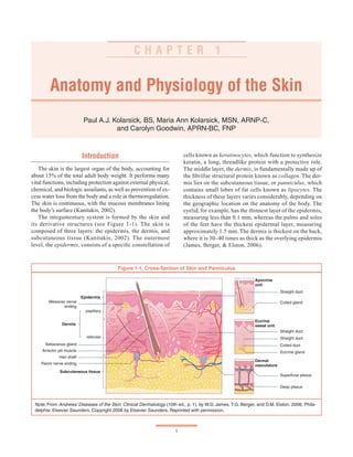
  • 1. 1 Anatomy and Physiology of the Skin Paul A.J. Kolarsick, BS, Maria Ann Kolarsick, MSN, ARNP-C, and Carolyn Goodwin, APRN-BC, FNP C H A P T E R 1 Introduction The skin is the largest organ of the body, accounting for about 15% of the total adult body weight. It performs many vital functions, including protection against external physical, chemical, and biologic assailants, as well as prevention of ex- cess water loss from the body and a role in thermoregulation. The skin is continuous, with the mucous membranes lining the body’s surface (Kanitakis, 2002). The integumentary system is formed by the skin and its derivative structures (see Figure 1-1). The skin is composed of three layers: the epidermis, the dermis, and subcutaneous tissue (Kanitakis, 2002). The outermost level, the epidermis, consists of a specific constellation of cells known as keratinocytes, which function to synthesize keratin, a long, threadlike protein with a protective role. The middle layer, the dermis, is fundamentally made up of the fibrillar structural protein known as collagen. The der- mis lies on the subcutaneous tissue, or panniculus, which contains small lobes of fat cells known as lipocytes. The thickness of these layers varies considerably, depending on the geographic location on the anatomy of the body. The eyelid, for example, has the thinnest layer of the epidermis, measuring less than 0.1 mm, whereas the palms and soles of the feet have the thickest epidermal layer, measuring approximately 1.5 mm. The dermis is thickest on the back, where it is 30–40 times as thick as the overlying epidermis (James, Berger, & Elston, 2006). Figure 1-1. Cross-Section of Skin and Panniculus Note. From Andrews’ Diseases of the Skin: Clinical Dermatology (10th ed., p. 1), by W.D. James, T.G. Berger, and D.M. Elston, 2006, Phila- delphia: Elsevier Saunders. Copyright 2006 by Elsevier Saunders. Reprinted with permission. Epidermis Meissner nerve ending Dermis papillary reticular Sebaceous gland Arrector pili muscle Hair shaft Pacini nerve ending Subcutaneous tissue Apocrine unit Straight duct Coiled gland Eccrine sweat unit Straight duct Straight duct Coiled duct Eccrine gland Dermal vasculature Superficial plexus Deep plexus
  • 2. SKIN CANCER 2 Epidermis The epidermis is a stratified, squamous epithelium layer that is composed primarily of two types of cells: keratinocytes and dendritic cells. The keratinocytes differ from the “clear” dendritic cells by possessing intercellular bridges and ample amounts of stainable cytoplasm (Murphy, 1997).The epidermis harborsanumberofothercellpopulations,suchasmelanocytes, Langerhans cells, and Merkel cells, but the keratinocyte cell type comprises the majority of the cells by far. The epidermis commonly is divided into four layers according to keratino- cyte morphology and position as they differentiate into horny cells, including the basal cell layer (stratum germinativum), the squamous cell layer (stratum spinosum), the granular cell layer (stratum granulosum), and the cornified or horny cell layer (stratum corneum) (James et al., 2006; Murphy) (see Figure 1-2).The lower three layers that constitute the living, nucleated cells of the epidermis are sometimes referred to as the stratum malpighii and rete malpighii (Murphy). The epidermis is a continually renewing layer and gives rise to derivative structures, such as pilosebaceous apparatuses, nails, and sweat glands. The basal cells of the epidermis un- dergo proliferation cycles that provide for the renewal of the outer epidermis. The epidermis is a dynamic tissue in which cells are constantly in unsynchronized motion, as differing individual cell populations pass not only one another but also melanocytes and Langerhans cells as they move toward the surface of the skin (Chu, 2008). Keratinocytes At least 80% of cells in the epidermis are the ectodermally derived keratinocytes.The differentiation process that occurs as the cells migrate from the basal layer to the surface of the skin results in keratinization, a process in which the kerati- nocyte first passes through a synthetic and then a degradative phase (Chu, 2008). In the synthetic phase, the cell builds up a cytoplasmic supply of keratin, a fibrous intermediate filament arranged in an alpha-helical coil pattern that serves as part of the cell’s cytoskeleton. Bundles of these keratin filaments converge on and terminate at the plasma membrane forming the intercellular attachment plates known as desmosomes. During the degradative phase of keratinization, cellular organelles are lost, the contents of the cell are consolidated into a mixture of filaments and amorphous cell envelopes, and the cell finally is known as a horny cell or corneocyte. The process of maturation resulting in cell death is known as terminal differentiation (James et al., 2006). Basal Layer The basal layer, also known as the stratum germinativum, contains column-shaped keratinocytes that attach to the base- ment membrane zone with their long axis perpendicular to the dermis. These basal cells form a single layer and adhere to one another as well as to more superficial squamous cells through desmosomal junctions (Murphy, 1997). Other dis- tinguishing features of the basal cells are their dark-staining oval or elongated nuclei and the presence of melanin pigment transferred from adjoining melanocytes (Murphy). The basal layer is the primary location of mitotically active cellsintheepidermisthatgiverisetocellsoftheouterepidermal layers. However, not all basal cells have the potential to divide (Jones, 1996; Lavker & Sun, 1982). Epidermal stem cells in the basallayerareclonogeniccellswithalonglifespanthatprogress through the cell cycle very slowly under normal conditions. Hy- perplasiogenic conditions, such as wounding, can increase the number of cycling cells in the epidermis by stimulating division of stem cells. DNA damage caused by carcinogenic agents may mutate cell proliferation machinery and can also affect the rate of cellular division. Migration of a basal cell from the basal layer to the cornified layer in humans takes at least 14 days, and the transit through the cornified layer to the outermost epidermis requires another 14 days (Chu, 2008). Squamous Cell Layer Overlying the basal cell layer is a layer of the epidermis that is 5–10 cells thick and known as the squamous cell layer Figure 1-2.Three Basic Cell Types in the Epidermis The three basic cell types in the epidermis include keratinocytes (some labeled K) and Langerhans cells (L) in the Malpighian layer and melanocytes (M) in the basal layer. Arrows point to the basement membrane zone, which separates the basal layer of the epidermis from the underlying dermis (D). Note. From Andrews’ Diseases of the Skin: Clinical Dermatology (10th ed., p. 4), by W.D. James, T.G. Berger, and D.M. Elston, 2006, Philadelphia: Elsevier Saunders. Copyright 2006 by El- sevier Saunders. Reprinted with permission. M M L LK K D
  • 3. CHAPTER 1. ANATOMY AND PHYSIOLOGY OF THE SKIN 3 or stratum spinosum (Murphy, 1997). The squamous layer is composed of a variety of cells that differ in shape, structure, and subcellular properties depending on their location. Supra- basal spinous cells, for example, are polyhedral in shape and have a rounded nucleus, whereas cells of the upper spinous layers are generally larger in size, become flatter as they are pushed toward the surface of the skin, and contain lamellar granules (Chu, 2008). Lamellar granules are membrane-bound organelles containing glycoproteins, glycolipids, phospholip- ids, free sterols, and a number of acid hydrolases, including lipases, proteases, acid phosphatases, and glycosidases. The abundance of hydrolytic enzymes indicates that the lamel- lar granules are a type of lysosome. Although the lamellar granules primarily are active in cells at the interface between the granular and cornified layers, they also function in cells of the upper spinous layer to deliver precursors of stratum corneum lipids into the intercellular space (Haake & Hol- lbrook, 1999). Intercellular spaces between spinous cells are bridged by abundant desmosomes that promote mechanical coupling be- tween cells of the epidermis and provide resistance to physical stresses. Organized concentrically around the nucleus, keratin filaments in the cytoplasm are bound to desmosomal plaques at one end and remain free at the end closer to the nucleus (Murphy, 1997). The desmosomal plaques are composed of six polypeptides found on the cytoplasmic side of the cell membrane that are important in the regulation of the calcium required for desmosomal assembly and maintenance (Fairley, Scott, Jensen, Goldsmith, & Diaz, 1991; Hennings & Holbrook, 1983; Lin, Mascaro, Liu, Espana, & Diaz, 1997).The spine-like appearance of the numerous desmosomes along cell margins is where the stratum spinosum derives its name (Chu, 2008). Gap junctions are another type of connection between epi- dermal cells. Essentially forming an intercellular pore, these junctions allow for physiologic communication via chemi- cal signals that is vital in the regulation of cell metabolism, growth, and differentiation (Caputo & Peluchetti, 1977). Granular Layer The most superficial layer of the epidermis containing living cells, the granular layer or stratum granulosum, is composed of flattened cells holding abundant keratohyaline granules in their cytoplasm. These cells are responsible for further synthesis and modification of proteins involved in keratinization (Chu, 2008).The granular layer varies in thick- ness in proportion to that of the overlying horny cell layer. For example, under thin cornified layer areas, the granular layer may be only 1–3 cell layers in thickness, whereas under the palms of the hands and soles of the feet the granular layer may be 10 times this thickness.A very thin or absent granular layer can lead to extensive parakeratosis in which the nuclei of keratinocytes persist as the cells move into the stratum corneum, resulting in psoriasis (Murphy, 1997). The keratohyaline granules are deeply basophilic and irregular in shape and size, and they are necessary in the formation of both the interfibrillary matrix that holds keratin filaments together and the inner lining of the horny cells. Enzymatic action of the keratohyaline granules results in the production of “soft” keratin in the epidermis by providing periodic cutting of keratin filaments. In contrast, the hair and nails do not contain keratohyaline granules, and the tonofibril filaments traversing the cell cytoplasm will harden because of the incorporation of disulfide bonds, producing “hard” keratin in those structures (Matoltsy, 1976; Schwarz, 1979). Lysosomal enzymes present only in small amounts in the stratum basalis and stratum spinosum are found at high lev- els in the stratum granulosum because the granular layer is a keratogenous zone of the epidermis. Here, the dissolution of cellular organelles is prepared as the cells of the granular layer undergo the abrupt terminal differentiation process to a horny cell of the cornified layer (Chu, 2008). Cornified Layer Horny cells (corneocytes) of the cornified layer provide mechanical protection to the underlying epidermis and a bar- rier to prevent water loss and invasion by foreign substances (Jackson, Williams, Feingold, & Elias, 1993). The corneo- cytes, which are rich in protein and low in lipid content, are surrounded by a continuous extracellular lipid matrix (Chu, 2008).The large, flat, polyhedral-shaped horny cells have lost their nuclei during terminal differentiation and technically are considered to be dead (Chu; Murphy, 1997).The physical and biochemical properties of cells in the cornified layer vary in accordance with position in order to promote desquama- tion moving outward. For instance, cells in the middle have a much higher capacity for water-binding than the deeper layers because of the high concentration of free amino acids found in the cytoplasm of middle layer cells. The deep cells also are more densely compact and display a greater array of intercellular attachments than the more superficial layers. Desmosomes undergo proteolytic degradation as the cells progress outward, contributing to the shedding of corneocytes during desquamation (Haake & Hollbrook, 1999). The Regulation of Epidermal Proliferation and Differentiation As a perpetually regenerating tissue, the epidermis must maintain a relatively constant number of cells as well as regu- late the interactions and junctions between epidermal cells. Adhesions between keratinocytes, the interactions of kerati- nocytes and immigrant cells, the adhesion between the basal lamina and the underlying dermis, and the process of terminal differentiation to produce corneocytes must be regulated as cells relocate during development as well as throughout life
  • 4. SKIN CANCER 4 (Haake & Hollbrook, 1999). Epidermal morphogenesis and differentiation is regulated in part by the underlying dermis, which also plays a critical role in the maintenance of postnatal structure and function.The epidermal-dermal interface is also a key site in the development of epidermal appendages. The maintenance of a constant epidermal thickness de- pends also on intrinsic properties of epidermal cells, such as the ability to undergo apoptosis, programmed cell death. Apoptosis follows an orderly pattern of morphologic and biochemical changes resulting in cell death without injury to neighboring cells, as is often the case in necrosis. This major homeostatic mechanism is regulated by a number of cellular signaling molecules including hormones, growth factors, and cytokines. In the skin, apoptosis is important in developmental remodeling, regulation of cell numbers, and defense against mutated, virus-infected, or otherwise damaged cells. Terminal differentiation is a type of apoptosis evolved to convert the ke- ratinocyte into the protective corneocyte (Haake & Hollbrook, 1999). The disruption of dynamic equilibrium maintaining constant epidermal thickness can result in conditions such as psoriasis, whereas the dysregulation of apoptosis is often seen in tumors of the skin (Kerr, Wyllie, & Currie, 1972). Nonkeratinocyte Cells of the Epidermis Melanocytes The melanocyte is a dendritic, pigment-synthesizing cell derived from the neural crest and confined in the skin pre- dominantly to the basal layer (Chu, 2008). Branching into more superficial layers, extensions of the melanocyte come into contact with keratinocytes but do not form cellular junc- tions. Melanocytes are responsible for the production of the pigment melanin and its transfer to keratinocytes. Melanin is produced in a rounded, membrane-bound organelle known as the melanosome via a series of receptor-mediated, hormone- stimulated, enzyme-catalyzed reactions (Haake & Hollbrook, 1999). Melanosomes are moved to the end of the melanocyte pro- cesses that lie closest to the skin surface and are transferred to keratinocytes (see Figure 1-3). In white skin, these mel- anosomes are aggregated into membrane-bound melanosome complexes containing two or three melanosomes, whereas melanosomes tend to be removed from these complexes more rapidly in keratinocytes of individuals with dark skin. Heavily pigmented skin can be attributed to the greater production of melanosomes in melanocytes, the higher degree of melaniza- tion in each melanosome, the larger size of melanosomes, the greater amount of dispersion of melanosomes in keratinocytes, and the slower rate of melanosome degradation in comparison to fair skin (Flaxman, Sosis, &Van Scott, 1973; Murphy, 1997; Olson, Nordquist, & Everett, 1970). Increased ultraviolet light exposure stimulates an increase in melanogenesis and a corresponding increase in melanosome transfer to keratinocytes where the melanosomes will aggre- gate toward the superficial side of the nucleus.This response, which results in tanning of the skin, increases the cell’s ability to absorb light and thus protect genetic information in the nucleus from damaging radiation. Merkel Cells Merkel cells are oval-shaped, slow-adapting, type I mechanoreceptors located in sites of high tactile sensitiv- ity that are attached to basal keratinocytes by desmosomal junctions. Merkel cells are found in the digits, lips, regions of the oral cavity, and outer root sheath of the hair follicle and are sometimes assembled into specialized structures known as tactile discs or touch domes (Moll, 1994). Rela- tively small deformations of adjoining keratinocytes are stimulus enough to cause Merkel cells to secrete a chemi- cal signal that generates an action potential in the adjoin- ing afferent neuron, which relays the signal to the brain. The high concentration of Merkel cells in certain regions such as the fingertips results in smaller and more densely Figure 1-3. Portion of a Melanocyte From Dark Skin Melanosomes are indicated by broad arrows. Thin arrows point to the basement membrane zone between the epidermis and the underlying dermis (D). Note. From Andrews’ Diseases of the Skin: Clinical Dermatology (10th ed., p. 4), by W.D. James, T.G. Berger, and D.M. Elston, 2006, Philadelphia: Elsevier Saunders. Copyright 2006 by El- sevier Saunders. Reprinted with permission. D
  • 5. CHAPTER 1. ANATOMY AND PHYSIOLOGY OF THE SKIN 5 packed receptive fields and thus higher tactile resolution and sensitivity. Langerhans Cells Langerhanscells areinvolvedinavarietyofT-cellresponses. Derivedfromthebonemarrow,thesecellsmigratetoasuprabasal position in the epidermis early in embryonic development and continue to circulate and repopulate the epidermis throughout life. The cells are dendritic and do not form cellular junctions with neighboring cells. Langerhans cells constitute 2%–8% of the total epidermal cell population and maintain nearly constant numbers and distributions in a particular area of the body. In the epidermis, the cells mainly are distributed among the squamous and granular layers with fewer cells in the basal layer. They are found in other squamous epithelia in addition to the epidermis, including the oral cavity, esophagus, and vagina, as well as in lymphoid organs and in the normal dermis (Chu, 2008). Langerhans cells must recognize and process soluble an- tigens found in epidermal tissue. When a membrane-bound antigen is ingested via endocytosis, cell granules are formed. The contents of these granules are delivered to phagolyso- somes in the cytoplasm containing hydrolytic enzymes similar to those found in macrophages. In the first stage of life, the Langerhans cells are weak stimulators of unprimedT cells but are able to ingest and process antigens. Later, once the cell has become an effective activator of naïve T cells, activation via contact with the antigen will not trigger phagocytosis but rather will stimulate cell migration (Udey, 1997). The Dermal-Epidermal Junction The interface between the epidermis and dermis is formed by a porous basement membrane zone that allows the exchange of cells and fluid and holds the two layers together (James et al., 2006). Basal keratinocytes are the most important components of structures of the dermal-epidermal junction; dermal fibro- blasts are also involved but to a lesser extent (Gayraud, Hopfner, Jassim, Aumailley, & Bruckner-Tuderman, 1997). The basal lamina is a layer synthesized by basal cells of the epidermis consisting mainly of type IV collagen as well as anchoring fibrils and dermal microfibrils. This includes an electron-lucent zone known as the lamina lucida as well as the lamina densa (Aumailley & Krieg, 1996; Lin et al., 1997; Masunaga et al., 1996;Wheelock & Jensen, 1992).The plasma membranes of basal cells are attached to the basal lamina by rivet-like hemidesmosomes that distribute tensile or shearing forces through the epithelium.The dermal-epidermal junction acts as support for the epidermis, establishes cell polarity and direction of growth, directs the organization of the cy- toskeleton in basal cells, provides developmental signals, and functions as a semipermeable barrier between layers (Stepp, Spurr-Michaud, Tisdale, Elwell, & Gipson, 1990). Epidermal Appendages The skin adnexa are a grouping of ectodermally derived appendages, including eccrine and apocrine glands, ducts, and pilosebaceous units that originate as downgrowths from the epidermis during development. After injury, all adnexal structures are capable of reepithelialization via the migration of keratinocytes from adnexal epithelium to the surface of the epidermis. Because areas such as the face and scalp contain a large quantity of pilosebaceous units, reepithelialization occurs more rapidly after injury in these areas than in areas with fewer adnexal structures, such as the back (James et al., 2006). Eccrine Sweat Glands Eccrine sweat glands are involved in the regulation of heat and are most abundant on the soles of the feet and least plentiful on the back (Murphy, 1997; Sato & Dobson, 1970). The sweat glands originate as a band of epithelial cells growing downward from the epidermal ridge (Mauro & Goldsmith, 2008). This tubular, or ductal, structure is modified during development to generate the three composite parts of the eccrine sweat unit, which are the intraepidermal spiral duct, the straight dermal portion, and the coiled secretory duct (see Figure 1-1) (James et al., 2006; Mauro & Goldsmith). The spiral duct opens onto the skin surface and is composed of dermal duct cells that have migrated upward. Cells undergo cornification within the duct, andthecorneocytesproducedultimatelywillbecomepartofthe cornified layer.The straight dermal segment connects the super- ficial spiral duct to the inner secretory portion of the gland. The secretory coil of the eccrine unit lies deep in the der- mis or within the superficial panniculus and is composed of glycogen-rich clear secretory cells, dark mucoidal cells, and myoepithelial cells specialized in contractile properties (James et al., 2006; Mauro & Goldsmith, 2008). Clear cells rest either on the basement membrane or on the myoepithelial cells and form intercellular canaliculi where two clear cells adjoin.The canaliculi open directly into the lumen of the gland (Mauro & Goldsmith). Large, glycogen-rich inner epithelial cells initi- ate the formation of sweat in response to a thermal stimulus. Initially an isotonic solution, the darker mucoidal cells in the secretory coil and in the dermal duct actively reabsorb sodium from sweat in the duct, thereby resulting in the extremely hypotonic solution that is emitted onto skin surface through the intraepidermal spiral duct.This response promotes cooling while conserving sodium (James et al.). Apocrine Sweat Glands Whereas eccrine glands are primarily involved in thermal regulation, apocrine glands are involved in scent release (Mur- phy, 1997). Apocrine sweat glands in humans are confined mainly to the regions of the axillae and perineum, and unlike eccrine and apoeccrine glands, they do not open directly to
  • 6. SKIN CANCER 6 the skin surface. Instead, the intraepithelial duct opens into pilosebaceous follicles, entering in the infundibulum above the sebaceous duct. The basal secretory coil of apocrine glands, which is normally located entirely in subcutaneous fat, differs from that of eccrine glands in that it is composed exclusively of secretory cells; no ductal cells are present (Murphy). Apocrine sweat glands develop their secretory portions and become active just before puberty, a response induced presumably by hormonal signals. The proteinaceous, viscous secretion has distinct odor and can function as a territorial marker, warning signal, and sexual attractant, but its sexual functions may now be vestigial in humans. Difficulties in acquiring pure samples of apocrine sweat have made it im- possible to determine the exact chemical composition of the secretion (Mauro & Goldsmith, 2008). Apoeccrine Sweat Glands The apoeccrine sweat gland (AEG) develops during puberty from eccrine-like precursors, opening directly unto the skin. Discovered during the isolation of human axillary sweat from patients with axillary hyperhidrosis, a condition characterized by abnormally increased rates of perspiration, the AEG is found in the adult axillae; its relative frequency varies from person to person. Like eccrine glands, the AEG opens directly to the skin surface. The AEG has a secretory rate as much as 10 times that of the eccrine gland and is there- fore thought to contribute to axillary hyperhidrosis (Mauro & Goldsmith, 2008). Hair Follicles Hair has many valuable biologic functions including protection from the elements and distribution of sweat-gland products. In addition, it has an important psychosocial role in society. Hair follicles vary considerably in size and shape, depending on their location, but they all have the same basic structure. The number and distribution of hair follicles over the body and the future phenotype of each hair is established during fetal development; no extra follicles are added after birth (Kratochwil, Dull, Farinas, Galceran, & Grosschedl, 1996; Millar, 1997; Paus & Cotsarelis, 1999; Paus, Foitzik, Welker, Bulfone-Paus, & Eichmuller, 1997; St-Jacques et al., 1998; Zhou, Byrne, Jacobs, & Fuchs, 1995). The particular spacing and allocation of the follicles are determined by genes that are expressed very early in the morphogenesis of the follicles (Paus & Cotsarelis; St-Jacques et al.). Mesenchymal cells in the fetal dermis aggregate below the basal layer of the epidermis during embryogenesis (James et al., 2006). The basophilic cells in the basal cell layer of the epidermis overlying these mesenchymal cell sites are induced to grow at a downward angle into the dermis (Murphy, 1997). The follicle continues to develop until finally widening at the base and forming a bulb around the group of mesenchymal cells from which the dermal papilla is formed (James et al.; Hashimoto, 1970a). Differentiation occurs at the lower portion of the hair fol- licle forming the hair cone and later the hair, the cuticle, and the two inner root sheaths. Differentiation also occurs in the upper segments of the follicle producing the hair canal in the upper dermis, through the epidermis, and opening to the surface prior to the time that the growing hair cone reaches the upper follicle (Hashimoto, 1970b). The sebaceous gland forms from a bud in the fetal hair follicle. Along the same side of the follicle but below the sebaceous gland, another bud develops into an attachment for the arrector pili muscle. The arrector pili (AP) are a smooth muscle bundle that attaches to the external root sheath of the follicle. The distal end of the AP muscle shows multiple branches at the level of the papillary dermis.The bulge, which is the zone of theAP muscle’s follicular attachment, is thought to contain epithelial stem cells responsible for regenerating follicles, a crucial role in the hair growth cycle (Cotsarelis, Sun, & Lavker, 1990). On the opposite side of the follicle, a third bud forms above the plane of the sebaceous gland and develops into the apocrine gland. The region of the follicle above the sebaceous gland is known as the infundibular segment, and the region between the sebaceous duct and AP attachment is known as the isthmus (James et al., 2006). The region below the isthmus is known as the inferior portion and contains the bottom of the follicle as well as the hair bulb.The inferior segment undergoes cycles of involution and regenera- tion throughout life, whereas the infundibular and isthmus portions remain permanently (James et al.). Rapidly proliferating cells in the hair bulb, called matrix cells, are responsible for the production of the hair shaft as well as the inner and outer root sheaths (James et al., 2006). Both the hair shaft and the inner root sheath progress upward as the hair grows until the inner sheath reaches the isthmus and sheds (James et al.). Matrix cells moving up the follicle are compressed as they enter the rigid inner root sheath (see Figure 1-4). The number of cells entering the sheath deter- mines the size of the hair, and the dimensions and curvature of the inner root sheath determine the shape of the hair (Paus & Cotsarelis, 1999). For example, the hairs on the scalp of Caucasians are round while pubic, facial, and eyelash hairs are oval. Scalp hairs on people of African descent also are oval, causing their hair to be curly (James et al., 2006). Hair color is determined by the distribution of melano- somes in the hair shaft. The hair bulb contains melanocytes that synthesize melanosomes and transfer them to the kera- tinocytes of the bulb matrix. People of African descent tend to have larger melanosomes than Caucasians, whose smaller melanosomes are amassed into membrane-bound complexes. Red hair contains spherical melanosomes.Aging causes a loss of melanocytes and a corresponding decrease in the produc- tion of melanosomes and results in graying hair (James et al., 2006).
  • 7. CHAPTER 1. ANATOMY AND PHYSIOLOGY OF THE SKIN 7 impressive effect are the androgens: testosterone and its active metabolite, dihydrotestosterone, which act through androgen receptors in the dermal papilla. These hormones increase the size of hair follicles in androgen-dependent areas such as the beard area during adolescence. Later in life, however, they can cause miniaturization of follicles in the scalp resulting in androgen alopecia (male pattern baldness) (Kaufman, 1996; Sawaya, 1994). Except for rare congenital hair defects caused by mutations in keratins or other structural proteins and scarring alopecias, hair loss and unwanted hair growth reflect deviations of hair follicle cycling and, therefore, are considered reversible events (Paus, 1996).The hair cycle can vary depending on a number of different physiologic factors. Pregnancy, for example, often results in a prolongation of the telogen phase and an increased number of scalp hairs in the anagen phase. When estrogen levels equilibrate after delivery, telogen hairs are lost while anagen hairs simultaneously are converted to telogen, and this great quantity of telogen hairs will be lost in three to five months.The synchronous termination of anagen or telogen is known as telogen effluvium and is often observed after trauma, such as childbirth, surgery, weight loss, and severe stress, and also is associated with drugs, endocrine disorders, anemia, and malnutrition (James et al., 2006). Regrowth typically follows, with the exception of any metabolic or nutritional deficiency (Headington, 1993; Paus & Cotsarelis, 1999). Sebaceous Glands Sebaceous glands are found in greatest number on the face and scalp but are present on nearly all other locations of the body with the exception of the tarsal plate of the eye- lids, the buccal mucosa and vermilion borders of the lip, the prepuce and mucosa lateral to the penile frenulum, the labia minora, and the female areola (James et al., 2006). Cells of the sebaceous glands contain abundant lipid droplets known as sebum in their cytoplasm and are arranged into lobules off the upper segment of the hair follicle. Basaloid germinative cells surrounding the lobule give rise to the lipid-filled cells, which are then expelled into the infundibular segment of the hair follicle via the sebaceous duct.The sebaceous glands are thought to be evolutionarily important in providing a second- ary lubrication during the passage through the birth canal. This extra lubrication covers the surfaces that come in direct contact with the birth canal including the vertex, anterior scalp over the forehead and nose to the lower jaw line, and the shoulders, chest, and upper aspect of arms posteriorly (Danby, 2005; Thiboutot, 2004). Nails Fingernails provide protection to the fingertips, enhance sensation, and allow small objects to be grasped. The un- derlying nail bed is part of the nail matrix containing blood Figure 1-4. Hair Follicle Structure Note. From Andrews’ Diseases of the Skin: Clinical Dermatology (10th ed., p. 8), by W.D. James, T.G. Berger, and D.M. Elston, 2006, Philadelphia: Elsevier Saunders. Copyright 2006 by El- sevier Saunders. Reprinted with permission. Hair growth occurs in a cyclical manner, but each follicle functions as an independent unit.The hair growth cell cycle is composed of three stages: anagen, catagen, and telogen (see Figure 1-5) (Millar, 1997; Paus, 1996; St-Jacques et al., 1998). Anagen, the active growth stage, typically lasts approximately three to five years on the scalp, during which hairs grow at a rate of about 0.33 mm per day.The length of the anagen phase decreases with age and decreases dramatically in individuals with alopecia (James et al., 2006). Catagen usually lasts about two weeks and is a period of involution resulting in club hair formation after many cells in the outer root sheath undergo apoptosis.The resting phase, telogen, lasts about three to five months on the scalp, and hairs in this stage are eventually pushed out by the growing anagen hair shaft. Other sites on the body tend to have shorter anagen and longer telogen phases, causing most body hair to be shorter and remain in place for longer periods of time (James et al., 2006). Two secreted molecules that may have important roles in hair follicle development and cycling are the insulin-like growth factor 1 and fibroblast growth factor 7. In mice, both are produced by the dermal papilla and have receptors pre- dominantly in overlying matrix cells (Danilenko, Ring, & Pierce, 1996). Hormonal factors controlling hair growth in- clude estrogens, thyroid hormones, glucocorticoids, retinoids, prolactin, and growth hormone. The hormones with the most
  • 8. SKIN CANCER 8 vessels, nerves, and melanocytes and has parallel rete ridges. The nail plate is formed from matrix keratinocytes (James et al., 2006). Fingernails grow at an average rate of 0.1 mm per day, two to three times faster than the rate of toenail growth. Because of the slow growth rate, toenails can provide information about toxic exposure or disease from many months in the past (James et al., 2006). For example, arsenic poisoning may cause a horizontal hypopigmentation across all nail plates known as Mees lines (Daniel & Scher, 1997). The Dermis The dermis is an integrated system of fibrous, filamen- tous, and amorphous connective tissue that accommodates stimulus-induced entry by nerve and vascular networks, epidermally derived appendages, fibroblasts, macrophages, and mast cells. Other blood-borne cells, including lympho- cytes, plasma cells, and other leukocytes, enter the dermis in response to various stimuli as well. The dermis comprises the bulk of the skin and provides its pliability, elasticity, and tensile strength. It protects the body from mechanical injury, binds water, aids in thermal regulation, and includes receptors of sensory stimuli. The dermis interacts with the epidermis in maintaining the properties of both tissues.The two regions collaborate during development in the morphogenesis of the dermal-epidermal junction and epidermal appendages and interact in repairing and remodeling the skin as wounds are healed. The dermis does not undergo an obvious sequence of differentiation that parallels epidermal differentiation, but the structure and organization of the connective tissue components are predictable in a depth-dependent manner. The matrix components, including collagen and elastic connective tissue, also vary in a depth-dependent manner and undergo turnover and remodeling in normal skin, in pathologic processes, and in response to external stimuli (Chu, 2008). The constituents of the dermis are mesodermal in origin except for nerves, which, like melanocytes, derive from the neural crest. Until the sixth week of fetal life, the dermis is merely a pool of dendritic-shaped cells full of acid-muco- polysaccharides, which are the precursors of fibroblasts. By the 12th week, fibroblasts are actively synthesizing reticu- lum fibers, elastic fibers, and collagen. A vascular network develops and fat cells have appeared beneath the dermis by the 24th week. Infant dermis is composed of small collagen bundles, whereas the adult dermis contains thicker bundles of collagen. Many fibroblasts are present in the infant dermis, but few persist in adulthood (James et al., 2006). The principal component of the dermis is collagen, a fi- brous family of proteins with at least 15 genetically distinct types in human skin. A major structural protein for the entire body, collagen is found in tendons, ligaments, the lining of bones, and the dermis. Collagen is a major stress-resistant ma- terial of the skin. Elastic fibers, on the other hand, play a role in maintaining elasticity but do very little to resist deformation and tearing of the skin. Collagen fibers exist in a constant state of flux, being degraded by proteolytic enzymes called spare collagenases and replaced by new fibers. Collagen represents 70% of the skin’s dry weight (James et al., 2006). Figure 1-5. Phases of Hair Growth Note. From Andrews’ Diseases of the Skin: Clinical Dermatology (10th ed., p. 9), by W.D. James, T.G. Berger, and D.M. Elston, 2006, Phila- delphia: Elsevier Saunders. Copyright 2006 by Elsevier Saunders. Reprinted with permission.
  • 9. CHAPTER 1. ANATOMY AND PHYSIOLOGY OF THE SKIN 9 Fibroblasts integrate the procollagen molecule, a specific helical polypeptide chain. Then, the cell secretes the fibro- blasts, and they assemble into collagen fibrils.The amino acids glysine, hydroxyproline, and hydroxylysine highly enrich collagen. The fibrillar collagens found in the skin comprise the major group and are the most abundant proteins in the body. The major constituent of the dermis is type I collagen. Loosely positioned collagen fibers are found in the papillary and adventitial dermis, whereas hefty collagen bundles are noted in the reticular dermis.Type IV collagen is found in the basement membrane zone, and the major structural component of anchoring fibrils is collagen type VII, which is produced primarily by keratinocytes (James et al., 2006). The elastic fiber differs both structurally and chemically from collagen and consists of two components: protein fila- ments and elastin, an amorphous protein.The fibroblast fuses elastic fiber to the extracellular matrix of the dermis, which is composed of glycosaminoglycans. The fibers are fine in the papillary dermis and coarse in the reticular dermis. Hyaluronic acid is a minor component of the normal dermis but is the major mucopolysaccharide that accumulates in pathologic states (James et al., 2006). Vasculature The dermal vasculature is made up of two intercom- municating plexuses: the subpapillary or superficial plexus composed of postcapillary venules found at the junction of the papillary and reticular dermis and the lower plexus at the der- mal-subcutaneous interface.The dermal papillae are supplied by capillaries, end arterioles, and venules of the superficial plexus. The deeper plexus is supplied by larger blood vessels and is more complex surrounding adnexal structures. Blood flow in human skin fluctuates significantly in response to thermal stress because of the regulation of the preoptic-anterior hypothalamus (Boulant, 2000). Vasodilation and increased skin blood flow, along with sweating, are crucial to heat dissipation during heat exposure and exercise. During exposure to cold, vasoconstriction in the skin decreases heat loss from the body to prevent hypothermia. Altered control of skin blood flow can considerably impair the ability to maintain normal body temperature (Charkoudian, 2003). For example, impairmentsincutaneousvascularcontrolnotedinpatientswith typeIIdiabetesmaycontributetotheincreasedincidenceofheat stroke and heat exhaustion during periods of elevated external temperatures. Similarly, menopausal hormones result in the occurrence of hot flashes (Brooks et al., 1997; Schuman, 1972; Semenza,McCullough,Flanders,McGeehin,&Lumpkin,1999; Tankersley, Nicholas, Deaver, Mikita, & Kenney, 1992). Muscles Involuntary or smooth muscle of the skin occurs as AP, tunica dartos of the external genitals, and the areolas around the nipples. The location of the nucleus in the center of the muscle cell and the absence of striation distinguishes smooth muscle from striated muscle (Murphy, 1997). The muscle fibers of the arrectores pilorum are located in the connective tissue of the upper dermis and are attached to the hair follicle below the sebaceous glands.They are situated at such an angle to the hair follicle that when contracted, the hair follicle is pulled into a vertical position, deforming the skin and causing “gooseflesh” (James et al., 2006). Different configurations make up small bundles of smooth muscle of the muscularis of veins and arteries. Glomus bodies are specialized aggregates of smooth muscle found between the arterioles and venules, which exist on the digits and lateral aspects of the palms and soles. They regulate body temperature and shunt blood (James et al.). Striated or voluntary muscle is found in the skin of the neck as platysma and in the skin of the face as muscle of expression. The superficial muscular aponeurotic system is an intricate network of muscle, fascia, and aponeuroses connecting muscles with the parts that they move (James et al., 2006). Nerves Nerve bundles, together with arterioles and venules, are found in great quantity in neurovascular bundles of the dermis (James et al., 2006). Meissner corpuscles, found in the dermal papillae, help to mediate touch and are found predominantly on the ventral sides of the hands and feet. Meissner corpuscles occur in greater abundance on the hands, with greatest con- centration in the fingertips. Vater-Pacini corpuscles are large nerve-end organs that generate a sense of pressure and are located in the deeper portion of the dermis of weight-bearing surfaces and genitalia. They also are found commonly in the nipple and anogenital region. Pain, temperature, and itch sensation are transmitted by unmyelinated nerve fibers that end around hair follicles and the papillary dermis (James et al.). Vasoconstriction is regulated by the postganglionic adren- ergic fibers of the autonomic nervous system. This system regulates the apocrine gland secretions and the contraction of AP muscles of hair follicles. Eccrine sweat secretions, on the other hand, are mediated by cholinergic fibers (James et al., 2006). Mast Cells Mast cells are specialized secretory cells derived from bone marrow and distributed in connective tissues through- out the body. Although present in greatest numbers in the papillary dermis, they also are present in the subcutaneous fat (Chu, 2008). In the normal dermis, mast cells appear as oval to spindle-shaped cells with a centrally located round to oval nucleus. Numerous mast cells are located around blood
  • 10. SKIN CANCER 10 vessels, especially postcapillary venules. Upon magnifica- tion, mast cells reveal numerous large and long villi at their periphery. Mast cell granules are round, oval, or angular membrane-bound structures containing histamine, heparin, serine proteinases, and certain cytokines (Murphy, 1997).The cell’s surface contains hundreds of thousands of glycoprotein receptor sites for immunoglobulin E.Type I or connective tis- sue mast cells are located in the dermis and submucosa. Type II or mucosal mast cells are located in the respiratory tract mucosa and in the bowel (James et al., 2006). Mast cells accumulate in the skin because of abnormal proliferation, migration, and failure of apoptosis when mas- tocytosis occurs. Traditionally associated with the allergic response, more recent studies suggest that these cells also may be capable of regulating inflammation, host defense, and in- nate immunity. Mast cells can undergo activation by antigens or allergens acting via the high-affinity receptor for immuno- globulin E, superoxides, complement proteins, neuropeptides, and lipoproteins.After activation, mast cells express histamine, leukotrienes, prostanoids, proteases, and many cytokines and chemokines. These mediators may be pivotal to the genesis of an inflammatory response. By virtue of their location and mediator expression, mast cells are thought to play an active role in many conditions such as allergy, parasitic diseases, atherosclerosis, malignancy, asthma, pulmonary fibrosis, and arthritis (Krishnaswamy, Ajitawi, & Chi, 2006). Subcutaneous Fat Embryologically, toward the end of the fifth month fat cells begin to develop in the subcutaneous tissue. These lobules of fat cells or lipocytes are separated by fibrous septa made up of large blood vessels and collagen. The panniculus var- ies in thickness depending on the skin site. Considered an endocrine organ, the subcutaneous tissue provides the body with buoyancy and functions as a storehouse of energy. Hor- mone conversion takes place in the panniculus, converting androstenedione into estrone by aromatase. Lipocytes produce leptin, a hormone that regulates body weight by way of the hypothalamus (James et al., 2006). Summary The three layers of the skin form an effective barrier to the external environment, allow the transmission of sensory information, and serve a significant role in maintaining ho- meostasis.The dynamic epidermis continually produces a pro- tective outer layer of corneocytes as cells undergo the process of keratinization and terminal differentiation. Collagen and elastic filaments of the dermal layer provide the underlying tensile strength of the skin, whereas the layer of subcutaneous fat provides a store of energy for the body. The high rate of cell proliferation in the epidermis and in epithelial tissue in general and the fact that this tissue is most frequently exposed to physical and chemical damage result in the exceedingly high rate of skin cancers found in humans as compared with other types of cancer. References Aumailley, M., & Krieg, T. (1996). Laminins: A family of diverse multifunctional molecules of basement membranes. Journal of Investigative Dermatology, 106(2), 209–214. Boulant, J.A. (2000). Role of the preoptic-anterior hypothalamus in thermoregulation and fever. Clinical Infectious Diseases, 31(Suppl. 5), S157–S161. Brooks, E.M., Morgan, A.L., Pierzga, J.M., Wladkowski, S.L., O’Gorman, J.T., Derr, J.A., et al. (1997). Chronic hormone re- placement therapy alters thermoregulatory and vasomotor func- tion in postmenopausal women. Journal of Applied Physiology, 83(2), 477–484. Caputo, R., & Peluchetti, D. (1977).The junctions of normal human epidermis: A freeze-fracture study. Journal of Ultrastructure Research, 61(1), 44–61. Charkoudian, N. (2003). Skin blood flow in adult human thermo- regulation: How it works, when it works, when it does not, and why. Mayo Clinic Proceedings, 78(5), 603–612. Chu, D.H. (2008). Overview of biology, development, and structure of skin. In K.Wolff, L.A. Goldsmith, S.I. Katz, B.A. Gilchrest,A.S. Paller, & D.J. Leffell (Eds.), Fitzpatrick’s dermatology in general medicine (7th ed., pp. 57–73). New York: McGraw-Hill. Cotsarelis, G., Sun, T.T., & Lavker, R.M., (1990). Label-retaining cells reside in the bulge of the pilosebaceous unit: Implications for follicular stem cells, hair cycle, and skin carcinogenesis. Cell, 61(7), 1329–1337. Danby, F.W. (2005). Why we have sebaceous glands. Journal of the American Academy of Dermatology, 52(6), 1071–1072. Daniel, R.C., & Scher, R.K. (1997). Nail changes secondary to systemic drugs and ingestants. In R.K. Scher & R.C. Daniel (Eds.), Nails:Therapy, diagnosis, surgery (2nd ed., pp. 251–258). Philadelphia: Saunders. Danilenko, D.M., Ring, B.D., & Pierce, G.F. (1996). Growth factors and cytokines in hair follicle development and cycling: Recent insights from animal models and the potentials for clinical therapy. Molecular Medicine Today, 2(11), 460–467. Fairley, J.A., Scott, G.A., Jensen, K.D., Goldsmith, L.A., & Diaz, L.A. (1991). Characterization of keratocalmin, a calmodulin-binding protein from human epidermis. Journal of Clinical Investigation, 88(1), 315–322. Flaxman, B.A., Sosis, A.C., & Van Scott, E.G. (1973). Changes in melanosome distribution in Caucasoid skin following topical application of nitrogen mustard. Journal of Investigative Derma- tology, 60(5), 321–326. Gayraud, B., Hopfner, B., Jassim, A., Aumailley, M., & Bruckner- Tuderman, L. (1997). Characterization of a 50-kDa component of epithelial basement membranes using GDA-J/F3 monoclonal antibody. Journal of Biological Chemistry, 272(14), 9531–9538. Haake, A.R., & Hollbrook, K. (1999). The structure and develop- ment of skin. In I. Freedberg, A. Eisen, K. Wolff, K. Austen, L. Goldsmith, S. Katz, et al. (Eds.), Fitzpatrick’s dermatology in general medicine (5th ed., pp. 70–111). New York: McGraw- Hill. Hashimoto, K. (1970a). The ultrastructure of the skin of human embryos V: The hair germ and perifollicular mesenchymal cells.
  • 11. CHAPTER 1. ANATOMY AND PHYSIOLOGY OF THE SKIN 11 Hair germ-mesenchyma interaction. British Journal of Dermatol- ogy, 83(1), 167–176. Hashimoto, K. (1970b). The ultrastructure of the skin of human embryos IX: Formation of the hair cone and intraepidermal hair canal. Archiv für Klinische und Experimentelle Dermatologie, 238(4), 333–345. Headington, J.T. (1993). Telogen effluvium: New concepts and review. Archives of Dermatology, 129(3), 356–363. Hennings, H., & Holbrook, K.A. (1983). Calcium regulation of cell-cell contact and differentiation of epidermal cells in culture: An ultrastructural study. Experimental Cell Research, 143(1), 127–142. Jackson, S.M.,Williams, M.L., Feingold, K.R., & Elias, P.M. (1993). Pathobiology of the stratum corneum. Western Journal of Medi- cine, 158(3), 279–285. James, W.D., Berger, T.G., & Elston, D.M. (2006). Andrews’ dis- eases of the skin: Clinical dermatology (10th ed.). Philadelphia: Elsevier Saunders. Jones, P.H. (1996). Isolation and characterization of human epidermal stem cells. Clinical Science, 91(2), 141–146. Kanitakis, J. (2002).Anatomy, histology and immunohistochemistry of normal human skin. European Journal of Dermatology, 12(4), 390–401. Kaufman, K.D. (1996). Androgen metabolism as it affects hair growth in androgenetic alopecia. Dermatologic Clinics, 14(4), 697–711. Kerr, J.F., Wyllie, A.H., & Currie, A.R. (1972). Apoptosis: A basic biological phenomenon with wide-ranging implication in tissue kinetics. British Journal of Cancer, 26(4), 239–257. Kratochwil, K., Dull, M., Farinas, I., Galceran, J., & Grosschedl, R. (1996). LeF1 expression is activated by BMP-4 and regulates inductive tissue interactions in tooth and hair development. Genes and Development, 10(11), 1382–1394. Krishnaswamy, G.,Ajitawi, O., & Chi, D.S. (2006).The human mast cell:An overview. In G. Krishnaswamy & D. Chi (Eds.), Molecular biology:Vol. 315. Mast cells: Methods and protocols (pp. 13–34). Totowa, NJ: Humana Press. Lavker, R.M., & Sun, T.T. (1982). Heterogeneity in basal kerati- nocytes: Morphological and functional correlations. Science, 215(4537), 1239–1241. Lin, M.S., Mascaro, J.M., Jr., Liu, Z., Espana, A., & Diaz, L.A. (1997). The desmosome and hemidesmosome in cutaneous au- toimmunity. Clinical and Experimental Immunology, 107(Suppl. 1), 9–15. Masunaga, T., Shimizu, H., Ishiko, A., Tomita, Y., Aberdam, D., Ortonne, J.P., et al. (1996). Localization of laminin-5 in the epidermal basement membrane. Journal of Histochemistry and Cytochemistry, 44(11), 1223–1230. Matoltsy,A.G. (1976). Keratinization. Journal of Investigative Der- matology, 67(1), 20–25. Mauro, T., & Goldsmith, L. (2008). Biology of eccrine, apocrine, and apoeccrine sweat glands. In K. Wolff, L.A. Goldsmith, S.I. Katz, B.A. Gilchrest,A.S. Paller, & D.J. Leffell (Eds.), Fitzpatrick’s dermatology in general medicine (7th ed., pp. 713–720). New York: McGraw-Hill. Millar, S. (1997). The role of patterning genes in epidermal differ- entiation. In P. Cowin & M.W. Klymkowsky (Eds.), Cytoskeletal- membrane interactions and signal transduction. Molecular biology intelligence unit (pp. 87–102). Austin, TX: Landes Bioscience. Moll, I. (1994). Merkel cell distribution in human hair follicles of the fetal and adult scalp. Cell and Tissue Research, 277(1), 131–138. Murphy, G.F. (1997). Histology of the skin. In D. Elder, R. Elenitsas, C. Jaworsky, & B. Johnson, Jr. (Eds.), Lever’s histopathology of the skin (8th ed., pp. 5–45). Philadelphia: Lippincott Williams & Wilkins. Olson, R.L., Nordquist, J., & Everett, M.A. (1970). The role of epidermal lysosomes in melanin physiology. British Journal of Dermatology, 83(1), 189–199. Paus, R. (1996). Control of the hair cycle and hair diseases as cycling disorders. Current Opinion in Dermatology, 3, 248–258. Paus, R., & Cotsarelis, G. (1999). The biology of hair follicles. New England Journal of Medicine, 341(7), 491–497. Paus, R., Foitzik, K., Welker, P., Bulfone-Paus, S., & Eichmuller, S. (1997). Transforming growth factor beta receptor type I and type II expression during murine hair follicle development and cycling. Journal of Investigative Dermatology, 109(4), 518–526. Sato, K., & Dobson, R.L. (1970). Regional and individual varia- tions in the function of the human eccrine sweat gland. Journal of Investigative Dermatology, 54(6), 443–449. Sawaya, M.E. (1994). Biochemical mechanisms regulating human hair growth. Skin Pharmacology, 7(1–2), 5–7. Schuman, S.H. (1972). Patterns of urban heat wave deaths and impli- cations for prevention: Data from NewYork and St. Louis during July 1966. Environmental Research, 5(1), 59–75. Schwarz, E. (1979). Biochemie der epidermalen keratinisation. InA. Marchionini (Ed.), Handbuch der hautund geschlectskrankheiten (Vol. I). Berlin: Springer-Verlag. Semenza, J.C., McCullough, J.E., Flanders, W.D., McGeehin, M.A., & Lumpkin, J.R. (1999). Excess hospital admissions during the July 1995 heat wave in Chicago. American Journal of Preventive Medicine, 16(4), 269–277. St-Jacques, B., Dassule, H.R., Karavanova, I., Botchkarev,V.A., Li, J., Danielian, P.S., et al. (1998). Sonic hedgehog signaling is essential for hair development. Current Biology, 8(19), 1058–1068. Stepp, M.A., Spurr-Michaud, S., Tisdale, A., Elwell, J., & Gipson, I.K. (1990). Alpha 6 beta 4 integrin heterodimer is a component of hemidesmosomes. Proceedings of the National Academy of Sciences of the United States of America, 87(22), 8970–8974. Tankersley, C.G., Nicholas, W.C., Deaver, D.R., Mikita, D., & Ken- ney, W.L. (1992). Estrogen replacement in middle-aged women: Thermoregulatory responses to exercise in the heat. Journal of Applied Physiology, 73(4), 1238–1245. Thiboutot, D. (2004). Regulation of human sebaceous glands. Journal of Investigative Dermatology, 123(1), 1–12. Udey, M.C. (1997). Cadherins and Langerhans cell immunobiology. Clinical and Experimental Immunology, 107(Suppl. 1), 6–8. Wheelock, M.J., & Jensen, P.J. (1992). Regulation of keratino- cyte intercellular junction organization and epidermal mor- phogenesis by E-cadherin. Journal of Cell Biology, 117(2), 415–425. Zhou, P., Byrne, C., Jacobs, J., & Fuchs, E. (1995). Lymphoid en- hancer factor 1 directs hair follicle patterning and epithelial cell fate. Genes and Development, 9(6), 700–713.