Congestive heart failure pathophysiology
•Download as PPT, PDF•
23 likes•1,280 views

Heart failure occurs when the heart cannot pump enough blood to meet the body's needs. The weakened heart muscle cannot contract effectively, reducing cardiac output and blood flow. This causes a build up of fluid in tissues like the lungs and legs, known as congestion. Over time, the strain on the heart further damages the myocardium and the condition worsens in a vicious cycle. Treatment focuses on managing symptoms with diuretics and vasodilators, and improving heart function long-term with medications like ACE inhibitors, beta-blockers, and ARBs.

Recommended
More Related Content
What's hot
What's hot (20)
Similar to Congestive heart failure pathophysiology
Similar to Congestive heart failure pathophysiology (20)
Recently uploaded
Recently uploaded (20)
Congestive heart failure pathophysiology
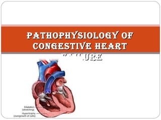
- 1. PATHOPHYSIOLOGY OFPATHOPHYSIOLOGY OF CONGESTIVE HEARTCONGESTIVE HEART FAILUREFAILURE
- 2. INTRODUCTION: Blood goes out of the heart when the heart muscle contracts (called systole) and comes into the heart when the muscle relaxes (called diastole). Heart failure is a disorder in which the heart pumps blood inadequately, leading to reduced blood flow which further weaken the heart. It can result from any structural or functional cardiac disorder that impairs ability of ventricle to fill with or eject blood. Most people have no symptoms at first - shortness of breath and fatigue develop gradually over days to months. It can occur in any age, even in young children. But more common among older people. About 5,00,000 new cases occur each year. Worldwide, about 23
- 3. Heart failure is called CCF/CHF because blood may build up in the tissues causing congestion in those tissues. Accumulation of blood coming into the left side of the heart causes congestion in the lungs, making breathing difficult. Accumulation of blood coming into the right side of the heart causes congestion and fluid accumulation in other parts of the body, such as the legs. Heart failure → ↓ CO → ↓ kidney perfusion → stim. Of RAA system
- 4. Types of heart failure:
- 6. ETIOLOGY:
- 7. SYMPTOMS:
- 8. PATHOPHYSIOLOGY: Heart failure is caused by any condition which reduces the efficiency of the myocardium or heart muscle, through damage or overloading of the ventricle, leads to reduced force of contraction. In a healthy heart, increased filling of the ventricle results in increased force of contraction (by the Frank–Starling law of the heart) and thus a rise in cardiac output. In heart failure this mechanism fails because heart muscle contraction becomes less efficient, due to reduced ability to cross-link actin and myosin filaments in over-stretched heart muscle. Decreased Systolic volume is caused by reduced contractility. Decreased diastolic volume results from impaired ventricular filling. Car diac out put (CO) = Heart Rat e (HR) x St roke Vol.(SV)
- 9. As the heart works harder to meet normal metabolic demands, the amount cardiac output can increase in times of oxygen demand (e.g. exercise) is reduced. This is failed during heart failure. Sympathetic activity may also cause potentially fatal arrhythmias. Hypertrophy (Enlargement) of the myocardium occur which result in increased stiffness and decreased ability to relax during diastole. The increase in ventricular volume also causes a reduction in stroke volume due to mechanical and contractile inefficiency. This increases the risk of cardiac arrest (specifically due to ventricular dysrhythmias) and reduces blood supply to the rest of the body. This destimulates baroreceptors in the carotid sinus and aortic arch which link to the nucleus tractus solitarii (center in the brain increases sympathetic activity, releasing catecholamines into the blood stream).
- 12. Reduced perfusion of muscle causes atrophy of the muscle fibres. This can result in weakness, increased fatigueability and decreased peak strength – all contributing to exercise intolerance. As a result, arm and leg muscles may tire more quickly and the kidneys may not function normally. The kidneys filter fluid and waste products from the blood into the urine, but when the heart cannot pump adequately, the kidneys malfunction and cannot remove excess fluid from the blood. As a result, the amount of fluid in the bloodstream increases, and the workload of the failing heart increases, creating a vicious circle. Thus, heart failure becomes even worse.
- 13. The increased peripheral resistance and greater blood volume place load on the heart and accelerates the process of damage to the myocardium. Vasoconstriction and fluid retention produce an increased hydrostatic pressure in the capillaries. This results in edema in the tissue. In lungs – this is called cardiogenic pulmonary edema. This reduces capacity for ventilation, causes stiffening of the lungs and reduces the efficiency of gas exchange by increasing the distance between the air and the blood causes dyspnea.
- 16. DIAGNOSIS ECG Echocardiography Chest X-rays Blood tests - electrolytes (sodium, potassium), measures of renal function, liver function tests, thyroid function tests, a complete blood count, elevated BNP. Angiography
- 17. TRAETMENT: Immediate treatments - Vasodilators + diuretics such as furosemide with ventilation. First-line therapy – Digoxin, ACE inhibitors, Diuretics, Beta-blockers, AT receptor blockers, Vasodilators, Parenteral iron if anaemia is found Immunosuppressive drugs to prevent rejection