
Recommended
More Related Content
What's hot
What's hot (20)
Similar to Ccf
Similar to Ccf (20)
More from Sowmya Shetty
More from Sowmya Shetty (12)
Recently uploaded
Recently uploaded (20)
Ccf
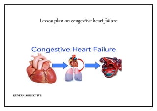
- 1. Lesson plan on congestive heart failure GENERALOBJECTIVE:
- 2. student will be able to gain knowledge regarding CCF, acquire skills, develop desirable attitude and able to practice in all health care settings globally. SPECIFIC OBJECTIVE: Student will be able to, define CCF enlist the causes ofCCF narrate the patho physiology discuss the types of CCF enlist the diagnostic procedures brief out the management of CCF
- 3. S.NO TIME SPECIFIC OBJECTIVE CONTENT TEACHING ACTIVITY LEARNE R ACTIVIT Y AV AIDS 1 1min introduce the topic BRINGING BACK INTO LIFE The heart is a hollow, muscular organ located in the centre of the thorax, where it occupies the space between the lungs (mediastinum) and rests on the diaphragm. It weighs approximately 300g (10.6 oz); heart weight and size are influenced by age, gender, body weight, extent of physical exercise and conditioning, and heart disease. The heart pumps blood to the tissues, supplying them with oxygen and other nutrients. Congestive heart failure occurs when the heart is not strong enough to pump blood efficiently around the body, causing fluid to collect in the lungs and body tissue, which leads to congestion. Heart failure becomes increasingly common with age. Congestive heart failure does not mean that the heart stops. It is a long-term condition that can be kept under control for many years with medication and lifestyle changes Asking questions Listening White board
- 4. 2 2min explain the anatomy and physiology Heart failure affects more than 750,000 people in the UK.The condition can affect people of all ages, but it is more common in older people (more than half of all people with heart failure are over 75 years of age). Heart failure is associated with a number of other serious health conditions, including coronaryheart disease,heartattack and high blood pressure (hypertension). ANATOMY AND PHYSIOLOGY OF THE HEART: The heart is a hollow, muscular organ located in the centre of the thorax, where it occupies the space between the lungs (mediastinum) and rests on the diaphragm. It weighs approximately 300g (10.6 oz); heart weight and size are influenced by age, gender, body weight, extent of physical exercise and conditioning, and heart disease. The heart pumps blood to the tissues, supplying them with oxygen and other nutrients. The pumping action of the heart is accomplished by the rhythmic contraction and relaxation of its muscular wall. During systole (contraction of the muscle), the chambers of the heart become smaller as the blood is ejected. During diastole (relaxation of the muscle), the heart chambers fill with blood in preparation for subsequent ejection. A normal resting adult heart beats Asking questions Listening Chart
- 5. approximately 60 to 80 times per minute. Each ventricle ejects approximately 70 mL of blood per beat and has an output of approximately 5 L per minute. STRUCTURE OF THE HEART
- 6. Heart Chambers The four chambers of the heart constitute the right- and left-sided pumping systems. The right side of the heart, made up of the right atrium and right ventricle, distributes venous blood(Deoxygenated blood) to the lungs via the pulmonary artery (pulmonary circulation) for oxygenation. The right atrium receives blood returning from the superior vena cava (head, neck, and upper extremities), inferior vena cava (trunk and lower extremities), and coronary sinus (coronary circulation). The left side of the heart, composed of the left atrium and left ventricle, distributes oxygenated blood to the remainder of the body via the aorta (systemic circulation). The left atrium receives oxygenated blood from the pulmonary circulation via the pulmonary veins. The varying thicknesses of the atrial and ventricular walls relate to the workload required by each chamber. The atria are thin-walled because blood returning to these chambers generates low pressures. In contrast, the ventricular walls are thicker because they generate greater pressures during systole. The right ventricle contracts against low pulmonary vascular pressure
- 7. and has thinner walls than the left ventricle. The left ventricle, with walls two-and-a-half times more muscular than those of the right PHYSIOLOGY OF HEART: The heart itself is made up of 4 chambers, 2 atria and 2 ventricles. De- oxygenated blood returns to the right side of the heart via the venous circulation. It is pumped into the right ventricle and then to the lungs where carbon dioxide is released and oxygen is absorbed. The oxygenated blood then travels back to the left side of the heart into the left atria, then into the left ventricle from where it is pumped into the aorta and arterial circulation The pressure created in the arteries by the contraction of the left ventricle is the systolic blood pressure. Once the left ventricle has fully contracted it begins to relax and refill with blood from the left atria. The pressure in the arteries falls whilst the ventricle refills. This is the diastolic blood pressure. The atrio-ventricular septum completely separates the 2 sides of the heart. Unless there is a septal defect, the 2 sides of the heart never directly communicate. Blood travels from right side to left side via the
- 8. lungs only. However the chambers themselves work together. The 2 atria contract simultaneously, and the 2 ventricles contract simultaneously. Cardiac Electrophysiology The cardiac conduction system generates and transmits electrical impulses that stimulate contraction of the myocardium. Under normal circumstances, the conduction system firstStimulates contraction of the atria and then the ventricles. The synchronization of the atrial and ventricular events allows the ventricles to fill completely before ventricular ejection, thereby maximizing cardiac output. Three physiologic characteristics of two specialized electrical cells, the nodal cells and the Purkinje cells, provide this synchronization: Automaticity: ability to initiate an electrical impulse Excitability: ability to respond to an electrical impulse Conductivity: ability to transmit an electrical impulse from one cell to another Both the senatorial (SA) node and the atrioventricular (AV) node are composed of nodal cells. The SA node, the primary pacemaker of the heart, is located at the junction of the superior vena cava and the right atrium. The SA
- 9. 3 2 mts Student will be able to define CCF? node in a normal resting adult heart has an inherent firing rate of 60 to 100 impulses per minute, but the rate can change in response to the metabolic demands of the body. The electrical impulses initiated by the SA node are conducted along the myocardial cells of the atria via specialized tracts called intermodal pathways. The impulses cause electrical stimulation and subsequent contraction of the atria. The impulses are then conducted to the AV node, which is located in the right atrial wall near the tricuspid valve. The AV node Coordinates the incoming electrical impulses from the atria and after a slight delay (allowing the atria time to contract and complete ventricular filling) relays the impulse to the ventricles. DEFINITION OF CONGESTIVE CARDIAC FAILURE HF is the inability of the heart to pump sufficient blood to meet the needs of the tissues for oxygen and nutrients. In the past, HF was often referred to as congestive heartfailure (CHF), A/ c to Brunner and Suddarths Heart failure is an abnormal clinical condition involving impaired cardiac pumping .It results in the characteristic pathophysiologic changes of vasoconstriction and fluid retention. teaching listening Can you define ccf?
- 10. 4. 2MIN Student will be able to enumerate the causes of ccf A/c to Lewis Heart failure, often called congestive heart failure or congestive cardiac failure, occurs when the heart is unable to provide sufficient pump action to maintain blood flow to meet the needs of the body. ... A/c to Wikipedia Congestive heart failure is the inability of the heart to pump an adequate amount of blood to the systemic circulation at normal filling pressures to meet the metabolic demands of the body. CAUSES OF CCF Heart failure is often a long-term (chronic) condition, but it may come on suddenly. It can be caused by many different heart problems. The condition may affect only the right side or only the left side of the heart. More often, both sides of the heart are involved. Heart failure is present when: Your heart muscle cannot pump (eject) the blood out of the heart very well. This is called systolic heart failure. lecturing Note taking Any body can tell causes of ccf?
- 11. Your heart muscles are stiff and do not fill up with blood easily. This is called diastolic heart failure. As the heart's pumping becomes less effective, blood may back up in other areas of the body. Fluid may build up in the lungs, liver, gastrointestinal tract, and the arms and legs. This is called congestive heart failure. The most common causes ofheart failure are: Coronary artery disease (CAD), a narrowing of the small blood vessels that supply blood and oxygen to the heart. This can weaken the heart muscle over time or suddenly. High blood pressure that is not well controlled, leading to problems with stiffness, or eventually leading to muscle weakening. 1. Cardiac a) Congenital heart disease – coarctation of aorta, endocardial fibroelastosis, transposition of great vessels, aortic atresia and severe pulmonary stenosis b) Large intra cardiac shunts – (the physical effects increase the load of one or both ventricles) Large septal defects, transposition and truncus arteriosus c) Acquired heart diseases – Rheumatic fever producing mitral stenosis, mitral incompetence
- 12. d) Cardiac tamponade – Eg., pericardial effusion e) Myocarditis – Coxsackie B, exanthematous fever, diphtheria, other virus infections, scorpionsting, etc. f) Systemic hypertension – essential g) Cardiac arrhythmias 2. Non-cardiac a) Anemia – Due to oxygen lack b) Deficiencydiseases – Eg., Beri-beri c) Acute nephritis d) Sepsis – Septicemia e) Metabolic disorders – Hypoglycemia, hypo or hyperthyroidism, acidosis, glycogen storage disease f) Respiratory diseases – Acute bronchiolitis, cor pulmonale, pulmonary hypertension, pneumothorax, bronchopneumonia. Other heart problems that may cause heartfailure are: Congenital heart disease Heart attack Heart valves that are leaky or narrowed) Infection that weakens the heart muscle Some types of abnormal heart rhythms (arrhythmias)
- 13. Other diseases thatcan cause orcontribute to heart failure: Amyloidosis Emphysema Overactive thyroid Sarcoidosis Severe anemia Too much iron in the body Underactive thyroid Common causes ofheart failure include: Coronary artery disease Previous heart attack High blood pressure Valve disease Congenital heart disease Endocardities Myocarditis Diabetes
- 14. 5 10 mts Student will be able to discuss the pathophysi ology of ccf PATH PHYSIOLOGY Normally the heart is able to expel the volume of blood it receives from the various parts of the body. In congestive heart failure a clinical syndrome develops wherein the heart is not able to maintain the output at the optimum level or adequately manage with the venous return or combination of both. Result in development of the CCF Heart failure is often separatedinto three categories: Right sided failure, Left-sided failure, and Combination of right and left-sided failure. In right sided failure the right ventricle is unable to pump blood effectively into the pulmonary artery, resulting in increased pressure in the right atrium and systematic venous circulation. Systemic venous hypertension causes hepatosplenomegaly and occasionally edema. explaining Note taking Any body can tell patho physiology
- 15. In left-sided failure the left ventricle is unable to pump blood into the systemic circulation, resulting in increased pressure in the left atrium and pulmonary veins. The lungs become congested with blood, causing elevated pulmonary pressures and pulmonary edema. Although each type of heart failure produces different signs and symptoms, clinically it is unusual to observe solely (alone) right or left-sided failure in children. Because both sides of the heart depend on adequate function of the other side, failure of one chamber causes a reciprocal change in the opposite chamber. Compensatorymechanisms: The heart initially tries to meet the body’s demand for increased cardiac output through several compensatory mechanisms called the cardiac reserve. These include hypertrophy and dilation of the cardiac muscle and stimulation of the sympathetic nervous system. Hypertrophy and dilation of the cardiac muscle In response to the need to increase cardiac output, the cardiac muscle hypertrophies, developing greater tension. It is able to generate increased pressure within the ventricle, pumping blood out of the heart at a higher pressure. Also the cardiac muscle can dilate and increase the stretch of its fibers, which increases the force of contraction. However, both hypertrophy and dilation have potentially negative effects. Hypertrophy may result in decreased ventricular compliance over time. Decreased compliance requires a higher pressure to produce the same stroke volume. The increased muscle mass impairs oxygenation to the heart muscle. Beyond a certain amount of dilation, the force of contraction decreases and the heart fails.
- 16. Stimulation of the sympathetic nervous system When the cardiac output begins to fall, stretch receptors and baroreceptors in the blood vessels stimulate the sympathetic nervous system, releasing catecholamines. Catecholamines increase the force and rate of myocardial contraction, as manifested by tachycardia. They cause peripheral vasoconstriction, resulting in increased systemic vascular resistance, increased venous return, and reduced blood flow to the limbs, viscera, and kidneys. Sympathetic cholinergic fibers cause sweating. Although initially successful in increasing cardiac output, prolonged sympathetic stimulation also has negative effects. By shortening the diastolic period, tachycardia increases oxygen consumption by the heart muscle, eliminates the heart’s resting phase, and impairs coronary artery perfusion. A continues increase in systemic vascular resistance increases the afterload on the heart muscle, which requires extra work by the heart muscle and reduces systemic blood flow. The renal system is particularly sensitive to reduction in blood flow and renal perfusion, activating the rennin-angiotension-aldosterone mechanism. Rennin-angiotension secretion causes vasoconstriction and leads to an increase in aldosterone secretion, which causes retention of salt and water. Retention of salt and water causes an increase in preload. Although at first helpful to the failing heart, the sodium and water retention becomes excessive, resulting in signs of systemic venous congestion and fluid overload.
- 17. 6. 7. 7 mts 5 mts Student will be able to narrate the types of ccf. Student will be able to enlist the clinical manifestati Types of congestive cardiacfailure: There are two types of HF, which are identified by assessment of left ventricular functioning, usually by echocardiogram. The more common type is an alteration in ventricular contraction called systolic heart failure, which is characterized by aweakened heart muscle. The less common alteration is diastolic heart failure, which is characterized by a stiff and noncompliant heart muscle, making it difficult for the ventricle to fill. An assessment of the ejection fraction (EF) is performed to assist in determining the type of HF. EF, an indication of the volume of blood ejected with each contraction, is calculated by subtracting the amount of blood at the end of systole from the amount at the end of diastole and calculating the percentage of blood that is ejected. A normal EF is 55% to 65% of the ventricular volume; the ventricle does not completely empty between contractions. The EF is normal in diastolic HF but severely reduced in systolic HF. CLINICAL MANIFESTATIONS: The clinical manifestations produced by the different types of HF (systolic, diastolic, or both) are similar and therefore do not assist in differentiating the Lecture Asking questions Listening Answeri ng questions What are the types of ccf? Can you state the manife
- 18. ons of ccf types of HF. The signs and symptoms of HF are most often described in terms of the effect on the ventricles. Left-sided heart failure (left ventricular failure) causes different manifestations than right-sided heart failure (right ventricular failure). In chronic HF, patients may have signs and symptoms of both left and right ventricular failure. There are mostly two types of cardiac failure, they are Left side heart failure Right side heart failure Left Side Heart Failure: Left-sided CHF damages your left ventricle (the chamber that pumps blood to the body), and is the most common type of CHF. It can cause fluid to build up in your lungs, which makes breathing difficult. Pulmonary congestion occurs when the left ventricle cannot effectively pump blood out of the ventricle into the aorta and the systemic circulation. The increased left ventricular end-diastolic blood volume increases the left ventricular end-diastolic pressure, which decreases blood flow from the left atrium into the left ventricle during diastole. The blood volume and pressure in the left atrium increases, which decreases blood flow from the pulmonary vessels. Pulmonary venous stations of ccf
- 19. 8. 8min Student will be able to briefout the assessment Of heart failure blood volume and pressure increase, forcing fluid from the pulmonary capillaries into the pulmonary tissues and alveoli, causing pulmonary interstitial edema and impaired gas exchange. The clinical manifestations of pulmonary congestion include Dyspnoea, cough, pulmonary crackles, and low oxygen saturation levels. An extra heart sound, the S3, or ventricular “gallop,” may be detected on auscultation. It is caused by a large volume of fluid entering the ventricle at the beginning of diastole. Assessing for Heart Failure Be on the alert for the following signs and symptoms: General Pale, cyanotic skin (with decreased perfusion to extremities) Dependent edema (with increased venous pressure) Decreased activity tolerance Unexplained confusion or altered mental status Cardiovascular Apical impulse, enlarged and left lateral displacement (with cardiac Explainin g listening PPT
- 20. enlargement) Third heart sound (S3) Murmurs (with valvular dysfunction) Tachycardia Increased jugular venous dissention (JVD) Cerebrovascular Light headedness Dizziness Confusion Gastrointestinal Nausea and anorexia Enlarged liver Ascites Hepatojugular test, increased Renal Decreased urinary frequency during the day Nocturia Respiratory Dyspnea on exertion
- 21. Orthopnea Paroxysmal nocturnal Dyspnea Bilateral crackles that do not clear with cough Cough on exertion or when supine Right Side Heart Failure: Right-sided CHF may accompany left-sided CHF, but does not always. Right-sided CHF is when the right ventricle has difficulty pumping blood to the lungs. Blood builds up in your blood vessels, which causes fluid retention in your lower extremities, abdomen, and other vital organs.When the right ventricle fails, congestion in the peripheral tissues and the viscera predominates. This occurs because the right side of the heart cannot eject blood and cannot accommodate all the blood that normally returns to it from the venous circulation. Increased venous pressure leads to JVD and increased hydrostatic pressure throughout the venous system.The systemic clinical manifestations include edema of the lower extremities (dependent edema), hepatomegaly (enlargement of the liver), ascites (accumulation of fluid in the peritoneal cavity), anorexia and nausea, and weakness and weight gain due to retention of fluid.
- 22. Edema usually affects the feet and ankles and worsens when the patient stands or dangles the legs. The edema decreases when the patient elevates the legs. The edema can gradually progress up the legs and thighs and eventually into the external genitalia and lower trunk. Edema in the abdomen, as evidenced by increased abdominal girth, may be the only edema present. Sacral edema is not uncommon in patients who are on bed rest, because the sacral area is dependent. Pitting edema, in which indentations in the skin remain after even slight compression with the fingertips, is obvious only after retention of at least 4.5 kg (10 lb) of fluid (4.5 L).Hepatomegaly and tenderness in the right upper quadrant of the abdomen result from venous engorgement of the liver. The increased pressure may interfere with the liver's ability to function (secondary liver dysfunction). As hepatic dysfunction progresses, increased pressure within the portal vessels may force fluid into the abdominal cavity, a condition known as ascites. Ascites may increase pressure on the stomach and intestines and cause gastrointestinal distress. Hepatomegaly may also increase pressure on the diaphragm, causing respiratory distress. Anorexia (loss of appetite) and nausea or abdominal pain results from the venous
- 23. engorgement and venous stasis within the abdominal organs. The weakness that accompanies right-sided HF Symptoms of Heart Failure: In the early stages of CHF, you most likely will not notice any changes in your health. But, as your condition gets worse, you will experience gradual changes in your body. You may feel more tired than usual, or experience noticeable weight gain even when your dietary habits have not changed. Symptoms you may notice first: fatigue swelling in your ankles, feet, and legs weight gain increased need to urinate, especially at night Symptoms that indicate your condition has worsened: irregular heartbeat a cough that develops from congested lungs wheezing
- 24. 9. 3min List out the diagnostic procedures Symptoms that indicate a severe heartcondition that requires immediate medical attention: chest pain that radiates through the upper body(this can also be a sign of a heart attack) rapid breathing skin that appears blue (from lack of oxygen in your lungs) fainting Diagnostic Findings: It is mainly based on the 1. Presence of clinical symptoms (or) clinical history 2. Physical examination 3. Chest x-ray – to determine cardiomegaly and increased pulmonary vascular marking due to increased pulmonary blood flow 4. Electrocardiography 5. Echocardiography 6. Laboratory studies includes arterial blood gas values, serum electrolytes levels, complete blood count, sedimentation rate, serum glucose, and calcium levels, urinalysis. Discussio n Answeri ng Flash cards
- 25. These tests may include: magnetic resonance imaging (MRI), which takes pictures of your heart stress tests to see how well your heart performs under different levels of stress blood tests to check for abnormal blood cells and infections An echocardiogram is usually performed to confirm the diagnosis of HF, help identify the underlying cause, and determine the EF, which helps identify the type and severity of HF. This information may also be obtained noninvasively by radionuclideventriculography or invasively by ventriculography as part of a cardiac catheterization procedure. A chest x-ray and an electrocardiogram (ECG) are obtained to assist in the diagnosis and to determine the underlying cause of HF. Laboratory studies usually completed in the initial workup include serum electrolytes, blood urea nitrogen (BUN), creatinine, thyroid stimulating hormone, complete blood cell count, B-type natriuretic peptide (BNP), and routine urinalysis. The BNP level is a key diagnostic indicator of HF.
- 26. 10. 10 mts Student will be Discuss the manageme nt of CCF Management: Therapeutic Management: The goals of the treatment are to 1. Improve cardiac function (increase contractility and decrease after load) 2. Remove accumulated fluid and sodium (decrease preload) 3. Decrease cardiac demands 4. Improve tissue oxygenation and decrease oxygen consumption 5. Medical management is based on the type, severity, and cause of HF. Specific objectives of medical management include the following: For most infants diagnosed with CHF, the cause is CHD. Infants are stabilized on medical therapy and then for surgical repair. For children newly diagnosed with CHF, the cause may be worsening ventricular function after a previous cardiac repair, cardiomyopathy, arrhythmia, or other causes. In addition to management of CHF, the underlying cause is treated if possible. To improve cardiac function Two groups of drugs are used to enhance myocardial performance in CHF. 1. Digitalis glycosides, which improve contractility. (Eg., Digoxin) 2. Angiotensin-converting enzyme (ACE) inhibitors, which reduce the afterload on the heart, making it easier for the heart to pump. (Eg., Captopril, Enalapril, and lisinopri) To remove accumulated fluid and sodium Treatment consists of diuretics, possible fluid and sodium restriction. Diuretics are mainstay of therapy to eliminate excess water and salt to prevent teaching listening
- 27. reaccumulation. The most common used agents are, Furosemide (Lasix) Chlorothiazide (Diuril) Spironolactone (Aldactone) Bumetanide (Bumex) Metolazone (Zaroxolyn) Decrease cardiac demands To decrease cardiac demands To lessen the workload on the heart, metabolic needs are minimized by 1. Providing a neutral thermal environment to prevent cold stress in infants 2. Treating any existing infections 3. Reducing the effort of breathing (semi-fowler position) 4. Using medication to sedate an irritable child 5. Providing rest and decreasing environmental stimuli. To improve tissue oxygenation Supplemental cool humidified oxygen may be administered to increase the amount available oxygen during inspiration. Oxygen administration is especially helpful in patients with pulmonary edema, inter-current respiratory infections, and increased pulmonary vascular resistance (oxygen is a vasodilator that decreases pulmonary vascular resistance). An oxygen hood is preferred with young infants to provide increased concentration of the gas. A nasal cannula or face tent may be useful with older infants and children.
- 28. PharmacologicTherapy Several medications are routinely prescribed for systolic HF, including ACE inhibitors, beta-blockers, diuretics, and digitalis. Medications for diastolic failure depend on the underlying condition, such as hypertension or valvular dysfunction Nutritional Therapy A low-sodium (2 to 3 g/day) diet and avoidance of excessive amounts of fluid are usually recommended. Dietary restriction of sodium reduces fluid retention and the symptoms of peripheral and pulmonary congestion. The purpose of sodium restriction is to decrease the amount of circulating blood volume, which would decrease the need for the heart to pump that volume. A balance needs to be achieved between the ability of the patient to comply with the diet and the recommended dietary restriction. Any change in diet needs to be made with consideration of good nutrition as well as the patient's likes, dislikes, and cultural food patterns. Patient compliance is important because dietary indiscretions may result in severe exacerbations of HF requiring hospitalization. However, behavioral changes in this area are difficult for many patients (Sneed & Paul, 2003).
- 29. Nursing Management Despite advances in medical and surgical approaches to HF, mortality remains high. Nurses can make a major difference in promoting positive outcomes. In both inpatient and outpatient settings, nursing interventions for the patient with HF include the following: Administering medications and assessing the patient's response to the pharmacologic regimen Assessing fluid balance, including intake and output, with a goal of optimizing volume status Weighing the patient daily at the same time and on the same scale, usually in the morning after urination; monitoring for a 2- to 3-lb gain in a day or 5-lb gain in a week Auscultation lung sounds to detect an increase or decrease in pulmonary crackles Determining the degree of jugular venous distension (JVD) Identifying and evaluating the severity of dependent edema Monitoring pulse rate and blood pressure; checking for postural hypotension due to dehydration
- 30. Examining skin turgor and mucous membranes for signs of dehydration Assessing for symptoms of fluid overload (eg, orthopnea, PND, DOE) Surgeries If medications are not effective on their own, surgery may be required. Angioplasty, a procedure to open up blocked arteries, is one option. Your cardiologist may also consider heart valve repair surgery to help your valves open and close properly. NURSING DIAGNOSIS: Decreased cardiac output related to, myocardial dysfunction as evidence by dyspnoea. Ineffective breathing pattern related to pulmonary congestion as evidence by observation. Activity intolerance and fatigue related to imbalance between oxygen supply and demand because of decreased CO Excess fluid volume related to excess fluid or sodium intake, and retention of fluid related to the HF syndrome Anxiety related to breathlessness and restlessness from inadequate oxygenation as evidenced by observation. Powerlessness related to inability to perform role responsibilities because of chronic illness and hospitalizations.
- 31. Imbalanced nutritional status less than body requirement related to nausea as evidenced by verbalization. Sleep pattern disturbance related to fear and anxiety as evidenced by verbalization. Knowledge deficit related to treatment regimen as evidenced by verbalization. Nursing Process Assessment: Subjective data: Patient complaints of dyspnoea, dry hacking cough Objective data: breathlessness, sweating, discomfort Diagnosis: Decreased cardiac output related to, myocardial dysfunction as evidence by dyspnoea. Goal: To improve the cardiac output Planning Rationale Monitor the patient condition and vital signs of patient Place the patient in semi-fowlers position. Helps to know the patient condition Helps to relive the dyspnoea.
- 32. Maintain the hourly urine output Administer medications as prescribed by the physician Assess the condition with lab investigations. Provide psychological support to the patient To know the output of the patient Helps to relive the condition Helps to know the patient condition To reduce the anxiety and tension of the patient. ExpectedOutcome: Patients cardiac output may be improved. Assessment: Objective data: Patient is having dyspnea,chesttightness,anxiety,sweating Diagnosis: Ineffective breathing pattern related to pulmonary congestion as evidence by observation. Goal: To improve the breathing pattern Planning Rationale Place the patient in a comfortable position Monitor the vital signs To relive the dyspnoea To know the patient condition
- 33. Administer oxygen to the patient Administer medications as prescribed by the physician Avoid tight clothing of the chest Avoid visitors Helps to improve the breathing pattern Helps to improve the patient condition To relive from chest discomfort To prevent infection Expectedoutcome: Patients breathing pattern may be improved Assessment Subjective data: Patient complaints of chest pain, weakness, fatigue, anxiety Objective data: Fear, weakness, unable to perform the activities Diagnosis: Activity intolerance and fatigue related to imbalance between oxygen supply and demand because of decreased CO Goal: To improve the patient’s activities Planning Intervention
- 34. Assess the patient condition Monitor the vital signs of the patient Assess the patient in doing the daily activities. Administer the medications as prescribed by the physician. Monitor daily weight ,intake and urine output of the patient Provide the Psychological supportto the patient. Educate the patient and relatives regarding the condition. To know the baseline data of the patient Helps to know about the patient To help the patient in doing the daily activities. Helps to improve the patient condition To know about the patient condition Helps to relive the anxiety level of the patient Helps to know about the condition and gains knowledge Expectedoutcome: Patients activity may be improved. 4)Nursing diagnosis:Imbalanced nutrition more than bodyrequirements may be related to Excessive intake in relation to metabolic need as
- 35. evidenced by nutritional assessment. Goal:maintain normal nutritional pattern. Nursing diagnosis Rationale Assess patient understanding of direct relationship between hypertension and obesity. Obesity is an added risk with high blood pressure because of the disproportionbetween fixed aortic capacity and increased cardiac output associated with increased bodymass. Reduction in weight may obviate the need for drug therapy or decrease the amount of medication needed for control of BP.Faulty eating habits contribute to atherosclerosis and obesity, which predisposeto hypertension and subsequent complications, e.g., stroke, kidney disease, heart failure. Discuss necessity for decreased caloric intake and limited intake of fats, salt, and sugar as indicated. Excessive salt intake expands the intravascular fluid volume and may damage kidneys, which can further aggravate hypertension. Note: One study showed that sodium reduction reduced the need for medication by 31%. Weight loss lowered the need for medication by 36% and the combination of the two by 53%. Determine patient’s desire to lose Motivation for weight reduction is
- 36. weight. internal. The individual must want to lose weight, or the program most likely will not succeed. Review usual daily caloric intake and dietary choices. Identifies current strengths/weaknesses in dietary program. Aids in determining individual need for adjustment/teaching. 5)Nursing diagnosis:Knowledge deficient regarding condition, treatment plan, self-care and discharge needs. Goal:patient will gain knowledge regarding the condition. Nursing diagnosis Rationale Assess readiness and blocks to learning. Include significant other (SO). Misconceptions and denial of the diagnosis because of long-standing feelings of well-being may interfere with patient/SO willingness to learn about disease, progression, and prognosis. If patient does not accept the reality of a life-threatening condition requiring continuing treatment, lifestyle/behavioral changes will not be initiated/sustained. Define and state the limits of desired BP. Explain hypertension and its Provides basis for understanding elevations of BP, and clarifies
- 37. effects on the heart, blood vessels, kidneys, and brain. frequently used medical terminology. Understanding that high BP can exist without symptoms is central to enabling patient to continue treatment, even when feeling well. Avoid saying “normal” BP, and use the term “well-controlled” to describe patient’s BP within desired limits. Because treatment for hypertension is lifelong, conveying the idea of “control” helps patient understand the need for continued treatment/medication. BLOCK BASED LEARNING II Year BSc Nursing SUBJECT: medical and surgical nursing. UNIT:3 REFLECTIVE LEARNING: 5X1=5M 1. What is CCF 2. What are the types of CCF? 3. Write the clinical manifestations of CCF?
- 38. 4. Name some diagnostic procedures of CCF? 5. Write the medical management of CCF? RESEARCHBASED LEARNING Roberts SL (2017) PAE can occur as a consequence of heart failure. Depending upon the extent of left ventricular failure, pulmonary edema can vary widely in severity. To anticipate potential health care needs and, ultimately, to treat PAE, the critical care nurse identifies common nursing diagnoses. Once the nurse understands the physiologic or psychological mechanisms for the particular nursing diagnoses, he or she can serve as a reference point to identify expected nursing outcomes. INTERACTIVE LEARNING Students has been equally divides into two groups and conducted group discussionon etiological factors and patho physiology of CCF. INTERNET BASEDLEARNING https://en.wikipedia.org/wiki/pumonary edema https://www.emedicinehealth.com
- 39. CONTEXTUALLEARNING ASSIGNMENT 10M Write an assignment on the medical & surgical management of CCF CAPSTONE PROJECT Students has to write about CCF in 200 words in their own language SUPERVISED LEARNING Divided the students in to 4 groups, 10 students in each group And provided etiological factors, Pathophysiology, clinical manifestations and medical management for discussionfor each group and. SUMMARY Today we have discussed about the brief review of anatomy and physiology of lheart and the definition , incidence, etiology, pathophysiology, clinical manifestations and collaborative management of CCF in detail.
- 40. CONCLUSION Heart failure is an abnormal clinical condition involving impaired cardiac pumping .It results in the characteristic pathophysiologic changes of vasoconstriction and fluid retention. BIBLIOGRAPHY BOOK REFERENCES Brunner and Suddharth’s “textbook of medical surgical nursing”, 11th edition, volume 1, 2006 by Lippincott William Wilkins publications. Donna D Ignatavicius “medical surgical nursing-a nursing process approach”, 2nd edition, volume 2, 1995 by saunders company Lewis, Heitkemper’s “medical surgical nursing” , 6th edition,2006,by mosby’s publications. Luckmann’s “core principles and practice of medical surgical nursing” , 1996, by WB Saunders company. Mosby’s “clinical nursing“ 4th edition, 1997 by mosby publications, Missouri. Phipps “medical surgical nursing” 8th edition, 2007 by mosby elesevier publications.
- 41. Prescilla Lemone’s “medical surgical nursing-critical thinking in client care” 4th edition by Saunders publications. Tortora , Grabowski’s “Anatomy and physiology” , 10th edition 2003 by wiley & sons,inc