Caroticocavernous fistula CCF
•Download as PPTX, PDF•
46 likes•12,129 views

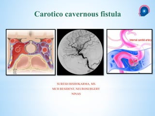
The caroticocavernous fistula is a specific type of dural arteriovenousfistula characterized by abnormal arteriovenous shunting within the cavernous sinus. A caroticocavernous fistula results in high-pressure arterial blood entering the low-pressure venous cavernous sinus. This interferes with normal venous drainage patterns and compromises blood flow within the cavernous sinus and the orbit.

Recommended
More Related Content
What's hot
What's hot (20)
Similar to Caroticocavernous fistula CCF
Similar to Caroticocavernous fistula CCF (20)
More from suresh Bishokarma
More from suresh Bishokarma (20)
Recently uploaded
Recently uploaded (20)
Caroticocavernous fistula CCF
- 1. cka SURESH BISHOKARMA, MS MCH RESIDENT, NEUROSURGERY NINAS Carotico cavernous fistula
- 2. Middle cranial fossa lateral to the body of the sphenoid bone APPLIED ANATOMY LOCATION OF CS
- 4. VENOUS SYTEM The two sinuses are connected by intercavernous sinuses which are anterior and posterior to the hypophysis
- 5. Numerous trabeculae cross the interior of the sinus.
- 6. Content of CS Artery inside CS The internal carotid artery enters the sinus from its base, runs forward and superiorly and then exits at the superior wall of the sinus. Nerve related to CS CNs: III, IV, V1,V2, VI, Sympathetic
- 7. * The caroticocavernous fistula is a specific type of dural arteriovenousfistula characterized by abnormal arteriovenous shunting within the cavernous sinus. * A caroticocavernous fistula results in high-pressure arterial blood entering the low-pressure venous cavernous sinus. * This interferes with normal venous drainage patterns and compromises blood flow within the cavernous sinus and the orbit. INTRODUCTION
- 8. • Caroticocavernous fistulas represent approximately 12% of all dural arteriovenous fistulas. • Direct CCFs are often secondary to trauma: head trauma: Youngs: • Presentation: acute/rapid. • indirect CCFs : Post menopause: insidious. Epidemiology
- 9. • Two main types: 1. Direct 2. Indirect CLASSIFICATION
- 10. CLASSIFICATION • Another method is to classify according to four main types: • Type A • Type B • Type C • Type D Barrow's Classification of CCF s.
- 11. 1. Type A: direct connection between the intracavernous ICA and CS 2. Type B: dural shunt between intracavernous branches of the ICA and CS 3. Type C: dural shunts between meningeal branches of the ECA and CS 4. Type D: B + C CLASSIFICATION
- 12. • Traumatic or spontaneous fistulas. • Flow: • Direct high flow • Indirect low flow fistula OTHER CLASSIFICATION
- 13. Venous Drainage–Based Classification System for Carotid Cavernous Fistulae
- 14. Direct: type A: ICACS Indirect: Br of ICA/ECS CS; types B, C, D The most frequent among indirect is type C, with meningeal branches of the ECA forming the fistula. Pathophysiology Direct CCF: • Trauma • Ruptured intracavernous carotid aneurysms • Collagen deficiency syndromes arterial dissection • Fibromuscular dysplasia • Direct surgical trauma Indirect CCF: Cause often unknown: • Pregnancy • Sinusitis • Trauma • Surgical procedures • Cavernous sinus thrombosis They are postulated to occur secondary to cavernous sinus thrombosis with revascularisation
- 15. • Their symptoms range from benign to extremely severe ophthalmologic or neurologic complications. • Clinical presentation is consequence of the elevated intracavernous pressure. • In direct, high-flow CCF ́s, symptoms appear suddenly. • Symptoms caused by CCFs are related to their size, duration, location, adequacy and route of venous drainage, and presence of arterial and venous collaterals Clinical presentation
- 16. • Pulsatile exophthalmos: ~75% • Chemosis and subconjunctival haemorrhage • Proptosis • Progressive visual loss: 25-32% • Pulsatile tinnitus (usually objective) • Raised intracranial pressure • Cranial nerve (III, IV, Vc, VI) palsies CLINICAL PRESENTATION The Dandy’s triad: pulsatile exophthalmos, bruit and chemosis
- 17. • Moreover, other factors like dominant pattern of venous drainage the size and location of CCF and the presence of collateral vessels (arterial or venous) are important in this setting. • Diplopia, pain, cephalic bruit, ophtalmoplegia, visual loss (Ophth. vein) • Intracranial haemorrhage : sphenoparietal sinus and deep middle cerebral vein) • External haemorrhage: Otorrhagia, epistaxis (Pterygoid plexus)
- 18. CT • Proptosis Enlarged superior ophthalmic veins • Extraocular muscles may be enlarged • Orbital oedema • May show SAH/ICH from a ruptured cortical vein Angiography (DSA) • Rapid shunting from ICA to CS • Enlarged draining veins • Retrograde flow from CS, most commonly into the ophthalmic veins Ultrasound • Arterialised ophthalmic veins may be seen on Doppler study Radiographic features
- 19. MRI Magnetic Resonance Angiogram (MRA) image demonstrating an enlarged superior ophthalmic vein MRA demonstrating a right carotid cavernous fistul
- 20. Three dimensional reconstructed image showing the CC fistula
- 21. DSA a. Digital angiogram of carotid circulation confirming carotid-cavernous fistula b. Digital angiogram of vertebral circulation showing right ophthalmic vein ingurgitated. c. Digital angiogram with final image after treatment of the traumatic CCF
- 22. • Treatment and prognosis • The natural history of CCF is highly varied, ranging from spontaneous closure to rapidly progressive symptoms. • Poor treatment outcome indicators include feeding vessel aneurysms (indirect CCF) and retrograde filling of cortical veins (increased risk of haemorrhage). • Direct fistulas have a relatively high spontaneous rate of haemorrhage (8.4%). • subarachnoid, intracerebral or external haemorrhage (epistaxis, or otorrhagia). • Subconjunctival haemorrhage is also common but does not carry the same poor prognosis
- 23. • Direct CCF: Occlude the tear between ICAand CS , preserving the patency of ICA • Indirect CCF : Interrupt fistulous communications/reduce CS pressure GOAL OF TREATMENT
- 25. • Contralateral hand: 10sec: 4-6/hr: Reduces AV shunting + Increase outlet venous pressures Thrombosis. • Most useful in the treatment of indirect fistulas resulting in spontaneous closure in up to 30% of cases. Carotid compression therapy
- 26. Options: • Ligation of the CC • Surgical trapping of the fistula, and • Surgical transvenous packing. Both direct and indirect CCFs: Disadv: Cranial nerve deficits and residual fistulous communications. Indications for surgical repair include 1. Compromised proximal arterial access that prevents endovascular repair or causes it to fail. 2. Salvage:failed endovascular treatments. Surgery
- 27. • Arterial sacrifice may be required as a life-saving emergency treatment • Indication: Difficult case: • Extensive traumatic vessel wall damage • Active hemorrhage or • A rapidly expanding hematoma of the soft tissues PARENT ARTERY OCCLUSION
- 28. • TOC: Symptomatic direct CCF. • If not possible, detachable coils may be use • Both arterial and venous access (including superior ophthalmic vein) • Indirect fistulas typically require a combined transarterial (closure of feeders) and transvenous (closure of cavernous sinus) approach. • Indirect types are more difficult to treat and have a higher rate of spontaneous closure Transarterial balloon embolisation
- 29. • This procedure requires that the CS must be large enough to put the balloon for embolization and the size of fistula must be smaller than the inflated balloon, but large enough to allow a deflated balloon. • The balloon has the advantage of being able to be flow-directed through the fistula and CS, and must be inflated to a volume larger than the fistula orifice to prevent its retrograde migration into ICA. • Angiography is repeated to ensure closure of the fistula and patency of the ICA. Balloon Occlusion
- 30. • Mainstay of treatment in high-flow direct CCF ́s. • It's an alternative when residual AV shunt remains in dural CCF. • Embolization can be made with detachable platinum coils and liquid embolic agents (n-butyl cyanoacrylate, ethylene-vinyl alcohol copolymer); • Coils are preferred because of their reliable and controlled deployment into CS. • Complications of this procedure includes thromboembolus and ICA dissection Transarterial embolization
- 31. • Recent Advance: poly flurotetraethylene-covered stents • Traumatic arterial damage • immediate obliteration of a direct CCF, while preserving ICA patency • Disadv: • Longitudinal flexibility: difficult navigation: tortuosity of the intracranial vasculature. • Vasospasms: Intra-arterial nimodipine and papaverine infusion • Endoleak, coverage of vital perforators, dissection and rupture Covered stent graft placement
- 32. • Is the current method of choice in treatment of indirect CCF’s. • The goal of this technique is to catheterize the abnormal CS superselectively and occlude the fistula without re-routing venous drainage to cortical structures.. • Several routes: Most: inferior petrosal sinus (IPS Transvenous embolization
- 33. • Indirect CCFs. • Gamma knife radiosurgery can be used either alone or as an adjunct therapy before/after endovascular intervention. • Preliminary data : safe and effective alternative treatment • Drawback: 22-mo average lag RADIOSURGERY
- 34. Fistulous point located at left CS, with ICA supply by meningo-hipofisary trunks (red arrow) and ECA supply by middle meningeal artery (blue arrow)and clivus branches from ascendent pharyngeal artery. Venous drainage to superior ophtalmic vein (yellow arrow) and to inferior petrous sinus.
- 35. Transarterial embolization Coil embolization of the fistula (red arrows) was performed via middle meningeal artery
- 36. Thank you NATIONAL INSTITUTE OF NEUROLOGICAL AND ALLIED SCIENCES, BANSBARI, KATHMANDUNATIONAL INSTITUTE OF NEUROLOGICAL AND ALLIED SCIENCES, BANSBARI, KATHMANDU CAROTICO CAVERNOUS FISTULA
- 37. Approach to vascular lesion brain
Editor's Notes
- The cavernous sinuses lie in the middle cranial fossa lateral to the body of the sphenoid bone.
- The two sinuses are connected by intercavernous sinuses which are anterior and posterior to the hypophysis. The sinus receives blood from the superior and inferior ophthalmic veins, the cerebral veins, the sphenoparietal sinus, sylvian vein and the central vein of the retina. They drain into the internal jugular vein and the transverse sinus. The cavernous sinus drains posteriorly through the inferior petrosal sinus (IPS) and superior petrosal sinus to the jugular bulb, inferiorly through the pterygoid plexus via emissary veins, and contralaterally through the contralateral cavernous sinus. The revised venous drainage of the CCFs may head toward the ophthalmic venous system anteriorly; the superior petrosal sinus, the IPS, or the basilar plexus posteriorly; the sphenoparietal sinus laterally; the intercavernous sinus contralaterally; the pterygoid plexus via the vein of the foramen rotundum and the vein of the foramen ovale inferiorly. Most often, the direction of the venous drainage is multidirectional
- The carotid artery gives rise to several branches in the sinus. The sixth cranial nerve runs through the sinus. The third and fourth cranial nerves, and the ophthalmic and maxillary divisions of the trigeminal nerve lie in the lateral wall of the sinus
- Caroticocavernous fistulas represent approximately 12% of all dural arteriovenous fistulas. Direct CCFs are often secondary to trauma: head trauma: Youngs: The presentation is acute and symptoms develop rapidly. I n contrast, indirect CCFs have a predilection for the postmenopausal female patient and the onset of symptoms is often insidious. Other conditions that predispose to increased risk include:
- Direct: direct communication between intracavernous ICA and Cavernous sinus. Indirect: communication exists via branches of the carotid circulation (ICA or ECA )
- Type A: direct connection between the intracavernous ICA and CS Type B: dural shunt between intracavernous branches of the ICA and CS Type C: dural shunts between meningeal branches of the ECA and CS Type D: B + C
- Carotid cavernous fistulae (CCFs) are most commonly classified based on arterial supply. Symptomatology and treatment approach, however, are largely influenced by venous drainage. OBJECTIVE: To propose an updated classification system using venous drainage
- A CCF allows highly pressurized arterial blood to be transmitted directly into the cavernous sinus and the draining veins, leading to venous hypertension. The clinical presentation of CCF is a direct consequence of elevation in intracavernous pressure and revised flow patterns. The revised venous drainage of the CCFs may head toward the ophthalmic venous system anteriorly; the superior petrosal sinus, the IPS, or the basilar plexus posteriorly; the sphenoparietal sinus laterally; the inter- cavernous sinus contralaterally; the pterygoid plexus via the vein of the foramen rotundum and the vein of the foramen ovale inferiorly. Most often, the direction of the venous drainage is multidirectional
- Digital substraction angiography (DSA) is used to obtain the following information: Size and location of the fistula. Characterize them as direct or indirect. To identify associated cavernous carotid aneurysms. Presence of complete or partial steal phenomena. Identification and confirmation of patency of outflow pathways of the CS. Assessment of cortical arterial circulation and collateral flow through circle of Willis. Identification of high-risk features (cortical venous drainage, pseudoaneurysm, CS varix). To depict venous drainage patterns, therapeutic route, associated vascular injuries and evaluation of carotid bifurcation before compression therapy.
- The patient is instructed to compress the carotid artery and jugular vein with the contralateral hand for a period of 10 s while sitting or lying down, four to six times each hour[7]. The aim of the compression therapy is the transient reduction of arteriovenous shunting by decreasing arterial inflow while simultaneously increasing the outlet venous pressure, thereby promoting spontaneous thrombosis within the fistula[25]. Use of the contralateral hand ensures that if ischemia develops, the symptomatic arm will fall away from the neck, thus allowing cortical revascularization
- When endovascular occlusion of a direct CCF with preservation of the ICA is not feasible due to extensive traumatic vessel wall damage, active hemorrhage or a rapidly expanding hematoma of the soft tissues.
- Detachable balloon occlusion: After Prolo and Hanberry described the use of a fixed balloon catheter to block a CCF in 1971, Serbinenko et al[29] reported the rst case of successful embolization of a CCF from an en- dovascular approach using a detachable silicone balloon with preservation of the ICA[13]. The use of detachable balloon catheters has ushered a new age in the treatment of type A direct CCFs. Transarterial balloon detachment has been accepted as the endovascular treatment of choice for direct CCFs since the 1980s. The small-diameter vessels that often make up dural fistulas usually do not allow the introduction of a balloon. However, the large carotid defect commonly present in type A CCFs frequently permits transarterial balloon occlusion of the stula with preservation of the ICA
- Coil and material embolization: Transarterial em- bolization with coils or other embolic material now is the mainstay of endovascular treatment for high-flow direct CCFs, given the limited availability of detach- able balloons[7]. Transarterial CCF embolization can be performed with the same technique as aneurysmal em- bolization. Embolization can be achieved with detach- able platinum coils, silk and liquid embolic agents such as n-butyl cyanoacrylate (n-BCA), and ethylene-vinyl alcohol copolymer (EVOH)[5]. The standard transarterial approach consists of placing a guiding catheter in the cervical ICA. Next, a microcatheter is superselectively advanced into the cavernous segment of the ICA and through the tear into the cavernous sinus. Through this microcatheter, embolic material is placed into the cav- ernous sinus[7].
- Recent advances in endovascular techniques such as placement of poly flurotetraethylene-covered stents have created alternatives to ICA sacrifice in traumatic arterial damage, especially in the setting of an unsuccessful balloon test occlusion study. Covered stent grafts can be extremely useful for the immediate obliteration of a direct CCF, while preserving ICA patency (Figure 2). Additionally, they may decrease the risk of ischemic stroke by preserving the involved artery while simultaneously sealing the site of the fistula[5,7,35]. Covered stent grafts have the technical disadvantage of limited longitudinal flexibility, making it difficult to navigate them through the tortuosity of the intracranial vasculature. Furthermore, the irritation caused by the stiffness of covered stents may frequently lead to periprocedural vasospasms, especially at the ends of the stent (Figure 2). Intra-arterial nimodipine and papaverine infusion can be used for the prevention and resolution of these vasospasms[36,37]. The complications of this procedure include endoleak, coverage of vital perforators, dissection and rupture
- There are other alternative routes, including facial vein and SOV, trans-contralateral CS, superficial middle cerebral vein and sphenoparietal sinus, pterygoid plexus and direct transorbital puncture of CS via the superior orbital fissure. rans- venous techniques have precedence over transarterial methods because of their simplicity, lower ischemic risk, higher success rates and capability to cure the stula in a single session.
- Drawback: 22-mo average lag between treatment and complete symptom relief.
- indirect type D CCF. Lateral digital substraction angiogram with right ICA (left) and right ECA (right) injection. Fistulous point located at left CS, with ICA supply by meningo-hipofisary trunks (red arrow) and ECA supply by middle meningeal artery (blue arrow)and clivus branches from ascendent pharyngeal artery. Venous drainage to superior ophtalmic vein (yellow arrow) and to inferior petrous sinus.
- Coronal (left) and lateral (right) digital substraction angiogram of the previous patient. Coil embolization of the fistula (red arrows) was performed, from the middle meningeal artery supply. Further, onyx was used to embolize from the middle meningeal artery to CS.