Interpretation of ecg_in_pulmonary_disease
•Download as PPTX, PDF•
11 likes•1,616 views

interpretation of ecg in pulmonary disease. cor pulmonale .. pul hypertension

Recommended
More Related Content
What's hot
What's hot (20)
Viewers also liked
Similar to Interpretation of ecg_in_pulmonary_disease
Similar to Interpretation of ecg_in_pulmonary_disease (20)
Recently uploaded
Recently uploaded (20)
Interpretation of ecg_in_pulmonary_disease
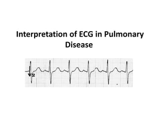
- 1. Interpretation of ECG in Pulmonary Disease
- 6. Normal PR interval and QRS complex • The normal P interval is 120-220 ms represented by 3-5 small squares. • Most of this time is taken up by delay in the AV node. • If the PR interval is very short either the atria have been depolarized from close to the AV node or there is abnormally fast conduction from the atria to the ventricles. • The duration of QRS complex is normally 120ms ,represented by 3 small squares. • QT interval is a measure of the time between the start of the Q wave and the end of the T wave in the heart's electrical cycle.
- 8. The ECG in Chronic Obstructive Pulmonary Disease ECG changes occur in COPD due to: 1.The presence of hyperexpanded emphysematous lungs within the chest. 2.The long-term effects of hypoxic pulmonary vasoconstriction upon the right side of the heart, causing pulmonary hypertension and subsequent right atrial and right ventricular hypertrophy (i.e. cor pulmonale).
- 9. • 3.Lung hyper expansion causes external compression of the heart and lowering of the diaphragms, with consequent elongation and vertical orientation of the heart. • 4.Due to its fixed attachments to the great vessels, the heart undergoes clockwise rotation in the transverse plane, with movement of the right ventricle anteriorly and displacement of the left ventricle posteriorly. • 5.The presence of increased air between the heart and recording electrodes has a dampening effect, leading to reduced amplitude of the QRS complexes.
- 10. Lung hyper expansion and vertical orientation of the heart
- 11. The most typical ECG findings in emphysema are: •Rightward shift of the P wave axis with prominent P waves in the inferior leads and flattened or inverted P waves in leads I and aVL. •Rightward shift of the QRS axis towards +90 degrees (vertical axis) or beyond (right axis deviation). •Exaggerated atrial depolarisation causing PR and ST segments that “sag” below the TP baseline.
- 12. •Low voltage QRS complexes, especially in the left precordial leads (V4-6). •Clockwise rotation of the heart with delayed R/S transition point in the precordial leads +/- persistent S wave in V6. There may be complete absence of R waves in leads V1-3 (the “SV1-SV2-SV3″ pattern). •Right atrial enlargement (P pulmonale) •Right ventricular hypertrophy
- 15. •Rightward QRS axis (+90 degrees). •Peaked P waves in the inferior leads > 2.5 mm (P pulmonale) with a rightward P-wave axis (inverted in aVL) •Clockwise rotation of the heart with a delayed R/S transition point (transitional lead = V5). •Absent R waves in the right precordial leads (SV1-SV2-SV3 pattern). •Low voltages in the left-sided leads (I, aVL, V5-6).
- 16. Right Atrial Enlargement Right atrial enlargement produces a peaked P wave (P pulmonale) with amplitude: •> 2.5 mm in the inferior leads (II, III and AVF) •> 1.5 mm in V1 and V2
- 17. • Causes • The principal cause is pulmonary hypertension due to: • Chronic lung disease (cor pulmonale) • Tricuspid stenosis • Congenital heart disease (pulmonary stenosis, Tetralogy of Fallot) • Primary pulmonary hypertension
- 18. Right atrial enlargement: P wave amplitude > 2.5mm in leads II, III and aVF Right atrial enlargement: P wave amplitude > 1.5 mm in V2
- 19. Right Ventricular Hypertrophy Diagnostic criteria •Right axis deviation of +110° or more. •Dominant R wave in V1 (> 7mm tall or R/S ratio > 1). •Dominant S wave in V5 or V6 (> 7mm deep or R/S ratio < 1). •QRS duration < 120ms (i.e. changes not due to RBBB).
- 20. Supporting criteria •Right atrial enlargement (P pulmonale). •Right ventricular strain pattern = ST depression / T wave inversion in the right precordial (V1-4) and inferior (II, III, aVF) leads. •S1 S2 S3 pattern = far right axis deviation with dominant S waves in leads I, II and III. •Deep S waves in the lateral leads (I, aVL, V5-V6). Other abnormalities caused by RVH •Right bundle branch block (complete or incomplete).
- 21. Causes •Pulmonary hypertension •Mitral stenosis •Pulmonary embolism •Chronic lung disease (cor pulmonale) •Congenital heart disease (e.g. Tetralogy of Fallot, pulmonary stenosis) •Arrhythmogenic right ventricular cardiomyopathy
- 23. Right Axis Deviation •QRS axis between + 90 and + 180 degree •Normal QRS axis is between – 30 and + 90 •QRS is positive (dominant R wave) in leads III and aVF •QRS is negative (dominant S wave) in leads I and aVL
- 25. Causes •Left posterior fascicular block •Lateral myocardial infarction •Right ventricular hypertrophy •Acute lung disease (e.g. PE) •Chronic lung disease (e.g. COPD) •Ventricular ectopy •Hyperkalaemia •Sodium-channel blocker toxicity •WPW syndrome
- 26. RIGHT VENTRICULAR HYPERTROPHY 1.In RBBB, activation of the right ventricle is delayed as depolarisation has to spread across the septum from the left ventricle. 2.The left ventricle is activated normally, meaning that the early part of the QRS complex is unchanged. 3.The delayed right ventricular activation produces a secondary R wave (R’) in the right precordial leads (V1- 3) and a wide, slurred S wave in the lateral leads.
- 27. 4.Delayed activation of the right ventricle also gives rise to secondary repolarization abnormalities, with ST depression and T wave inversion in the right precordial leads. 5.In isolated RBBB the cardiac axis is unchanged, as left ventricular activation proceeds normally via the left bundle branch.
- 29. Tall R' wave in V1 ("M" pattern) with wide, slurred S wave in V6 ("W" pattern) ECG changes in RBBB Diagnostic Criteria •Broad QRS > 120 ms •RSR’ pattern in V1-3 (‘M-shaped’ QRS complex) •Wide, slurred S wave in the lateral leads (I, aVL, V5-6) Associated Features ST depression and T wave inversion in the right precordial leads (V1-3) T
- 30. Typical pattern of T-wave inversion in V1-3 with RBBB Typical RSR' pattern ('M'-shaped QRS) in V1
- 31. •Right ventricular hypertrophy / cor pulmonale •Pulmonary embolus •Ischaemic heart disease •Rheumatic heart disease •Myocarditis or cardiomyopathy •Degenerative disease of the conduction system •Congenital heart disease (e.g. atrial septal defect) Causes of RBBB
- 32. •Incomplete RBBB is defined as an RSR’ pattern in V1-3 with QRS duration < 120ms. •It is a normal variant, commonly seen in children (of no clinical significance).
- 33. Right Ventricular Strain Repolarisation abnormality due to right ventricular hypertrophy or dilatation. Electrocardiographic Features •ST depression and T wave inversion in the leads corresponding to the right ventricle, i.e •The right precordial leads: V1-3, often extending out to V4 •The inferior leads: II, III, aVF, often most pronounced in lead III as this is the most rightward-facing lead.
- 34. Causes Associated with increased pulmonary artery pressures in the setting of acute or chronic right ventricular hypertrophy or dilatation: •Pulmonary hypertension •Mitral stenosis •Pulmonary embolism •Chronic lung disease (cor pulmonale) •Congenital heart disease (e.g. Tetralogy of Fallot, pulmonary stenosis) •Arrhythmogenic right ventricular cardiomyopathy
- 35. Typical right ventricular strain pattern: ST depression and T-wave inversion in V1-4 (plus lead III), in this case due to right ventricular hypertrophy.
- 36. Right ventricular strain pattern involving both the precordial and inferior leads: T-wave inversions are seen in the right precordial (V1-4) and inferior leads (III, aVF) in this patient with acute right ventricular dilatation due to massive pulmonary embolism.
- 37. The ECG in Pulmonary Embolism • Sinus tachycardia – the most common abnormality; seen in 44% of patients. • Complete or incomplete RBBB – associated with increased mortality; seen in 18% of patients. • Right ventricular strain pattern – T wave inversions in the right precordial leads (V1-4) ± the inferior leads (II, III, aVF). This pattern is seen in up to 34% of patients and is associated with high pulmonary artery pressures. • Right axis deviation – seen in 16% of patients. Extreme right axis deviation may occur, with axis between zero and - 90 degrees, giving the appearance of left axis deviation (“pseudo left axis”).
- 38. • Dominant R wave in V1 – a manifestation of acute right ventricular dilatation. • Right atrial enlargement (P pulmonale) – peaked P wave in lead II > 2.5 mm in height. Seen in 9% of patients. • SI QIII TIII pattern – deep S wave in lead I, Q wave in III, inverted T wave in III. This “classic” finding is neither sensitive nor specific for pulmonary embolism; found in only 20% of patients with PE. • Clockwise rotation – shift of the R/S transition point towards V6 with a persistent S wave in V6 (“pulmonary disease pattern”), implying rotation of the heart due to right ventricular dilatation.
- 39. 1. Atrial tachyarrhythmias – AF, flutter, atrial tachycardia. Seen in 8% of patients. 2. Non-specific ST segment and T wave changes, including ST elevation and depression. Reported in up to 50% of patients with PE.
- 40. •RBBB •Extreme right axis deviation (+180 degrees) •S1 Q3 T3 •T-wave inversions in V1-4 and lead III •Clockwise rotation with persistent S wave in V6 •Sinus tachycardia. •Simultaneous T-wave inversions in the anterior (V1-4) and inferior leads (II, III, aVF). •Non-specific ST changes – slight ST elevation in III and aVF.
- 41. A 23 year-old man is brought in by paramedics after an episode of syncope at home. On arrival he is in severe respiratory distress, extremely pale and dripping with sweat. BP is 125/70, HR is 80bpm, RR 40, SaO2 95% on 15L O2 via NRB. Portable CXR is normal. His ECG is presented below:
- 42. Describe the ECG
- 43. • Sinus rhythm at 80 bpm • Left axis deviation (-70 degrees) • RBBB with wide QRS (150ms), tall R wave in V1, RSR’ complexes in leads V1-3 • Right ventricular strain pattern with deep T wave inversions in V1-3 • Widespread ischaemic changes with 1-2mm ST elevation and early Q waves in the anterior leads (V2-5) and 1-2 mm ST depression in inferior leads (II, III and aVF). There is also significant ST elevation in leads aVR and aVL with formation of Q waves. • .
- 44. • Q2. What is your interpretation of the ECG findings given the clinical context? • The combination of new RBBB with signs of right ventricular strain (deep T wave inversions in V1-3) in a patient presenting with dyspnoea and syncope is strongly suggestive of acute pulmonary hypertension secondary to massive PE. • Widespread myocardial ischaemia/infarction is likely related to severe global tissue hypoxia from obstructive shock (VBG in this patient showed venous saturations of 10%, venous PO2 10mmHg with a lactate of 8mmol/L). • Left axis deviation may represent left anterior fascicular block
- 45. THANK YOU