2. Objectives
1. Discuss the ultra-structure of different cell organelles and correlate
with function.
2. Identify the microscopic characteristic of each organelle.
2
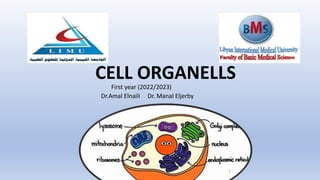
3. Cell organelles
❑ Membranous organelles ( bounded
by membranes ) such as
mitochondria, Golgi complex,
nucleus, endoplasmic reticulum,
lysosomes & peroxisomes.
❑ Non- membranous organelles (not
bounded by membranes) such as
ribosomes, centrioles &
cytoskeleton.
3
4. Mitochondria
• They are found where metabolic activity is high
such as those of liver and skeletal muscle, thus
termed the powerhouse of the cell.
• Found in all cells except mature red blood cells.
• Mitochondria are very mobile, moving around
the cell by means of microtubules, a component
of the cytoskeleton
4
5. • Under Light microscope(L/M ): Thread-like, rod-shaped organelles.
• Under Electron microscope (E/M):
1. They have an outer smooth and inner irregular folded membrane
(cristae).
2. The cristae, which project into the matrix and greatly increase the
membrane’s surface area
The number of cristae in mitochondria also corresponds to the energy needs
of the cell.
L/M E/M
5
6. ❑ The space found in between the two
membranes is called intermembranous space.
❑Inside the inner membrane is the Matrix Space.
❑The matrix space consists of:
I. Circular molecular of DNA and three varieties of
RNA.
II. Rounded electron dense granules rich in Ca+2.
III. Enzymes for the citric acid (Krebs)cycles.
6
7. Endoplasmic reticulum
• A system of interconnected tubules and vesicles whose lumen is
termed cisternae
• It is either : rough or smooth.
• It may be covered by ribosomes ( rER )
• May have NO ribosomes (sER)
7
8. Rough endoplasmic reticulum
• Increase in cells having high protein
secretion (e.g. Fibroblast).
• Sometimes continuous with the outer
nuclear membrane.
• Functions:
1. Synthesis of proteins (via ribosomes)
2. Storage and transport of proteins.
8
9. Smooth endoplasmic reticulum
• The SER is made up of tubules and vesicles that
branch out to form a network.
• NO attached ribosomes.
• Its cisternae are more tubular.
• Functions:
1. The principal functions of smooth endoplasmic
reticulum are lipid biosynthesis (e.g steroid
hormones) and membrane synthesis and repair.
2. Calcium ion storage
3. Drug detoxification
9
11. Golgi apparatus
• The organelle was named after histologist
Camillo Golgi who discovered it in 1898.
• Composed of series of flattened, slightly
curved cisternae (Golgi stack).
• Has two surfaces:
• Cis-face: immature & close to RER
• Trans-face: mature & toward the cell
membrane
• The periphery of each cisterna is dilated
and show vesicles that are fusing with or
budding off.
11
13. Function of Golgi Apparatus
It modifies proteins that have been delivered in transport vesicles from the RER.
The Golgi apparatus modifies, sorts, and packages different substances for secretion
out of the cell, or for use within the cell.
13
14. Lysosomes
• Usually spherical membranous vesicles formed by the Golgi
apparatus .
• They contain more than 40 different degradative enzymes including
proteases, lipases and nucleases.
• These are collectively known as acid hydrolases because they are
optimally active at a pH of about 5.0
• Increase in cells with high phagocytic activity (e.g. macrophages).
14
15. Lysosomes
• Primary lysosomes : which are newly formed from Trans-face
of Golgi
• Secondary lysosomes: which are formed from the fusion of
primary lysosomes with other substances.
• Functions:
1. Digestion of certain substances such as solid material,
fluids and dead organelles.
2. Cell metabolism: Lysosomes are important in breakdown
of intracellular glycogen e.g. in liver cells.
3. Phagocytosis of bacteria and viruses so contribute in
defense mechanism
15
17. Ribosomes
• Non-membranous cell organelles, about
20 nm in diameter.
• They are formed in nucleus and then pass
to the cytoplasm to perform their
functions.
• The main function are proteins synthesis.
• Each ribosome is composed of a large and
a small subunit
17
18. • Each subunit consists of a strand of
RNA (ribosomal RNA, rRNA) with
associated ribosomal proteins
forming a globular structure.
• Ribosomes are often found attached
to mRNA molecules in small spiral-
shaped aggregations called
polyribosomes or polysomes
• Ribosomes and polyribosomes may
be free or attached to the surface of
endoplasmic reticulum.
18
19. Cytoskeleton
• A network of protein filaments.
• Responsible for keeping the cell
morphology, help in cellular
motion. Consist of :
1.Microtubules.
2.Thin filaments(microfilaments).
3.Intermediate filaments.
19
20. 1) Microtubules
• Tubular structures consist of α and β tubulin.
• Their synthesis is controlled by the microtubule organizing centers.
• Keep the cell shape.
• Transport of organelles and vesicles, such as secretory granules.
20
21. 2) Intermediate filaments
• They are intermediate in size with an average diameter of 10-12 nm.
• Types of intermediate filaments :
A. Keratins are found in epithelium.
B. Desmin is found in smooth muscle skeletal and cardiac muscle.
C. Vimentin filaments are found in the cells of mesemchymal origin
(e.g. fibroblast).
D. Glial filaments in the astrocytes.
E. Neurofilaments consist of several polypeptides in the nerve cells.
21
22. 3) Microfilaments
• Mainly contractile thin (actin) and
thick (myosin) filaments in the skeletal
muscle.
• They form a thin sheath under plasma
membrane called the cell cortex .
• Help in moving cytoplasmic
components.
• Help in cleavage of mitotic cells.
22
23. Centrioles
• They are non membranous organelles.
• By E.M.: Non membranous small paired structures
arranged at right angle to each other.
• One pair of centrioles is called a centrosome. Each
centriole is cylinder in shape.
• In cross section, the wall of the centriole is formed of
9 bundles of microtubules and each bundle is formed
of 3 microtubules (triplet), embedded in fibrillar
material.
• Function: They are essential for cell division to
form mitotic spindle.
23
24. Cell Inclusions
• Non-living components of the cell.
• The most common inclusions are glycogen, lipid droplet and
pigments.
• Glycogen is very common, abundant in cells of muscle and liver.
Pigments: hemoglobin of red blood cells and melanin manufactured
by melanocytes.
• Lipids: stored mainly in specialized cells, adipocytes in the form of
triglycerides. They work for energy reserve
24
29. References
• Mescher, A. L., Mescher, A. L., & Junqueira, L. C. U. (2016).
Junqueira's basic histology: Text and atlas (14th ed.). New York:
McGraw-Hill Education.
29