
Recommended
More Related Content
What's hot
What's hot (20)
Similar to CELL PHYSIOLOGY
Similar to CELL PHYSIOLOGY (20)
Recently uploaded
Recently uploaded (20)
CELL PHYSIOLOGY
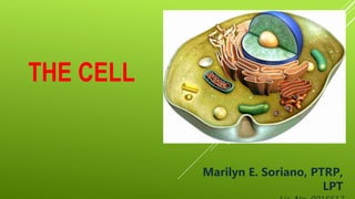
- 1. THE CELL Marilyn E. Soriano, PTRP, LPT
- 2. Three main parts of the Cell: 1. plasma membrane, 2. cytoplasm, 3. nucleus. 1. The plasma membrane forms the cell’s flexible outer surface, separating the cell’s internal environment from the external environment a selective barrier that regulates the flow of materials into and out of a cell. • This selectivity helps establish and maintain the appropriate environment for normal cellular activities plays a key role in communication among cells and between cells and their external environment
- 3. 3. The nucleus is a large organelle that houses most of a cell’s DNA. Within the nucleus, a chromosome- a single molecule of DNA associated with several proteins, contains thousands of hereditary units called genes that control most aspects of cellular structure and function. 2. The cytoplasm consists of all the cellular contents between the plasma components: cytosol and organelles. 1. Cytosol (intracellular fluid)- the fluid portion of cytoplasm, contains water, dissolved solutes, and suspended particles. 2. Organelles • Each type of organelle has a characteristic shape and specific functions: cytoskeleton, ribosomes, endoplasmic reticulum, Golgi complex, lysosomes, peroxisomes, and mitochondria.
- 5. a flexible yet sturdy barrier that surrounds and contains the cytoplasm of a cell. is best described by using a structural model called the fluid mosaic model. THE PLASMA MEMBRANE
- 6. The Lipid Bilayer The basic structural framework of the plasma membrane is the lipid bilayer, two back-to- back layers made up of three types of lipid molecules— phospholipids, cholesterol, and glycolipids. • phospholipids- 75%. • cholesterol- 20%, • glycolipids- 5%, THE PLASMA MEMBRANEStructure of the Plasma Membrane
- 7. Lipids are amphipathic molecules, which means that they have both polar and nonpolar parts. • In phospholipids: o The polar part is the phosphate- containing “head,” which is hydrophilic. • hydrophilic heads facing outward • the heads face a watery fluid on either side—cytosol on the inside and extrafluid on the outside o The nonpolar parts are the two long fatty acid “tails,” which are hydrophobic • hydrophobic tails in each half of the bilayer point toward one another • nonpolar, hydrophobic region in the membrane’s interior THE PLASMA MEMBRANE
- 8. Cholesterol molecules are weakly amphipathic and are interspersed among the other lipids in both layers of the membrane. o the only polar region of cholesterol is the tiny —OH group • it forms hydrogen bonds with the polar heads of phospholipids and glycolipids. o The nonpolar are the stiff steroid rings and hydrocarbon tail of cholesterol • they fit among the fatty acid tails of the phospholipids and glycolipids. THE PLASMA MEMBRANE
- 9. The carbohydrate groups of glycolipids. o A polar “head” o A nonpolar their fatty acid “tails”. o Glycolipids appear only in the membrane layer that faces the extracellular fluid, which is one reason the two sides of the bilayer are asymmetric, or different. THE PLASMA MEMBRANE
- 10. MEMBRANE PROTEINS o icebergs in the lipid sea, whereas others are anchored specific locations like islands. o allow passage of several types of lipid-soluble molecules o act as a barrier to the entry or exit of charged or polar substances. o act as signal receptors or as molecules that link the plasma membrane to intracellular or extracellular proteins. THE PLASMA MEMBRANE
- 11. Membrane proteins are classified as: I. Integral- proteins extend into or through the lipid bilayer and are firmly embedded which are called transmembrane proteins. Many integral proteins are glycoproteins attached to the that protrude into the extracellular fluid. Amphipathic: • Their hydrophilic regions protrude into either the watery extracellular fluid or the cytosol. • their hydrophobic regions extend among the fatty acid tails. THE PLASMA MEMBRANEArrangement of Membrane Proteins
- 12. II. Peripheral- are not as firmly embedded in the membrane. They are attached to the polar heads of membrane lipids or to integral proteins at the inner or outer surface of the membrane. THE PLASMA MEMBRANE
- 13. A. INTEGRAL PROTEINS 1. Ion channels- pores or holes that specific ions can flow through to get into or out of the cell. Most ion channels are selective; they allow only a single type of ion to pass through. 2. Carriers (transporters) selectively moving a polar substance or ion from one side of the membrane to the other. Functions of Membrane Proteins THE PLASMA MEMBRANE
- 14. 3. Receptors- serve as cellular recognition sites: recognizes and binds a specific type of molecule. Insulin receptors bind the hormone insulin. 4. Enzymes- catalyze specific chemical reactions at the inside or outside surface of the cell. *Peripheral proteins Functions of Membrane Proteins THE PLASMA MEMBRANE
- 15. 5. Linkers- anchor proteins in the plasma membranes of neighboring cells to one another or to protein filaments inside and outside the cell. *Peripheral proteins B. GLYCOPROTEINS 1. Cell identity markers-enable a cell to (1) recognize other cells of the same kind during tissue formation or (2) recognize and respond to potentially dangerous foreign cells. Functions of Membrane Proteins THE PLASMA MEMBRANE
- 16. Functions of Membrane Proteins C. PERIPHERAL PROTEINS- help support the plasma membrane, anchor integral proteins, and participate in mechanical activities such as moving materials and organelles within cells, changing cell shape in dividing and muscle cells, and attaching cells to one another. THE PLASMA MEMBRANE
- 17. THE CYTOPLASM
- 18. Cytoplasm consists of all the cellular contents between the plasma membrane and the nucleus, and has two components: 1. the cytosol 2. organelles, tiny structures that perform different functions in the cell. CYTOPLASMTHE CYTOSOL (intracellular fluid) is the fluid portion of the cytoplasm that surrounds organelles constitutes about 55% of total cell volume 75–90% water plus various dissolved and suspended components: ions, glucose, amino acids, fatty acids, proteins, lipids, ATP, and waste products; organic molecules: lipid droplets that contain triglycerides, and clusters of glycogen molecules called glycogen granules the site of many chemical reactions required for a cell’s existence.
- 19. The cytoskeleton is a network of protein filaments that extends throughout the cytosol. Three types of filaments contribute to the cytoskeleton’s structure, as well as the structure of other organelles A. microfilaments, B. Intermediate filaments, C. Microtubules
- 20. A. Microfilaments are the thinnest elements of the cytoskeleton composed of the proteins actin and myosin and are most prevalent at the edge of a cell CYTOSOL have two general functions: 1. help generate movement: muscle contraction, cell division, and cell locomotion (migration of embryonic cells during development, the invasion of tissues by white blood cells to fight infection, or the migration of skin cells during wound healing) 2. provide mechanical support: responsible for the basic strength and shapes of cells anchor the cytoskeleton to integral proteins in the plasma membrane. provide mechanical support for cell extensions called microvilli which are abundant on cells involved in absorption, such as the epithelial cells that line the small intestine
- 21. B. Intermediate Filaments thicker than microfilaments but thinner than microtubules. found in parts of cells subject to mechanical stress they help stabilize the position of organelles such as the nucleus and help attach cells to one another. CYTOSOLC. Microtubules the largest, long, unbranched hollow tubes composed mainly of the protein tubulin. Assembly begins in the centrosome which grow outward from the centrosome toward the periphery of the cell help determine cell shape function in the movement of organelles
- 22. specialized structures within the cell that have characteristic shapes, and they perform specific functions in cellular growth, maintenance, and reproduction cooperate to maintain homeostasis ORGANELLES
- 23. 1. The centrosome located near the nucleus consists of two components: i. a pair of centrioles ii. pericentriolar material- Surrounding the centrioles, which contains hundreds of ring-shaped complexes composed of the protein tubulin ORGANELLES
- 24. i. Cilia- numerous, short, hairlike projections that extend from the surface of the cell • Each cilium contains a core of 20 microtubules surrounded by plasma membrane. 2. Cilia and Flagella motile projections of the cell surface ORGANELLES
- 25. ii. Flagella are similar in structure to cilia but are typically much longer. Flagella usually move an entire cell. A flagellum generates forward motion along its axis by rapidly wiggling in a wavelike pattern. ORGANELLES
- 26. 3. Ribosomes the sites of protein synthesis high content of one type of ribonucleic acid (ribosomal RNA, or rRNA), but each one also includes more than 50 proteins consists of two subunits, one about half the size of the other attached to the outer surface of the nuclear membrane and to an extensively folded membrane called the endoplasmic reticulum. Other ribosomes are “free” or unattached to other cytoplasmic structures. ORGANELLES
- 27. 4. Endoplasmic Reticulum is a network of membranes in the form of flattened sacs or tubules. extends from the nuclear envelope to which it is connected and projects throughout the cytoplasm contain two distinct forms of ER, which differ in structure and function. Rough ER Smooth ER ORGANELLES
- 28. a. Rough ER is continuous with the nuclear membrane and usually is folded into a series of flattened sacs. • outer surface of rough ER is studded with ribosomes, the sites of protein synthesis. • proteins synthesized by ribosomes attached to rough ER enter spaces within the ER for processing and sorting. • rough ER produces secretory proteins, membrane proteins, and many organellar proteins b. Smooth ER extends from the rough ER to form a network of membrane tubules. does not have ribosomes on the outer surfaces of its membrane. synthesize fatty acids and steroids, such as estrogens and testosterone. In liver cells, enzymes of the smooth ER help release glucose into the bloodstream and inactivate or detoxify lipid-soluble drugs or potentially harmful substances, such as alcohol, pesticides, and carcinogens (cancer-causing agents). In liver, kidney, and intestinal cells a smooth ER enzyme removes the phosphate group from glucose-6-phosphate, which allows the “free” glucose to enter the bloodstream. In muscle cells, the calcium ions (Ca2) that trigger contraction are released from the sarcoplasmic reticulum, a form of smooth ER.
- 29. 5. Golgi Complex proteins synthesized by ribosomes attached to rough ER are ultimately transported to other regions of the cell. The first step in the transport pathway is through an organelle called the Golgi complex. consists of 3 to 20 cisternae, small, flattened membranous sacs with bulging edges that resemble a stack of pita bread. The cisternae are often curved, giving the Golgi complex a cuplike shape. The cisternae at the opposite ends of a Golgi complex differ from each other in size, shape, and enzymatic activity. o The convex entry (cis) face is a cisterna that faces the rough ER. o The concave exit (trans) face is a cisterna that faces the plasma membrane. o Sacs between the entry and exit faces are called medial cisternae. ORGANELLES
- 30. Proteins arriving at, passing through, and exiting the Golgi complex do so through maturation of the cisternae and exchanges that occur via transfer vesicles: 1. Proteins synthesized by ribosomes on the rough ER are surrounded by a piece of the ER membrane, which eventually buds from the membrane surface to form transport vesicles. 2. Transport vesicles move toward the entry face of the Golgi complex. 3. Fusion of several transport vesicles creates the entry face of the Golgi complex and releases proteins into its lumen (space). 4. The proteins move from the entry face into one or more medial cisternae. Enzymes in the medial cisternae modify the proteins to form glycoproteins, glycolipids, and lipoproteins. Transfer vesicles that bud from the edges of the cisternae move specific enzymes back toward the entry face and move some partially modified proteins toward the exit face. ORGANELLES
- 31. 5. The products of the medial cisternae move into the lumen of the exit face. 6. Within the exit face cisterna, the products are further modified and are sorted and packaged. 7. Some of the processed proteins leave the exit face and are stored in secretory vesicles. These vesicles deliver the proteins to the plasma membrane, where they are discharged by exocytosis into the extracellular fluid. 8. Finally, some processed proteins leave the exit face in transport vesicles that will carry the proteins to another cellular destination. For instance, transport vesicles carry digestive enzymes to lysosomes. ORGANELLES
- 32. 6. Lysosomes are membrane-enclosed vesicles that form from the Golgi complex. contain as many as 60 kinds of powerful digestive and hydrolytic enzymes that can break down a wide variety of molecules Because lysosomal enzymes work best at an acidic pH, the lysosomal interior has a pH of 5, which is 100 times more acidic than the pH of the cytosol (pH 7) help recycle worn-out cell structures. The lysosomal membrane also includes transporters that move the final products of digestion, such as glucose, fatty acids, and amino acids, into the cytosol. lysosome can engulf another organelle, digest it, and return the digested components to the cytosol for reuse. ORGANELLES
- 33. Autophagy is the process by which entire worn-out organelles are digested: i. in which the organelle to be digested is enclosed by a membrane derived from the ER to create a vesicle called an autophagosome; ii. the vesicle then fuses with a lysosome. • Autophagy is also involved in • cellular differentiation, • control of growth, • tissue remodeling, • adaptation to adverse environments, • cell defense. Autolysis is the process to destroy the entire cell that contains them. responsible for the tissue deterioration that occurs immediately after death. Fertilization: the head of a sperm cell releases lysosomal enzymes that aid its penetration of the oocyte by dissolving its protective coating in a process called the acrosomal reaction. ORGANELLES
- 34. 7. Peroxisomes (microbodies) similar in structure to lysosomes but smaller contain several oxidases: that can oxidize (remove hydrogen atoms) various organic substances. example: amino acids and fatty acids are oxidized, and toxic substances such as alcohol. very abundant in the liver where detoxification of alcohol and other damaging substances occurs. 8. Proteasomes tiny barrel-shaped structures consisting of four stacked rings of proteins around a central core functions as continuous destruction of unneeded, damaged, or faulty proteins example: proteins that are part of metabolic pathways need to be degraded after they have accomplished their function. ORGANELLES
- 35. 9. Mitochondria “powerhouses” of the cell because they generate most of the ATP through aerobic respiration. Active cells that use ATP at a high rate— such as those found in the muscles, liver, and kidneys—have a large number of mitochondria. play an important and early role in apoptosis: orderly, genetically programmed death of a cell. • Large numbers of destructive free radicals • DNA damage • Growth factor deprivation • Lack of oxygen and nutrients ORGANELLES
- 36. A mitochondrion consists of: 1. Outer mitochondrial membrane 2. Inner mitochondrial membrane: contains a series of folds called mitochondrial cristae. 3. Central fluid-filled space between in the mitochondrial matrix ORGANELLES
- 37. The nucleus is a spherical or oval-shaped structure that usually is the most prominent feature of a cell. I. A double membrane called the nuclear envelope: which are lipid bilayers similar to the plasma membrane is continuous with rough ER and resembles it in structure NUCLEUS
- 38. II. Nuclear pores extend through the nuclear envelope which consists of a circular arrangement of proteins surrounding a large central opening that is about 10 times wider than the pore of a channel protein in the plasma membrane. control the movement of substances between the nucleus and the cytoplasm. III. Nucleoli function in producing ribosomes simply a cluster of protein, DNA, and RNA not enclosed by a membrane sites of synthesis of rRNA and assembly of rRNA and proteins into ribosomal subunits NUCLEUS
- 39. IV. Genes cell’s hereditary units: control cellular structure and direct cellular activities man somatic cells: 46 chromosomes, 23 inherited from each parent. o Each chromosome is a long molecule of DNA that is coiled together with several proteins. o Chromatin: complex of DNA, proteins, and some RNA. o chromatin has a beads-on-a- string structure. • Each bead is a nucleosome that consists of double-stranded DNA wrapped twice around a core of eight proteins called histones, which help organize the coiling and folding of DNA. • The string between the beads is called linker DNA, which holds adjacent nucleosomes together. NUCLEUS