Perianal Diseases Guide
•
1 like•21 views

1. The document describes various perianal diseases including their anatomy, etiology, clinical features, diagnosis, and treatment. It covers anal fissure, hemorrhoids, anorectal abscess, fistula, pilonidal sinus, and anal warts. 2. The anal canal is approximately 4 cm long and contains the dentate line, anal columns, anal sinuses, and internal and external anal sphincters which are innervated differently. 3. Common perianal diseases include anal fissure which is a tear in the anal lining, hemorrhoids which are dilated cushions in the anal canal, and anorectal abscess or fistula from infected anal glands

Recommended
More Related Content
Similar to Perianal Diseases Guide
Similar to Perianal Diseases Guide (20)
More from عباس مشتاق
More from عباس مشتاق (19)
Recently uploaded
Recently uploaded (20)
Perianal Diseases Guide
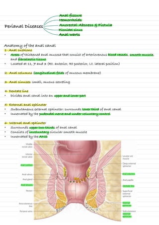
- 1. Perianal Diseases Anal fissure Hemorrhoids Anatomy of the anal canal 1- Anal cushions • Areas of thickened anal mucosa that consist of arteriovenous blood vessels, smooth muscle, and fibroelastic tissue • Located at 11, 7 and 3 (Rt. anterior, Rt posterior, Lt. lateral position) 2- Anal columns (longitudinal folds of mucous membrane) 3- Anal sinuses: small, mucus-secreting 4- Dentate line • Divides anal canal into an upper and lower part 5- External anal sphincter • Subcutaneous external sphincter: surrounds lower third of anal canal • Innervated by the pudendal nerve and under voluntary control 6- Internal anal sphincter • Surrounds upper two-thirds of anal canal • Consists of involuntary circular smooth muscle • Innervated by the ANS Anorectal Abscess & Fistula Pilonidal sinus Anal warts
- 2. Perianal Diseases Anatomy • The surgical anal canal is about 4 cm in length • Dentate line is the junction between the superior 2/3 and inferior1/3 anal canal • The dentate line is surrounded by longitudinal mucosal folds External sphincter (Red) • Bulk of the anal sphincter complex • Voluntary muscle • Innervated by Pudendal Nerve Internal sphincter (white) • Thickened distal • Involuntary • Innervated by ANS Intersphincteric plane • A potential space between ES & IS. it contains intersphincteric anal glands Anal fissure A longitudinal tear in the anoderm (epithelial) of the distal anal canal posterior midline in >85% of cases, anterior midline(10-15%) Symptoms • Pain at defecation • Bleeding (fresh) “Hematochezia” • Pruritus and discharge • Constipation Signs Acute: • Anal skin is puckered Chronic: • Skin tag(sentinel tag) • Hypertrophied anal papilla Etiology Primary (Local Trauma) • Chronic constipation or diarrhea • Anal sex Secondary (Underlying disease) • IBD (Crohn disease) Diagnostics • History • Clinical examination . ~ is 1 A. 10-15%. 9 - - 3 P 8s Acute fissure cronich fissure arouich fissure #yperbrophied And Papilla ⑧ E Skin Tag Skin tag op
- 3. Treatment • Perianal ulcer • Anal fistula or abscess • Anal carcinoma Differential diagnoses Conservative • Dietary improvement (Increase dietary fiber and water) • Stool softeners (e.g., docusate) • Anti‑inflammatory and analgesic creams and/or suppositories (e.g., 2% lidocaine) • Sitz baths • Topical vasodilator therapy (calcium channel blocker gel) Outpatient procedures • Bot. toxin A (botox) injection into the internal anal sphincter Surgical (lateral internal sphincterotomy) • when conservative treatment is unsuccessful • Disadvantage is risk of fecal incontinence Hemorrhoids Are dilated submucosal vascular cushions within the anal canal • Excessive straining (Chronic constipation) • Extended periods of sitting • Pregnancy • Older age Etiology Classificatio n 1-Internal hemorrhoids • Develop above the dentate line, which is not innervated by cutaneous nerves; distension does not cause pain. 2- External hemorrhoids • Develop below the dentate line, which is innervated by cutaneous nerves; distention of this innervated skin due to thrombosis results in severe pain. Grading of internal hemorrhoids Grade Palpation findings I Hemorrhoids bleed but do not prolapse. II Prolapse when straining, but spontaneously reduce at rest III Prolapse when straining; only reducible manually IV Irreducible prolapse; may be strangulated and thrombosed with possible ulceration
- 4. Clinical features 1- Internal hemorrhoids • Painless • Bright red bleeding at the end of defecation • Perianal mass in the event of prolapse • Pruritus 2- External hemorrhoids • Bright red bleeding • pruritus • perianal mass • perianal pain Diagnostics 1- History 2- physical Examination • Perianal examination • Digital rectal examination • Anoscopy • Proctoscopy • Anal skin tags (Polyps) • Hypertrophied anal papillae (Anal fissures) • Anal carcinoma • Anorectal varices DDx Treatment 1- Conservative treatment (Grade I II) • Dietary improvement (Increase dietary fiber and water) • Stool softeners (e.g., docusate) • Anti‑inflammatory and analgesic creams and/or suppositories (e.g., 2% lidocaine) • Sitz baths 2- Non-Surgery (procedures) (Grade I , II) • Rubber band ligation • Sclerotherapy • Infrared coagulation 3- Surgery (Grade III IV + Unsuccessful treatment) • Hemorrhoidectomy ASK GOOgIe NOT ME ASK GOOgIe NOT ME ASK GOOgIe NOT ME ⑤ II I I= I 'f -many means
- 5. Is a pus-filled develops from an infected anal gland following obstruction and bacterial overgrowth Anorectal Abscess Etiology • Obstruction and infection of the anal crypt glands (90%) • IBD (Crohn's disease, ulcerative colitis) • Malignancy (colorectal cancer) Classification Anal abscesses and fistulae • Perianal (most common) ◦ Abscess under the perianal skin ◦ Does not transverse the external sphincter • Ischiorectal: abscess below the levator ani muscle • Intersphincteric: abscess between the internal and external sphincters • Supralevator (least common): abscess above the levator ani muscle 2- Fistulas (Park's classification) • Intersphincteric (Park's Type I) • Transsphincteric (Park's Type II) • Suprasphincteric (Park's Type III) • Extrasphincteric (Park's Type IV) • Subcutaneous 1- Abscesses Most Common Clinical features • pruritus • Erythematous • subcutaneous mass • Pain Diagnostics • CT • MRI • Endoscopy and U/S Treatment 1- Abscesses • Surgical incision and drainage • Postoperative ◦ Sitz baths ◦ Analgesics and stool softeners ◦ Antibiotics 2- Fistulae • Fistulotomy (standard approach) • Possible seton placement (enables adequate drainage and fibrosis) • Possible fibrin glue or fistula plug • Antibiotics fi 0 - ⑮ . . . . . . )>)=>)* .js an
- 6. Pilonidal sinus Is a skin condition caused by local inflammation of the superior midline gluteal cleft, which may progress to a local abscess or fistula. • Young men with excessive body hair • Obesity • Deep gluteal cleft • Poor anal hygiene Risk factors • fever • painful • Erythematous swelling • Discharge (Possible purulent discharge) Clinical features Differential diagnoses • Anal fistula (e.g., due to Crohn disease) • Hidradenitis suppurativa • Anorectal abscess • Sacrococcygeal teratoma Treatment Conservative treatment • Indications ◦ Asymptomatic patients ◦ Postsurgical care of symptomatic patients • Approach ◦ Improved local hygiene Surgical treatment • Indication ◦ symptomatic patients • Procedures 1- Acute pilonidal cyst: incision and drainage, with secondary wound closure 2- Chronic or recurrent pilonidal sinus: surgical resection ▪ Primary wound closure ▪ Secondary wound closure
- 7. Anal warts • HPV types 6 and 11 They spread by ❖ Anal Sex ❖ Directly from the genitals • Exophytic • Pruritus • Discharge • Bleeding in rare cases • Pain Infections of the skin and mucous membranes by HPV, HPV may lead to anal cancer Etiology Clinical features Diagnostics • Application of 5% acetic acid turns lesions white • biopsy Treatment 1- Pharmacotherapy • local cytostatic treatment (podophyllin) • Immune response modifiers (imiquimod) 2- Surgical excision