1. Scand J Med Sci Sports 2000: 10: 114–116 COPYRIGHT C MUNKSGAARD 2000 ¡ ISSN 0905-7188
Printed in Denmark ¡ All rights reserved
Case Report
Anterior cruciate ligament injury in indoor ball games
J. F. Ebstrup, F. Bojsen-Møller
Laboratory for Functional Anatomy, Biomechanics and Motor Control, University of Copenhagen, Denmark
Corresponding author: F. Bojsen-Møller, Department of Medical Anatomy, University of Copenhagen, Blegdamsvej 3,
2200 Copenhagen N, Denmark
Accepted for publication 3 September 1999
Three videorecorded incidents of knee injuries inflicted
during indoor ball games are reported. Injuries and espe-
cially anterior cruciate ligament ruptures seemed to be
triggered in varus loaded knees by femural external ro-
tation, or in valgus loaded knees by femural internal ro-
Knee injuries, and especially anterior cruciate liga-
ment (ACL) injuries, are very common in European
team handball and basketball and are of great con-
cern to the players, their instructors, and the sports
associations (Myklebust et al. 1993, Yde & Nielsen
1990). To elucidate the course of the injury, video-
recordings of actual episodes resulting in knee in-
juries were collected from a series of Danish indoor
ball games throughout a winter season. A total of 15
episodes of combined or isolated ACL injury were
sent in for analysis from 15 different handball arenas.
The course of the injury from three typical cases are
reported here.
Case 1
A right wing player catches the ball while passing
across the court along the three meter line. In order
to make a left hand shot through an opening in the
defense line, she crouches and makes a sudden turn
to the right with the left leg stretched out and the
foot internally rotated. With the knee in varus, her
body is suddenly forced into an external rotation by
contact with an opponent (Fig. 1). Clinically, this was
an ACL rupture, but arthroscopy was not performed.
Case 2
A female handball player at the elite level is part of
a fast break offensive. She receives the ball from the
left while running at top speed directly towards the
goal while the keeper comes forward straight at her.
Just before the three meter line she takes off in a jump
with her left foot while making a right arm shot. She
114
tation with the pivot shifted to the lateral femurotibial
compartment. The observations suggest that it may be to
the players’ advantage to be trained in not letting their
knees sag medially or laterally during side-stepping or sud-
den changes in speed.
lands on her left foot and proceeds forward at high
speed. In order to avoid collision with the keeper, she
shifts her direction to the right. During this shift the
player’s loaded left foot seems to lock to the ground
and with the knee in valgus the thigh shifts from an
initially external rotation to an internal rotation. An
ACL rupture was diagnosed by arthroscopy.
Case 3
A female basketball player at the elite level runs for-
ward following an opponent in an attempt to get a
passing ball. During this maneuver she has to stop
and turn left. With her right leg extended to the right
and the knee in valgus, the body with the thigh makes
an internal rotation in the knee. With arthroscopy
the player was later diagnosed with an ACL rupture
and a tear in the lateral meniscus.
Discussion
Fast and high jumps are frequent in modern indoor
ball games. During touchdowns and take-offs the
supporting leg experiences peak loads of several body
weights which create large frictional forces between
foot and floor with the consequence that for several
hundreds of milliseconds the foot is unable to make a
rotation. Rotations necessary for simultaneous side-
stepping or shifts in direction are consequently trans-
ferred to the knee.
With the knee flexed, the femur can make internal
and external rotations relative to the tibia. In doing
so the lateral meniscus can move 12–14 mm antero-
posteriorly on the tibial plateau, while the medial one
2. ACL injury in indoor ball games
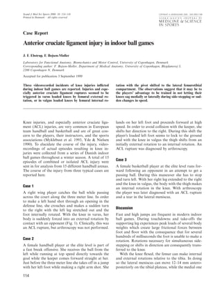
Fig. 1AπB. Case no. 1 captured with two cameras. With the
left foot internally rotated, the dark player crouches to make a
left hand shot but is forced into external rotation and varus
load of the knee by the contact with a defender. A moment
later the knee gives away with great pain.
only moves 4 mm. The center of rotation is therefore
located somewhere medially to the intercondylar emi-
nence (Matsumoto & Seedholm 1993). Because of the
directions and distances between the ligaments and
the pivot, external rotations of the femur initially
tend to tighten the ACL and relax the posterior cru-
ciate ligament. To equalize this, a small sliding of the
femur on the tibia must be expected. However, if the
knee is loaded in varus and especially if it is loaded
with several body weights as in the stand-phase of a
jump, the medial condyle of the femur can be ex-
pected to be retained in the most concave area of the
tibial condyle with no sliding possible. In such situ-
ations the pivot will be centerd in this area and the
brunt of a femural external rotation will be taken by
the ACL together with the biceps and the iliotibial
115
tract muscles (Fig. 2). A fatigue or coordination fail-
ure of these lateral muscles will expose the ACL to
heavy loads and a consequent risk of rupturing.
In situations where the feet are set apart as in cases
1 and 2, the knee tends to be loaded in valgus with
the thrust line passing through the lateral femurotib-
ial joint. According to statics of loaded columns, the
medial compartment will hereby be unloaded and the
medial supporting structures even put under tension
(Grood et al. 1981, Pauwels 1948). In such cases the
pivot will shift to the lateral joint and the ACL will
Fig. 2. Diagram of left knee seen from above with the contours
of the femural condyles marked in four positions of external
rotation. With varus load, the pivot is maintained in the medial
joint compartment and the ACL becomes taut by the shown
rotation.
Fig. 3. Diagram of left knee seen from above with the contours
of the femural condyles marked in four positions of internal
rotation. With valgus load, the pivot is shifted to the lateral
joint compartment and the ACL becomes taut by the shown
rotation.
3. Ebstrup & Bojsen-Møller
restrain a femural internal rotation, and thus be en-
dangered by shifts in this direction (Fig. 3).
The knee is usually flexed when loaded with a
consequent pronounced contraction of the quadri-
ceps muscle, which is also known to put an anteriorly
directed traction on the tibia. This is counteracted by
a co-contraction of the hamstring muscles which have
a posteriorly directed force component on the tibia.
The difference between the anteriorly and posteriorly
directed components must be taken up by the sup-
porting structures of the knee and among these the
ACL. The degree of hamstring muscle contraction is,
however, mainly governed by the need for creating an
extensor moment in the hip joint controlling the
upper body. The position and the inertia of the upper
body will therefore influence the loadings of the cru-
ciate ligaments. The directions and magnitudes of
these were not possible to judge from our videos.
Cross and colleagues (1989) studied the direction
and degree of tibial rotation in 11 male subjects who
performed a controlled side-step cutting maneuver.
References
Cross MJ, Gibbs NJ, Bryant GJ. An of the tibia in the normal and Pauwels P. The principles of construction
analysis of the sidestep cutting ligament-deficient knee. A study using of the locomotor system. Their
manoeuvre. Am J Sports Med 1989: biplanar photography. Proc Instn significance for the stressing of the
17: 363–66. Mech Engrs 1993: 207: 175–84. tubular bones. Z Anat Entwickl Gesch
Grood ES, Noyes FR, Butler DL, Suntay Myklebust G, Strand T, Engebretsen L, 1948: 114: 129–66.
WJ. Ligamentous and capsular Nilsson S, Hegermann C, Mæhlum S. Yde J, Nielsen AB. Sports injuries in
restraints preventing straight medial Registration of anterior cruciate adolescents’ games: soccer, handball,
and lateral laxity in intact human ligament injuries in the 3 upper and basketball. Br J Sports Med 1990:
cadaver knees. J Bone Joint Surg 1981: divisions in Norwegian team handball: 24: 51–4.
63A: 1257–69. a prospective study. Scand J Med Sci
Matsumoto H, Seedholm BB. Rotation Sport 1993: 3: 194 (abstract).
116
During the take-off phase they noted only submaxi-
mal internal rotations of the femur, together with the
knee in valgus, and concluded that the ACL injuries
were unlikely to occur here. In our study, the move-
ments were uncontrolled with the players stressed e.g.
by an opponent. The risk may therefore be different.
All our video episodes seemed to show that the in-
juries happened in uncontrolled situations at high
speeds and when attempting to avoid collisions, or
in defense situations with brakings and changes in
direction. Knee injuries, and especially ACL rup-
tures, seemed to be triggered in varus loaded knees
by femural external rotation, or in valgus loaded
knees with the pivot shifted to the lateral compart-
ment by femural internal rotation. It seems reason-
able to train the players in not letting their knees sag
medially or laterally.
Key words: anterior cruciate ligament injury; knee in-
jury; team handball and basketball; indoor ball
games.