SlideShare a Scribd company logo
1 of 27
Circulatory systems in the living world ( part 2)-
Human Heart , its functioning and circulatory circuits
Smita Amit – M.sc (botany ) M.ed
Teaching experience as P.G.T biology in K.V and
other leading plus two schools , as well as
Faculty for pre medical entrance preparation
Circulatory systems in the living world ( part 2)-
Human Heart , its functioning and circulatory circuits
What we will learn
1 Parts of a circulatory system – Outline
2 Heart –internal & External structure
3 Position, surface, borders, sulci, shape
4 Covering of heart
5 Musculature
6 Valves of the heart
7 Great vessels of the heart
8 Chambers of the heart
9 Innervation of the heart
10 Cardiac Cycle
11 Regulation of heart beat
Heart Blood vessels
Blood
• Heart- It is like a house
• A. Walls- Made of cardiac muscles.
• B. Chambers (rooms)- , 2 auricles and 2
ventricles, demarcated externally and
separated internally
• C. Valves (doors).
• D. Blood vessels (plumbing).
• E. Electrical conduction system
(electricity).
Blood vessels
• Arteries- Carry blood from
heart to body parts
• Veins- Carry blood from body
parts to heart
• Capillaries- Very small & thin ,
microscopic , connect veins and
arteries in the tissue to form a
network. Diffusion of all
materials takes place here
Blood
Body contains 4-6 L of blood
Consists of
Water
Red Blood Cells
Plasma
White blood cells and platelets
Parts of a circulatory system
External & INTERNAL
FEATURES
HEART
Apex
• ‘apex’-Inferior
• Located at the bottom of the heart , it is angled downward and
forward and is slightly directed towards the left at its sharpest
point.
• Formed by the left ventricle lying in the fifth intercostal space
9 cms from the median plane
Base
• The base is the heart’s superior aspect , it also connects the
heart to suspensory tissues and thus retains a support function
Orientation
The longitudinal line from the center point of the base of the
heart to the point of the apex is called the anatomical axis.
The heart is orientated as if the pyramid has fallen onto one of
its sides so that its base faces the posterior thoracic wall, and its
apex is pointed toward the anterior thoracic wall.
Base :
Faces the posterior
thoracic wall.
Apex:
Pointed toward the anterior thoracic wall.
Anatomical axis
Anatomical axis
Position : It sits between the right and left
lungs (the left lung is slightly smaller to
make room for the heart in the left chest.
The ribcage protects it.
SHAPE AND POSITION
Sternocostal Surface -
Formed by right auricle ,
right ventricle , left
auricle and left ventricle.
Sternocostal Surface -
Formed by right auricle ,
right ventricle , left
auricle and left ventricle.
1.Left Surface- Formed
by left auricle and left
ventricle
1.Diaphragmic Surface-
2/3rd is formed by left
ventricle and 1/3rd by
right ventricle
Surface
Borders
Superior –
Formed by
the two
atria
Right –
Formed by
the right
atrium
Inferior- Mainly
by right ventricle
and near the
apex by the left
ventricle
Left –
Formed by
left auricle
and left
ventricle
SURFACE & BORDERS
• Interatrial Sulcus
• Sulcus Terminalis
Preicardium
Serous- inner , helps to
lubricate the heart
Visceral Parietal
Fibrous - outer layer, made
from thick connective tissue ,
attached to the diaphragm. It
holds the heart in place in the
chest cavity and protects from
infections
Pericardium -A two-layered serous sac which
covers & protects the heart from mechanical
injury and checks its overstretching and
overfilling with blood
Has Pericardial fluid (50 ml
approx.). It protects it from injury.,
keeps it from rubbing against other
organs, keeps the heart moist,
allows its free movement and
reduces the friction between the
heart wall and the surrounding
tissues when the heart beats.
Musculature of Heart
Epicardium-last part of
pericardium , loose
connective tissue and
adipose tissue,
Myocardium-middle,
thick, thickest at LV,
made of cardiac muscle
fibres or
cardiomyocytes,
Contractile
cardiomyocytes
Endocrine
cardiomyocytes-
ANP
Cardiomyocytes of
the conduction
system
Endocardium-
innermost, single celled,
loose connective tissue,
aids in contraction
Epicardium
• It is also known as
visceral pericardium as
it forms the inner layer
of the pericardium.
• Protects the inner heart
layers and also assists
in the production of
pericardial fluid ( fills
the pericardial cavity
and helps to reduce
friction between
pericardial membranes)
• Has the coronary blood
vessels, which supply
the heart wall with
blood.
Myocardium
• The myocardium functions as a syncytium functioning to
1. Contraction of the heart in a synchronized manner.
2. Conduction of electrical stimuli
3. Provide a scaffold for heart chambers
4. Made up of cardiomyocytes
a. Contractile : The ordinary cardiomyocytes that form the
majority of the myocardium of the atria and ventricles. Their
main function is contraction
b. Endocrine cardiomyocytes: Found in the atria, particularly
the right atrium. Have fewer myofibrils. They have numerous
electron-dense secretory granules containing the atrial
natriuretic peptide which has a role in the control of blood
pressure and electrolyte balance
c. Cardiomyocytes of the conduction system- These are
specialized in the initiation and propagation of the waves of
depolarization through the myocardium faster than the
contractile cardiac muscle fibers.
Endocardium
• Lines the inner heart chambers and covers the heart
valves.
• It prevents the blood from sticking to the inner walls,
thereby preventing potentially fatal blood clots.
• Comprised of loose connective tissue and simple
squamous epithelial tissue
• Is similar in its composition to the endothelium
which lines the inside of blood vessels.
• It regulates contractions and aids
cardiac embryological development.
Cardiac Muscle
Myofibrils-Rod-like units , consisting of long chains of sarcomeres( the
fundamental contractile units ). They branch and anastomose thus they
are not perfectly in register with one another and hence cardiac
muscle fibers appear faintly striated.
The pattern of cross striations and the designation of the A, I, M, H-
bands, and Z-lines are identical to that of skeletal muscle.
The myofibrils diverge around the nucleus, leaving the sarcoplasm at
the nuclear poles containing various organelles e.g. numerous
mitochondria, and inclusions e.g. glycogen granules and lipofuscin
pigments.
T tubules-Represent inward extensions of the
extracellular space at the sarcolemma. They are large
& numerous surrounding the myofibrils at the Z line.
Sarcoplasmic reticulum: it is less organized than
the skeletal muscle with absent terminal cisternae.
It consists of narrow anastomosing sarcotubules,
in close apposition with the T tubules forming
diads
Intercalated discs-These are the specialized junctions of cell
membranes of adjacent cardiomyocytes. They extend across
the fiber at the level of the Z lines in a stepwise manner.
Structure Location Function/Description
chordae
tendineae
connecting the cusps
of AV valves with the
papillary muscles
"heart strings" aid in holding AV valves closed to prevent valve
from flipping open during ventricular systole
fossa ovalis
indentation in the
right atrium along the
interatrial septum
location where a hole used to be during fetal development
(foramen ovale); since fetal lungs are non-functional, the
pulmonary circuit of circulation is extraneous and therefore
blood is allowed to pass through foramen ovale from the right
atrium to the left atrium; a flap closes over foramen ovale after
birth and eventually seals up
interatrial
septum
tissue between the
right and left atria
separate the right and left atria; keeps oxygen-poor blood on
the right side of the heart from mixing with oxygen-rich blood
on the left side of the heart
interventricular
septum
tissue between the
right and left
ventricles
separate the right and left ventricles; keeps oxygen-poor blood
on the right side of the heart from mixing with oxygen-rich
blood on the left side of the heart
papillary
muscles
muscles on the
internal heart wall
connected to chordae
tendineae
aid in holding AV valves closed to prevent valve from flipping
open during ventricular systole (prolapse)
trabeculae
carneae
ridges along the inside
wall of the ventricles
aid in holding AV valves closed to prevent valve from flipping
open during ventricular systole (prolapse)
INTERNAL STRUCTURE
Right Atrium
1. Smaller than the left atrium.
2. Receives deoxygenated blood from the systemic
circulation via the superior and inferior vena cava &
moves it into the heart’s right ventricle.
3. Locations for sinoatrial and atrioventricular nodes;
three internal surfaces (venous, vestibular,
auricular).
4. Has the Right auricle , also called the right atrial
appendage (RAA) .It’s a small, cone-shaped pouch,
highly muscular ,lined with small muscles on its
surface which comes out from the upper and front
part of the atrium and overlaps the root of the aorta
Atrium:
• Upper chambers
• Thinner and less muscular walls
• receive blood from great veins.
• Ear-like muscular pouch arising from each atrium called Auricle.(The name auricle comes from the
Latin word auricula, which means “ear” and refers to the floppy dog-ear shape of the auricle)
Left Atrium
Receives oxygenated blood from the lungs via the pulmonary veins (4
ostia) and pumps it to the left ventricle.
Bigger with thicker wall
Has the left auricle also known as the left atrial appendage (LAA). It is
a flap of heart wall on the anterior surface of the left atrium and
plays an important role in the pumping of blood within the heart.
Landmarks - T5 - T8 (supine), T6 - T9 (erect)
Ventricle
• Lower ,
• Larger and more muscular chambers responsible for pumping and pushing blood out into
circulation.
Right Ventricle
Smaller than left ventricle, has Lower pressure gradient, Limited by
the tricuspid and pulmonary valves
Three prominent papillary muscles (anterior, posterior, and septal)
Septomarginal trabecula (moderator band)
Pumps blood into the pulmonary circuit
Left Ventricle
Larger, most muscular of all chambers, Has Higher pressure gradient
Limited by the mitral and aortic valves
Pumps blood into the systemic circulation
The Chambers of the heart
Valves
Atrioventricular (AV)
Separate atrium from ventricles , prevent backflow of blood
from ventricle to atrium
Semilunar
Separate ventricles from great vessels and prevent backflow of
blood from vessels to heart
• They act like doors.
• They incorporate two or three leaflets /cusps (thin but strong flaps of
tissue) around the AV orifices and the roots of great vessels.
• They open and close to let blood flow from one area of the heart to
another, at the right time and in the correct direction.
• As the valves open and close, they create two sounds, which are the
heartbeat.
Pulmonary semilunar valve
It is between the right ventricle and the opening
of the pulmonary trunk. It has three semilunar
cusps/leaflets: anterior/non-adjacent, left/left
adjacent, and right/right adjacent. It allows blood
to pump from the right ventricle to the
pulmonary artery. This artery leads to the lungs.
It prevents blood from going backward from the
pulmonary artery to the right ventricle.
Aortic semilunar valve
It is between the left ventricle and the
opening of the aorta. It has three semilunar
cusps/leaflets: left/left coronary, right/right
coronary, and posterior/non-coronary. They
open to let blood flow from the heart’s left
ventricle to the aorta and prevent backward
flow from the aorta into the left ventricle.
Mitral valve-
The left atrioventricular/bicuspid valve
has two cusps cusps/leaflets:
anterior/aortic and posterior/mural .
It resembles a miter in shape. It is
between the left atrium and left ventricle
and prevents backward flow from the left
ventricle to the left atrium
Tricuspid valve./ The right AV
valve
Between the right atrium and right ventricle.
It has three cusps/leaflets: anterior/anterIo superior,
septal, and posterior/inferior.
It allows blood to flow from the right atrium to the
right ventricle and prevents blood from flowing
backward from the right ventricle to the right atrium
VALVES
Great
vessels
of
the
heart
Arteries
Carry blood from Heart to the
Body
Veins
Carry blood from the body to
the Heart
Pulmonary Artery
Begin as the pulmonary trunk, a thick
and short vessel, separated from the
right ventricle by the pulmonary valve
Aorta
Arises from the aortic orifice at the
base of the left ventricle, with inflow
via the aortic valve
Coronary-
Provide oxygenated blood to
heart muscles
Arch
Ascending
Descending
Brachiocephalic Trunk
Left Common Carotid
Left Subclavian
The left
pulmonary artery
supplies blood to
the left lung,
The right
pulmonary artery
is thicker , it
supplies blood to
the right lung,
Vena Cava
Pulmonary vein
Superior pulmonary
veins (2 in number)
return blood from the
upper lobes of each
lung
Inferior
pulmonary veins
(2 in number)one
for each lung
Return blood
from the lower
lobes of the lung
Superior
It receives blood from the upper
body (superior to the diaphragm,
excluding the lungs and heart), and
pours it to the right atrium
Inferior
The inferior vena cava
receives blood from the lower body
(all structures inferior to the
diaphragm). It drains into the
inferior portion of the right atrium
Conduction system and control
Sinoatrial (SA) node:
1. An area of sub-specialized cells
2. It sends the electrical impulses that start the heartbeat.
3. It is referred to as the pacemaker of the heart.
4. The SA node is able to spread its impulse to the rest of the right
and left atria through preferential conductive pathways.
Atrioventricular (AV) node:
1. It is located in an area known as the triangle of Koch (located
between the septal leaflet of the tricuspid valve, the coronary sinus
and the membranous portion of the interatrial septum).
2.It delays the SA node’s electrical signal by a consistent amount of
time (a fraction of a second) each time. The delay ensures that the
atria are empty of blood before the contraction stops.
Purkinje fibers
1. The Purkinje fibers are branches of specialized
nerve cells.
2. They send electrical signals very quickly to the
right and left heart ventricles that contract,
3. They are in the subendocardial surface of
ventricle walls.(a part of the endocardium, the inner
layer of tissue that lines the heart’s chambers).
Bachmann’s bundle (BB)
Also known as the interatrial bundle, is a muscular
bundle comprising of parallel aligned myocardial
strands connecting the right and left atrial walls and
is considered to be the main pathway of interatrial
conduction
Bundle of His:
Also called the atrioventricular bundle. It is a branch of fibers (nerve cells) that
extends from the AV node. It runs down the length of the interventricular septum, the
structure that separates the right and left ventricles. It has two branches that receive
the electrical signal from the AV node and carries it to the Purkinje fibers.
Left bundle branch sends electrical signals through the Purkinje fibers to the left
ventricle.
Right bundle branch sends electrical signals through the Purkinje fibers to the right
ventricle
HEART BEAT
The rhythmic contraction and relaxation of the heart
one complete heart beat consists of one systole and one diastole and last for about 0.8
seconds
NUEROGENIC HEART
• Heart beat is initiated
by a nerve impulse
coming from a nerve
ganglion situated
near the heart eg
some annelids,
arthropods
MYOGENIC HEART
• Heart beat is initiated
by a patch of
modified heart
muscle itself eg
molluscs , vertebrates
including man
1. SYSTOLE (CONTRACTION )
2. S.A node the Pacemaker of the heart (located in
muscle of right atrium )stimulates electrical impulses
3. The two atria contract and blood forced to ventricle
4. A_V node delays the impulse a bit so that atria empty
fully
5. Now impulse traves to A-V node BOH, PF and
ventricles contract
6. As they contract, blood flows from the right ventricle
to the pulmonary arteries and from the left ventricle
to the aorta
7. The PT send blood to lungs for oxygenation and the
aorta which is the body’s largest artery., sends blood
from heart to the rest oft he body.
DIASTOLE (RELAXATION )
1. Following contraction, the ventricles relax, and
pressure within them falls
2. The blood again flows in to the heart and it starts
filling via atriums( right atrium receives
deoxygenated blood from the superior and inferior
venae cavae and coronary sinus. and left atrium
receives oxygenated blood from pulmonary vein )
3. The whole process starts again with impulse starting
from SA node once again
CARDIAC CYCLE
Cardiac output
• The volume of blood flowing through the systemic or pulmonary circuit per minute
• Cardiac output= Heart rate * stroke volume =72 beats/min *.07 ltr/beat= 5.0 ltr/min
• Note- stroke volume is the blood ejected by each ventricle during each beat
Complete
ventricular diastole
Beginning of
Ventricular
Diastole
Atrial
Diastole
•Ventricles
relax
•Semilunar
valves close –
2nd heart
sound –dub’
•Tricuspid and
Mitral valve open
as pressure in
ventricles falls
•Blood from Atria
flows into
Ventricles
Atrioventricular valves
open
The atrial muscles relax ,
they start relaxing while
ventricle is still in diastole
All valves – the semilunar (
at the base of aorta and
pulmonary trunk ) are
closed
Blood returns to right
atrium from the vena cava (
deoxygenated).
Atria passively filling
•Atria contract as
wave of contraction
is stimulated at SA
node
•Blood is forced into
ventricles(
Oxygenated blood
into left and
deoxygenated into
right.)
Atrial Systole
•Stimulated by AV
node ventricles begin
to contract
•Mitral and Tricuspid
valves close
immediately
•1st heart sound
’Lubb’
Beginning of
Ventricular
Systole
•Ventricles complete
their contraction
•Blood flows into the
pulmonary trunk
(deoxygenated) and
Aorta (oxygenated)
Complete
Ventricular
Systole
CARDIAC CYCLE- Contd
First Heart Sound (LUBB)
Both the AV valves close at the beginning of
ventricular contraction
Generated by the vibration of blood and the
ventricular wall
Low pitched (Frequency of 25-45 Hz) and longer
(0.15 sec) than the second sound.
Second Heart Sound(DUPP)
Occurs when the aortic and pulmonary semilunar
valves close at beginning of ventricular dilation or
diastole(aorta valves close slightly before pulmonary
valve )
Generated by the vibration of blood against aorta
Shorter duration ( 0.10 sec) , high pitched and louder (
frequency 50 hz)
HEART SOUNDS
NEURAL REGULATION OF HEARTBEAT
• Autonomic nervous system
• The heart cycle is regulated completely
subconsciously by efferent autonomic
nerves that affect the rate and force
of heart contractions.
• a. Sympathetic fibres arise from the
cervical & upper thoracic (1, 2, 3, 4)
ganglia of sympathetic trunks. They
accelerate heart rate
• b. Parasympathetic fibres that arise from
the vagus nerves. They slow heart rate
(constriction of coronay arteries)
• Postganglionic fibres reach heart
along –
• (1) SAN(SINOATRIAL NODE )
• (2) AVN( ATRIOVENTICULAR NODE
)
• (3) nerve plexus around coronary
arteries
• (4)Cardiac plexus situated below arch
of aorta
Areas in brain that effect heart through ANS
Vasomotor centre- situated in the reticular formation of the
medulla oblongata and lower part of pons . It has 3 areas
i. Vasoconstrictor- Lateral portion found in the floor of the 4th
ventricle, reticular formation of medulla, increases heart rate
through SNS
ii. Vasodilator- Medial portion, found in the floor of the 4th
ventricle, reticular formation of medulla, decreases heart rate
through PNS
iii. Sensory- Posterior part lies in the nucleus of tractus
solitaries in medulla . Controls both the above portions.
HORMONAL REGULATION OF HEARTBEAT
• Endocrine gland and their hormones
• 1.Adrenal gland medulla- epinephrine
( accelerates heart beat under
emergency) and dopamine (and
ultimately, norepinephrine that
accelerates normally) are all involved
in the initiation of the “fight-or-flight”
response.
• 2.Thyroid-thyroxine , indirectly effects
increase of heart beat
• 3.Pituitary-vasopressin effects water
reabsorption and effects heart beat
RAAS * (will be discussed in detail later)
It includes
a. Renin (enzyme )-Kidney
b. Angiotensin II- Liver
c. Aldosterone- Adrenal Cortex
d. Atrial-natriuretic peptide- hormone secreted
from the cardiac atria
They are all involved in water reabsorption for the
purpose of blood pressure regulation that affects
the heart rate or cardiovascular reflexes
Systemic circulation ( from heart to the body and back )-In the
systemic circulation, the left ventricle pumps oxygen-rich blood into the
main artery (aorta). The blood travels from the main artery to larger
and smaller arteries and into the capillary network. There the blood
drops off oxygen, nutrients and other important substances and picks
up carbon dioxide and waste products. The blood, which is now low in
oxygen, is collected in veins and travels to the right atrium and into the
right ventricle.
Pulmonary circulation( from heart to the lung and back) The right
ventricle pumps low-oxygen blood into the pulmonary artery, which
branches off into smaller and smaller arteries and capillaries. The
capillaries form a fine network around the pulmonary vesicles (grape-
like air sacs at the end of the airways). This is where carbon dioxide is
released from the blood into the air inside the pulmonary vesicles, and
fresh oxygen enters the bloodstream. When we breathe out, carbon
dioxide leaves our body. Oxygen-rich blood travels through the
pulmonary veins and the left atrium into the left ventricle
Body
Double Circulation
Lungs
Body
the right
side of the
system
deals with
deoxygenat
ed blood.
the left
side of the
system
deals with
oxygenated
blood.
Pathway of Blood
• Deoxygenated blood passes from the right
atrium into the right ventricle and then goes
to the lungs’
• From the lungs, blood moves back toward
the heart into the left atrium to the left
ventricle and then passes into the aorta to
go to the rest of the body
Right atrium Left atrium
Right Ventricle Left Ventricle
Body Lungs
SOME HEART FACTS :
1. The heart is about the same size as a fist for a child , it is bigger between the size of one and
two fists for an adult.
2. The heart pumps blood 24/7, from birth to death. If we consider the average lifespan to be
76 years, with an average heart rate of 70-72 beats per minute, that works out to
2,796,192,000 pumping actions over the course of a lifetime. We use roughly 6,518,900
calories over our lifespan or about 235 calories each day to power the heart.
3. Heartbeat begins by alternating contraction and relaxation of myocardium
4. At REST, the heart pumps about 5 QUARTS of blood a minute.
5. During EXTREME EXERTION (exercise) it can pump 40 quarts a minute.
6. Heart is composed of special muscle fibres – cardiac muscle.
7. Wave of contraction (ACTION POTENTIAL) originates at SA node due to entrance of Ca++
ions into the myocytes.
8. Diastole is the longer of the two phases so that the heart can rest between contractions
9. Human heart beats incessantly on the whole with one part relaxing while the other
contracts.
10. There is a local SA / Pace maker of the heart.
11. The body has about 5.6 litres (6 quarts) of blood. This 5.6 litres of blood circulates through
the body three times every minute. In one day, the blood travels a total of 19,000 km
(12,000 miles)
12. Pulse – The rhythmic contraction and relaxation of the aorta and its main arteries. It is due
to the flow of blood from the heart and is dependent on heart beat.
13. The aorta, the largest artery in the body, is almost the diameter of a garden hose.
Capillaries, on the other hand, are so small that it takes ten of them to equal the thickness
of a human hair.
14. Give a tennis ball a good, hard squeeze. You're using about the same amount of force your
heart uses to pump blood out to the body. Even at rest, the muscles of the heart work
hard—twice as hard as the leg muscles of a person sprinting
15. Heart sounds can be heard using a stethescope
One mark questions
1. Why are the ventricle walls thicker than the atrium
2. What is the purpose of the pericardial fluid
Two mark Question
3. What is a cardiac cycle?
4. What is the pulse rate?
Three mark questions
5.Write short notes
a. Purkinje fibres b. AV node
6. How does the heart beat continuously.
Five mark question
7. Differentiate between
a. Atrium and Ventricle
b. Semilunar and AV valves
8. Differentiate between
a. Neurogenic and myogenic heart
b. L_U_U_B and D_U_B_B sounds
9. Differentiate between
a. systemic and pulmonary circulation
b. Pulmonary artery and Pulmonary vein.
10. Why is the metabolic rate higher in man in comparison to fishes?
Test your knowledge
• 1.The heart is placed in the
• (A). Abdominal cavity
• (B). Middle mediastinum
• (C) . Thoracic cavity
• (D). Superior mediastinum
• 2. The base of the heart lies:
• (A). Superiorly
• (B). Inferiorly
• (C) . Anteriorly
• (D). Posteriorly
• 3. The anterior interventricular groove in
the sternocostal surface lodges
• A. A branch of the right coronary artery
• B. A branch of the left coronary artery
• C. Both a and b
• D. None of the above
• 4.Which of the following options regarding
the given statements is correct
• i. Human heart is ectodermal derivative
• ii. Mitral valve guards the opening
between the right atrium and left ventricle
• iii. SAN is located in the upper left corner
of the right atrium
• iv. stroke volume* heart rate= Cardiac
output
• (A). i (B.) i & ii (C) . ii. &iii (D). iv only
• 5.In the given figure the durations of the
events of the cardiac cycle are given.
Identify the sequence of these
events(A,B,C) and select the correct option
A
C
B
(A)Auricular Systole, Joint Diastole, Ventricular Systole
(B)Ventricular systole, joint Diastole, Auricular systole
(C)Ventricular systole, Auricular Systole, Joint Diastole
(D)Joint Auricular systole, Ventricular systole
Multiple Choice Questions
For more question /answers and
detailed explanation
Visit my website and book your class.
Smita Amit

More Related Content

Similar to Circulatory systems in the living world - 2.pptx

anatomyofheart-160826060632 2.pdf
anatomyofheart-160826060632 2.pdfanatomyofheart-160826060632 2.pdf
anatomyofheart-160826060632 2.pdfraksha318928
 
anatomyofheart-160826060632.pdf
anatomyofheart-160826060632.pdfanatomyofheart-160826060632.pdf
anatomyofheart-160826060632.pdfraksha318928
 
anatomyofheart-160826060632.pdf
anatomyofheart-160826060632.pdfanatomyofheart-160826060632.pdf
anatomyofheart-160826060632.pdfDiamondKumar2
 
Heart (Its Structure & Function).pptx
Heart (Its Structure & Function).pptxHeart (Its Structure & Function).pptx
Heart (Its Structure & Function).pptxVipin Chandran
 
Anatomy of the heart, Schleich
Anatomy of the heart, SchleichAnatomy of the heart, Schleich
Anatomy of the heart, SchleichTariq Abdulla
 
cardiovascular system
cardiovascular systemcardiovascular system
cardiovascular systemAkashsagar22
 
Anatomy of the Heart chambers and valves of the heart.pdf
Anatomy of the Heart chambers and valves of the heart.pdfAnatomy of the Heart chambers and valves of the heart.pdf
Anatomy of the Heart chambers and valves of the heart.pdfsy6000217
 
Blood supply of head and neck.pdf
Blood supply of head and neck.pdfBlood supply of head and neck.pdf
Blood supply of head and neck.pdfParulGarg894243
 
ANATOMY AND PHYSIOLOGY OF THE HEART.pptx
ANATOMY AND PHYSIOLOGY OF THE HEART.pptxANATOMY AND PHYSIOLOGY OF THE HEART.pptx
ANATOMY AND PHYSIOLOGY OF THE HEART.pptxCharlesSsekawu
 
ANATOMY AND PHYSIOLOGY OF THE HEART.pptx
ANATOMY AND PHYSIOLOGY OF THE HEART.pptxANATOMY AND PHYSIOLOGY OF THE HEART.pptx
ANATOMY AND PHYSIOLOGY OF THE HEART.pptxCharlesSsekawu
 
Anatomy and physiology of the heart
Anatomy and physiology of the heartAnatomy and physiology of the heart
Anatomy and physiology of the heartCharlesSsekawu
 
Cardiovascular system by Yogesh patel
Cardiovascular system by Yogesh patelCardiovascular system by Yogesh patel
Cardiovascular system by Yogesh patelPrabhat Kumar
 
Cardiovascular system
Cardiovascular systemCardiovascular system
Cardiovascular systems shariff
 
Anatomy Of Heart by Dr Rahul Sharma
Anatomy Of Heart by Dr Rahul SharmaAnatomy Of Heart by Dr Rahul Sharma
Anatomy Of Heart by Dr Rahul SharmaRahulSharmaHindustan
 

Similar to Circulatory systems in the living world - 2.pptx (20)

anatomyofheart-160826060632 2.pdf
anatomyofheart-160826060632 2.pdfanatomyofheart-160826060632 2.pdf
anatomyofheart-160826060632 2.pdf
 
anatomyofheart-160826060632.pdf
anatomyofheart-160826060632.pdfanatomyofheart-160826060632.pdf
anatomyofheart-160826060632.pdf
 
Anatomy of heart
Anatomy of heartAnatomy of heart
Anatomy of heart
 
anatomyofheart-160826060632.pdf
anatomyofheart-160826060632.pdfanatomyofheart-160826060632.pdf
anatomyofheart-160826060632.pdf
 
Cardiovascular system
Cardiovascular systemCardiovascular system
Cardiovascular system
 
Heart (Its Structure & Function).pptx
Heart (Its Structure & Function).pptxHeart (Its Structure & Function).pptx
Heart (Its Structure & Function).pptx
 
Anatomy of the heart, Schleich
Anatomy of the heart, SchleichAnatomy of the heart, Schleich
Anatomy of the heart, Schleich
 
cardiovascular system
cardiovascular systemcardiovascular system
cardiovascular system
 
Anatomy of the Heart chambers and valves of the heart.pdf
Anatomy of the Heart chambers and valves of the heart.pdfAnatomy of the Heart chambers and valves of the heart.pdf
Anatomy of the Heart chambers and valves of the heart.pdf
 
Blood supply of head and neck.pdf
Blood supply of head and neck.pdfBlood supply of head and neck.pdf
Blood supply of head and neck.pdf
 
ANATOMY AND PHYSIOLOGY OF THE HEART.pptx
ANATOMY AND PHYSIOLOGY OF THE HEART.pptxANATOMY AND PHYSIOLOGY OF THE HEART.pptx
ANATOMY AND PHYSIOLOGY OF THE HEART.pptx
 
ANATOMY AND PHYSIOLOGY OF THE HEART.pptx
ANATOMY AND PHYSIOLOGY OF THE HEART.pptxANATOMY AND PHYSIOLOGY OF THE HEART.pptx
ANATOMY AND PHYSIOLOGY OF THE HEART.pptx
 
Anatomy and physiology of the heart
Anatomy and physiology of the heartAnatomy and physiology of the heart
Anatomy and physiology of the heart
 
heart.pptx
heart.pptxheart.pptx
heart.pptx
 
Cardiovascular system by Yogesh patel
Cardiovascular system by Yogesh patelCardiovascular system by Yogesh patel
Cardiovascular system by Yogesh patel
 
Heart by dr. armaan singh
Heart by dr. armaan singhHeart by dr. armaan singh
Heart by dr. armaan singh
 
Structure Of Heart
Structure Of HeartStructure Of Heart
Structure Of Heart
 
Cardiovascular system
Cardiovascular systemCardiovascular system
Cardiovascular system
 
Anatomy Of Heart by Dr Rahul Sharma
Anatomy Of Heart by Dr Rahul SharmaAnatomy Of Heart by Dr Rahul Sharma
Anatomy Of Heart by Dr Rahul Sharma
 
Anatomy (The Heart)
Anatomy (The Heart)Anatomy (The Heart)
Anatomy (The Heart)
 

Recently uploaded

Z Score,T Score, Percential Rank and Box Plot Graph
Z Score,T Score, Percential Rank and Box Plot GraphZ Score,T Score, Percential Rank and Box Plot Graph
Z Score,T Score, Percential Rank and Box Plot GraphThiyagu K
 
ICT Role in 21st Century Education & its Challenges.pptx
ICT Role in 21st Century Education & its Challenges.pptxICT Role in 21st Century Education & its Challenges.pptx
ICT Role in 21st Century Education & its Challenges.pptxAreebaZafar22
 
Unit-V; Pricing (Pharma Marketing Management).pptx
Unit-V; Pricing (Pharma Marketing Management).pptxUnit-V; Pricing (Pharma Marketing Management).pptx
Unit-V; Pricing (Pharma Marketing Management).pptxVishalSingh1417
 
Holdier Curriculum Vitae (April 2024).pdf
Holdier Curriculum Vitae (April 2024).pdfHoldier Curriculum Vitae (April 2024).pdf
Holdier Curriculum Vitae (April 2024).pdfagholdier
 
Seal of Good Local Governance (SGLG) 2024Final.pptx
Seal of Good Local Governance (SGLG) 2024Final.pptxSeal of Good Local Governance (SGLG) 2024Final.pptx
Seal of Good Local Governance (SGLG) 2024Final.pptxnegromaestrong
 
Accessible design: Minimum effort, maximum impact
Accessible design: Minimum effort, maximum impactAccessible design: Minimum effort, maximum impact
Accessible design: Minimum effort, maximum impactdawncurless
 
Measures of Dispersion and Variability: Range, QD, AD and SD
Measures of Dispersion and Variability: Range, QD, AD and SDMeasures of Dispersion and Variability: Range, QD, AD and SD
Measures of Dispersion and Variability: Range, QD, AD and SDThiyagu K
 
APM Welcome, APM North West Network Conference, Synergies Across Sectors
APM Welcome, APM North West Network Conference, Synergies Across SectorsAPM Welcome, APM North West Network Conference, Synergies Across Sectors
APM Welcome, APM North West Network Conference, Synergies Across SectorsAssociation for Project Management
 
psychiatric nursing HISTORY COLLECTION .docx
psychiatric  nursing HISTORY  COLLECTION  .docxpsychiatric  nursing HISTORY  COLLECTION  .docx
psychiatric nursing HISTORY COLLECTION .docxPoojaSen20
 
Russian Escort Service in Delhi 11k Hotel Foreigner Russian Call Girls in Delhi
Russian Escort Service in Delhi 11k Hotel Foreigner Russian Call Girls in DelhiRussian Escort Service in Delhi 11k Hotel Foreigner Russian Call Girls in Delhi
Russian Escort Service in Delhi 11k Hotel Foreigner Russian Call Girls in Delhikauryashika82
 
Activity 01 - Artificial Culture (1).pdf
Activity 01 - Artificial Culture (1).pdfActivity 01 - Artificial Culture (1).pdf
Activity 01 - Artificial Culture (1).pdfciinovamais
 
microwave assisted reaction. General introduction
microwave assisted reaction. General introductionmicrowave assisted reaction. General introduction
microwave assisted reaction. General introductionMaksud Ahmed
 
Advanced Views - Calendar View in Odoo 17
Advanced Views - Calendar View in Odoo 17Advanced Views - Calendar View in Odoo 17
Advanced Views - Calendar View in Odoo 17Celine George
 
Introduction to Nonprofit Accounting: The Basics
Introduction to Nonprofit Accounting: The BasicsIntroduction to Nonprofit Accounting: The Basics
Introduction to Nonprofit Accounting: The BasicsTechSoup
 
Sports & Fitness Value Added Course FY..
Sports & Fitness Value Added Course FY..Sports & Fitness Value Added Course FY..
Sports & Fitness Value Added Course FY..Disha Kariya
 
1029-Danh muc Sach Giao Khoa khoi 6.pdf
1029-Danh muc Sach Giao Khoa khoi  6.pdf1029-Danh muc Sach Giao Khoa khoi  6.pdf
1029-Danh muc Sach Giao Khoa khoi 6.pdfQucHHunhnh
 
Gardella_PRCampaignConclusion Pitch Letter
Gardella_PRCampaignConclusion Pitch LetterGardella_PRCampaignConclusion Pitch Letter
Gardella_PRCampaignConclusion Pitch LetterMateoGardella
 
fourth grading exam for kindergarten in writing
fourth grading exam for kindergarten in writingfourth grading exam for kindergarten in writing
fourth grading exam for kindergarten in writingTeacherCyreneCayanan
 

Recently uploaded (20)

Z Score,T Score, Percential Rank and Box Plot Graph
Z Score,T Score, Percential Rank and Box Plot GraphZ Score,T Score, Percential Rank and Box Plot Graph
Z Score,T Score, Percential Rank and Box Plot Graph
 
INDIA QUIZ 2024 RLAC DELHI UNIVERSITY.pptx
INDIA QUIZ 2024 RLAC DELHI UNIVERSITY.pptxINDIA QUIZ 2024 RLAC DELHI UNIVERSITY.pptx
INDIA QUIZ 2024 RLAC DELHI UNIVERSITY.pptx
 
ICT Role in 21st Century Education & its Challenges.pptx
ICT Role in 21st Century Education & its Challenges.pptxICT Role in 21st Century Education & its Challenges.pptx
ICT Role in 21st Century Education & its Challenges.pptx
 
Unit-V; Pricing (Pharma Marketing Management).pptx
Unit-V; Pricing (Pharma Marketing Management).pptxUnit-V; Pricing (Pharma Marketing Management).pptx
Unit-V; Pricing (Pharma Marketing Management).pptx
 
Holdier Curriculum Vitae (April 2024).pdf
Holdier Curriculum Vitae (April 2024).pdfHoldier Curriculum Vitae (April 2024).pdf
Holdier Curriculum Vitae (April 2024).pdf
 
Seal of Good Local Governance (SGLG) 2024Final.pptx
Seal of Good Local Governance (SGLG) 2024Final.pptxSeal of Good Local Governance (SGLG) 2024Final.pptx
Seal of Good Local Governance (SGLG) 2024Final.pptx
 
Accessible design: Minimum effort, maximum impact
Accessible design: Minimum effort, maximum impactAccessible design: Minimum effort, maximum impact
Accessible design: Minimum effort, maximum impact
 
Measures of Dispersion and Variability: Range, QD, AD and SD
Measures of Dispersion and Variability: Range, QD, AD and SDMeasures of Dispersion and Variability: Range, QD, AD and SD
Measures of Dispersion and Variability: Range, QD, AD and SD
 
APM Welcome, APM North West Network Conference, Synergies Across Sectors
APM Welcome, APM North West Network Conference, Synergies Across SectorsAPM Welcome, APM North West Network Conference, Synergies Across Sectors
APM Welcome, APM North West Network Conference, Synergies Across Sectors
 
psychiatric nursing HISTORY COLLECTION .docx
psychiatric  nursing HISTORY  COLLECTION  .docxpsychiatric  nursing HISTORY  COLLECTION  .docx
psychiatric nursing HISTORY COLLECTION .docx
 
Russian Escort Service in Delhi 11k Hotel Foreigner Russian Call Girls in Delhi
Russian Escort Service in Delhi 11k Hotel Foreigner Russian Call Girls in DelhiRussian Escort Service in Delhi 11k Hotel Foreigner Russian Call Girls in Delhi
Russian Escort Service in Delhi 11k Hotel Foreigner Russian Call Girls in Delhi
 
Activity 01 - Artificial Culture (1).pdf
Activity 01 - Artificial Culture (1).pdfActivity 01 - Artificial Culture (1).pdf
Activity 01 - Artificial Culture (1).pdf
 
microwave assisted reaction. General introduction
microwave assisted reaction. General introductionmicrowave assisted reaction. General introduction
microwave assisted reaction. General introduction
 
Advanced Views - Calendar View in Odoo 17
Advanced Views - Calendar View in Odoo 17Advanced Views - Calendar View in Odoo 17
Advanced Views - Calendar View in Odoo 17
 
Introduction to Nonprofit Accounting: The Basics
Introduction to Nonprofit Accounting: The BasicsIntroduction to Nonprofit Accounting: The Basics
Introduction to Nonprofit Accounting: The Basics
 
Sports & Fitness Value Added Course FY..
Sports & Fitness Value Added Course FY..Sports & Fitness Value Added Course FY..
Sports & Fitness Value Added Course FY..
 
1029-Danh muc Sach Giao Khoa khoi 6.pdf
1029-Danh muc Sach Giao Khoa khoi  6.pdf1029-Danh muc Sach Giao Khoa khoi  6.pdf
1029-Danh muc Sach Giao Khoa khoi 6.pdf
 
Gardella_PRCampaignConclusion Pitch Letter
Gardella_PRCampaignConclusion Pitch LetterGardella_PRCampaignConclusion Pitch Letter
Gardella_PRCampaignConclusion Pitch Letter
 
fourth grading exam for kindergarten in writing
fourth grading exam for kindergarten in writingfourth grading exam for kindergarten in writing
fourth grading exam for kindergarten in writing
 
Advance Mobile Application Development class 07
Advance Mobile Application Development class 07Advance Mobile Application Development class 07
Advance Mobile Application Development class 07
 

Circulatory systems in the living world - 2.pptx

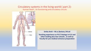
  • 1. Circulatory systems in the living world ( part 2)- Human Heart , its functioning and circulatory circuits Smita Amit – M.sc (botany ) M.ed Teaching experience as P.G.T biology in K.V and other leading plus two schools , as well as Faculty for pre medical entrance preparation
  • 2. Circulatory systems in the living world ( part 2)- Human Heart , its functioning and circulatory circuits What we will learn 1 Parts of a circulatory system – Outline 2 Heart –internal & External structure 3 Position, surface, borders, sulci, shape 4 Covering of heart 5 Musculature 6 Valves of the heart 7 Great vessels of the heart 8 Chambers of the heart 9 Innervation of the heart 10 Cardiac Cycle 11 Regulation of heart beat
  • 3. Heart Blood vessels Blood • Heart- It is like a house • A. Walls- Made of cardiac muscles. • B. Chambers (rooms)- , 2 auricles and 2 ventricles, demarcated externally and separated internally • C. Valves (doors). • D. Blood vessels (plumbing). • E. Electrical conduction system (electricity). Blood vessels • Arteries- Carry blood from heart to body parts • Veins- Carry blood from body parts to heart • Capillaries- Very small & thin , microscopic , connect veins and arteries in the tissue to form a network. Diffusion of all materials takes place here Blood Body contains 4-6 L of blood Consists of Water Red Blood Cells Plasma White blood cells and platelets Parts of a circulatory system
  • 5. Apex • ‘apex’-Inferior • Located at the bottom of the heart , it is angled downward and forward and is slightly directed towards the left at its sharpest point. • Formed by the left ventricle lying in the fifth intercostal space 9 cms from the median plane Base • The base is the heart’s superior aspect , it also connects the heart to suspensory tissues and thus retains a support function Orientation The longitudinal line from the center point of the base of the heart to the point of the apex is called the anatomical axis. The heart is orientated as if the pyramid has fallen onto one of its sides so that its base faces the posterior thoracic wall, and its apex is pointed toward the anterior thoracic wall. Base : Faces the posterior thoracic wall. Apex: Pointed toward the anterior thoracic wall. Anatomical axis Anatomical axis Position : It sits between the right and left lungs (the left lung is slightly smaller to make room for the heart in the left chest. The ribcage protects it. SHAPE AND POSITION
  • 6. Sternocostal Surface - Formed by right auricle , right ventricle , left auricle and left ventricle. Sternocostal Surface - Formed by right auricle , right ventricle , left auricle and left ventricle. 1.Left Surface- Formed by left auricle and left ventricle 1.Diaphragmic Surface- 2/3rd is formed by left ventricle and 1/3rd by right ventricle Surface Borders Superior – Formed by the two atria Right – Formed by the right atrium Inferior- Mainly by right ventricle and near the apex by the left ventricle Left – Formed by left auricle and left ventricle SURFACE & BORDERS
  • 7. • Interatrial Sulcus • Sulcus Terminalis
  • 8. Preicardium Serous- inner , helps to lubricate the heart Visceral Parietal Fibrous - outer layer, made from thick connective tissue , attached to the diaphragm. It holds the heart in place in the chest cavity and protects from infections Pericardium -A two-layered serous sac which covers & protects the heart from mechanical injury and checks its overstretching and overfilling with blood Has Pericardial fluid (50 ml approx.). It protects it from injury., keeps it from rubbing against other organs, keeps the heart moist, allows its free movement and reduces the friction between the heart wall and the surrounding tissues when the heart beats.
  • 9. Musculature of Heart Epicardium-last part of pericardium , loose connective tissue and adipose tissue, Myocardium-middle, thick, thickest at LV, made of cardiac muscle fibres or cardiomyocytes, Contractile cardiomyocytes Endocrine cardiomyocytes- ANP Cardiomyocytes of the conduction system Endocardium- innermost, single celled, loose connective tissue, aids in contraction Epicardium • It is also known as visceral pericardium as it forms the inner layer of the pericardium. • Protects the inner heart layers and also assists in the production of pericardial fluid ( fills the pericardial cavity and helps to reduce friction between pericardial membranes) • Has the coronary blood vessels, which supply the heart wall with blood. Myocardium • The myocardium functions as a syncytium functioning to 1. Contraction of the heart in a synchronized manner. 2. Conduction of electrical stimuli 3. Provide a scaffold for heart chambers 4. Made up of cardiomyocytes a. Contractile : The ordinary cardiomyocytes that form the majority of the myocardium of the atria and ventricles. Their main function is contraction b. Endocrine cardiomyocytes: Found in the atria, particularly the right atrium. Have fewer myofibrils. They have numerous electron-dense secretory granules containing the atrial natriuretic peptide which has a role in the control of blood pressure and electrolyte balance c. Cardiomyocytes of the conduction system- These are specialized in the initiation and propagation of the waves of depolarization through the myocardium faster than the contractile cardiac muscle fibers. Endocardium • Lines the inner heart chambers and covers the heart valves. • It prevents the blood from sticking to the inner walls, thereby preventing potentially fatal blood clots. • Comprised of loose connective tissue and simple squamous epithelial tissue • Is similar in its composition to the endothelium which lines the inside of blood vessels. • It regulates contractions and aids cardiac embryological development.
  • 10. Cardiac Muscle Myofibrils-Rod-like units , consisting of long chains of sarcomeres( the fundamental contractile units ). They branch and anastomose thus they are not perfectly in register with one another and hence cardiac muscle fibers appear faintly striated. The pattern of cross striations and the designation of the A, I, M, H- bands, and Z-lines are identical to that of skeletal muscle. The myofibrils diverge around the nucleus, leaving the sarcoplasm at the nuclear poles containing various organelles e.g. numerous mitochondria, and inclusions e.g. glycogen granules and lipofuscin pigments. T tubules-Represent inward extensions of the extracellular space at the sarcolemma. They are large & numerous surrounding the myofibrils at the Z line. Sarcoplasmic reticulum: it is less organized than the skeletal muscle with absent terminal cisternae. It consists of narrow anastomosing sarcotubules, in close apposition with the T tubules forming diads Intercalated discs-These are the specialized junctions of cell membranes of adjacent cardiomyocytes. They extend across the fiber at the level of the Z lines in a stepwise manner.
  • 11. Structure Location Function/Description chordae tendineae connecting the cusps of AV valves with the papillary muscles "heart strings" aid in holding AV valves closed to prevent valve from flipping open during ventricular systole fossa ovalis indentation in the right atrium along the interatrial septum location where a hole used to be during fetal development (foramen ovale); since fetal lungs are non-functional, the pulmonary circuit of circulation is extraneous and therefore blood is allowed to pass through foramen ovale from the right atrium to the left atrium; a flap closes over foramen ovale after birth and eventually seals up interatrial septum tissue between the right and left atria separate the right and left atria; keeps oxygen-poor blood on the right side of the heart from mixing with oxygen-rich blood on the left side of the heart interventricular septum tissue between the right and left ventricles separate the right and left ventricles; keeps oxygen-poor blood on the right side of the heart from mixing with oxygen-rich blood on the left side of the heart papillary muscles muscles on the internal heart wall connected to chordae tendineae aid in holding AV valves closed to prevent valve from flipping open during ventricular systole (prolapse) trabeculae carneae ridges along the inside wall of the ventricles aid in holding AV valves closed to prevent valve from flipping open during ventricular systole (prolapse) INTERNAL STRUCTURE
  • 12. Right Atrium 1. Smaller than the left atrium. 2. Receives deoxygenated blood from the systemic circulation via the superior and inferior vena cava & moves it into the heart’s right ventricle. 3. Locations for sinoatrial and atrioventricular nodes; three internal surfaces (venous, vestibular, auricular). 4. Has the Right auricle , also called the right atrial appendage (RAA) .It’s a small, cone-shaped pouch, highly muscular ,lined with small muscles on its surface which comes out from the upper and front part of the atrium and overlaps the root of the aorta Atrium: • Upper chambers • Thinner and less muscular walls • receive blood from great veins. • Ear-like muscular pouch arising from each atrium called Auricle.(The name auricle comes from the Latin word auricula, which means “ear” and refers to the floppy dog-ear shape of the auricle) Left Atrium Receives oxygenated blood from the lungs via the pulmonary veins (4 ostia) and pumps it to the left ventricle. Bigger with thicker wall Has the left auricle also known as the left atrial appendage (LAA). It is a flap of heart wall on the anterior surface of the left atrium and plays an important role in the pumping of blood within the heart. Landmarks - T5 - T8 (supine), T6 - T9 (erect) Ventricle • Lower , • Larger and more muscular chambers responsible for pumping and pushing blood out into circulation. Right Ventricle Smaller than left ventricle, has Lower pressure gradient, Limited by the tricuspid and pulmonary valves Three prominent papillary muscles (anterior, posterior, and septal) Septomarginal trabecula (moderator band) Pumps blood into the pulmonary circuit Left Ventricle Larger, most muscular of all chambers, Has Higher pressure gradient Limited by the mitral and aortic valves Pumps blood into the systemic circulation The Chambers of the heart
  • 13. Valves Atrioventricular (AV) Separate atrium from ventricles , prevent backflow of blood from ventricle to atrium Semilunar Separate ventricles from great vessels and prevent backflow of blood from vessels to heart • They act like doors. • They incorporate two or three leaflets /cusps (thin but strong flaps of tissue) around the AV orifices and the roots of great vessels. • They open and close to let blood flow from one area of the heart to another, at the right time and in the correct direction. • As the valves open and close, they create two sounds, which are the heartbeat. Pulmonary semilunar valve It is between the right ventricle and the opening of the pulmonary trunk. It has three semilunar cusps/leaflets: anterior/non-adjacent, left/left adjacent, and right/right adjacent. It allows blood to pump from the right ventricle to the pulmonary artery. This artery leads to the lungs. It prevents blood from going backward from the pulmonary artery to the right ventricle. Aortic semilunar valve It is between the left ventricle and the opening of the aorta. It has three semilunar cusps/leaflets: left/left coronary, right/right coronary, and posterior/non-coronary. They open to let blood flow from the heart’s left ventricle to the aorta and prevent backward flow from the aorta into the left ventricle. Mitral valve- The left atrioventricular/bicuspid valve has two cusps cusps/leaflets: anterior/aortic and posterior/mural . It resembles a miter in shape. It is between the left atrium and left ventricle and prevents backward flow from the left ventricle to the left atrium Tricuspid valve./ The right AV valve Between the right atrium and right ventricle. It has three cusps/leaflets: anterior/anterIo superior, septal, and posterior/inferior. It allows blood to flow from the right atrium to the right ventricle and prevents blood from flowing backward from the right ventricle to the right atrium VALVES
  • 14. Great vessels of the heart Arteries Carry blood from Heart to the Body Veins Carry blood from the body to the Heart Pulmonary Artery Begin as the pulmonary trunk, a thick and short vessel, separated from the right ventricle by the pulmonary valve Aorta Arises from the aortic orifice at the base of the left ventricle, with inflow via the aortic valve Coronary- Provide oxygenated blood to heart muscles Arch Ascending Descending Brachiocephalic Trunk Left Common Carotid Left Subclavian The left pulmonary artery supplies blood to the left lung, The right pulmonary artery is thicker , it supplies blood to the right lung, Vena Cava Pulmonary vein Superior pulmonary veins (2 in number) return blood from the upper lobes of each lung Inferior pulmonary veins (2 in number)one for each lung Return blood from the lower lobes of the lung Superior It receives blood from the upper body (superior to the diaphragm, excluding the lungs and heart), and pours it to the right atrium Inferior The inferior vena cava receives blood from the lower body (all structures inferior to the diaphragm). It drains into the inferior portion of the right atrium
  • 15. Conduction system and control Sinoatrial (SA) node: 1. An area of sub-specialized cells 2. It sends the electrical impulses that start the heartbeat. 3. It is referred to as the pacemaker of the heart. 4. The SA node is able to spread its impulse to the rest of the right and left atria through preferential conductive pathways. Atrioventricular (AV) node: 1. It is located in an area known as the triangle of Koch (located between the septal leaflet of the tricuspid valve, the coronary sinus and the membranous portion of the interatrial septum). 2.It delays the SA node’s electrical signal by a consistent amount of time (a fraction of a second) each time. The delay ensures that the atria are empty of blood before the contraction stops. Purkinje fibers 1. The Purkinje fibers are branches of specialized nerve cells. 2. They send electrical signals very quickly to the right and left heart ventricles that contract, 3. They are in the subendocardial surface of ventricle walls.(a part of the endocardium, the inner layer of tissue that lines the heart’s chambers). Bachmann’s bundle (BB) Also known as the interatrial bundle, is a muscular bundle comprising of parallel aligned myocardial strands connecting the right and left atrial walls and is considered to be the main pathway of interatrial conduction Bundle of His: Also called the atrioventricular bundle. It is a branch of fibers (nerve cells) that extends from the AV node. It runs down the length of the interventricular septum, the structure that separates the right and left ventricles. It has two branches that receive the electrical signal from the AV node and carries it to the Purkinje fibers. Left bundle branch sends electrical signals through the Purkinje fibers to the left ventricle. Right bundle branch sends electrical signals through the Purkinje fibers to the right ventricle
  • 16. HEART BEAT The rhythmic contraction and relaxation of the heart one complete heart beat consists of one systole and one diastole and last for about 0.8 seconds NUEROGENIC HEART • Heart beat is initiated by a nerve impulse coming from a nerve ganglion situated near the heart eg some annelids, arthropods MYOGENIC HEART • Heart beat is initiated by a patch of modified heart muscle itself eg molluscs , vertebrates including man
  • 17. 1. SYSTOLE (CONTRACTION ) 2. S.A node the Pacemaker of the heart (located in muscle of right atrium )stimulates electrical impulses 3. The two atria contract and blood forced to ventricle 4. A_V node delays the impulse a bit so that atria empty fully 5. Now impulse traves to A-V node BOH, PF and ventricles contract 6. As they contract, blood flows from the right ventricle to the pulmonary arteries and from the left ventricle to the aorta 7. The PT send blood to lungs for oxygenation and the aorta which is the body’s largest artery., sends blood from heart to the rest oft he body. DIASTOLE (RELAXATION ) 1. Following contraction, the ventricles relax, and pressure within them falls 2. The blood again flows in to the heart and it starts filling via atriums( right atrium receives deoxygenated blood from the superior and inferior venae cavae and coronary sinus. and left atrium receives oxygenated blood from pulmonary vein ) 3. The whole process starts again with impulse starting from SA node once again CARDIAC CYCLE Cardiac output • The volume of blood flowing through the systemic or pulmonary circuit per minute • Cardiac output= Heart rate * stroke volume =72 beats/min *.07 ltr/beat= 5.0 ltr/min • Note- stroke volume is the blood ejected by each ventricle during each beat
  • 18. Complete ventricular diastole Beginning of Ventricular Diastole Atrial Diastole •Ventricles relax •Semilunar valves close – 2nd heart sound –dub’ •Tricuspid and Mitral valve open as pressure in ventricles falls •Blood from Atria flows into Ventricles Atrioventricular valves open The atrial muscles relax , they start relaxing while ventricle is still in diastole All valves – the semilunar ( at the base of aorta and pulmonary trunk ) are closed Blood returns to right atrium from the vena cava ( deoxygenated). Atria passively filling •Atria contract as wave of contraction is stimulated at SA node •Blood is forced into ventricles( Oxygenated blood into left and deoxygenated into right.) Atrial Systole •Stimulated by AV node ventricles begin to contract •Mitral and Tricuspid valves close immediately •1st heart sound ’Lubb’ Beginning of Ventricular Systole •Ventricles complete their contraction •Blood flows into the pulmonary trunk (deoxygenated) and Aorta (oxygenated) Complete Ventricular Systole CARDIAC CYCLE- Contd
  • 19. First Heart Sound (LUBB) Both the AV valves close at the beginning of ventricular contraction Generated by the vibration of blood and the ventricular wall Low pitched (Frequency of 25-45 Hz) and longer (0.15 sec) than the second sound. Second Heart Sound(DUPP) Occurs when the aortic and pulmonary semilunar valves close at beginning of ventricular dilation or diastole(aorta valves close slightly before pulmonary valve ) Generated by the vibration of blood against aorta Shorter duration ( 0.10 sec) , high pitched and louder ( frequency 50 hz) HEART SOUNDS
  • 20. NEURAL REGULATION OF HEARTBEAT • Autonomic nervous system • The heart cycle is regulated completely subconsciously by efferent autonomic nerves that affect the rate and force of heart contractions. • a. Sympathetic fibres arise from the cervical & upper thoracic (1, 2, 3, 4) ganglia of sympathetic trunks. They accelerate heart rate • b. Parasympathetic fibres that arise from the vagus nerves. They slow heart rate (constriction of coronay arteries) • Postganglionic fibres reach heart along – • (1) SAN(SINOATRIAL NODE ) • (2) AVN( ATRIOVENTICULAR NODE ) • (3) nerve plexus around coronary arteries • (4)Cardiac plexus situated below arch of aorta Areas in brain that effect heart through ANS Vasomotor centre- situated in the reticular formation of the medulla oblongata and lower part of pons . It has 3 areas i. Vasoconstrictor- Lateral portion found in the floor of the 4th ventricle, reticular formation of medulla, increases heart rate through SNS ii. Vasodilator- Medial portion, found in the floor of the 4th ventricle, reticular formation of medulla, decreases heart rate through PNS iii. Sensory- Posterior part lies in the nucleus of tractus solitaries in medulla . Controls both the above portions.
  • 21. HORMONAL REGULATION OF HEARTBEAT • Endocrine gland and their hormones • 1.Adrenal gland medulla- epinephrine ( accelerates heart beat under emergency) and dopamine (and ultimately, norepinephrine that accelerates normally) are all involved in the initiation of the “fight-or-flight” response. • 2.Thyroid-thyroxine , indirectly effects increase of heart beat • 3.Pituitary-vasopressin effects water reabsorption and effects heart beat RAAS * (will be discussed in detail later) It includes a. Renin (enzyme )-Kidney b. Angiotensin II- Liver c. Aldosterone- Adrenal Cortex d. Atrial-natriuretic peptide- hormone secreted from the cardiac atria They are all involved in water reabsorption for the purpose of blood pressure regulation that affects the heart rate or cardiovascular reflexes
  • 22. Systemic circulation ( from heart to the body and back )-In the systemic circulation, the left ventricle pumps oxygen-rich blood into the main artery (aorta). The blood travels from the main artery to larger and smaller arteries and into the capillary network. There the blood drops off oxygen, nutrients and other important substances and picks up carbon dioxide and waste products. The blood, which is now low in oxygen, is collected in veins and travels to the right atrium and into the right ventricle. Pulmonary circulation( from heart to the lung and back) The right ventricle pumps low-oxygen blood into the pulmonary artery, which branches off into smaller and smaller arteries and capillaries. The capillaries form a fine network around the pulmonary vesicles (grape- like air sacs at the end of the airways). This is where carbon dioxide is released from the blood into the air inside the pulmonary vesicles, and fresh oxygen enters the bloodstream. When we breathe out, carbon dioxide leaves our body. Oxygen-rich blood travels through the pulmonary veins and the left atrium into the left ventricle Body Double Circulation Lungs Body the right side of the system deals with deoxygenat ed blood. the left side of the system deals with oxygenated blood.
  • 23. Pathway of Blood • Deoxygenated blood passes from the right atrium into the right ventricle and then goes to the lungs’ • From the lungs, blood moves back toward the heart into the left atrium to the left ventricle and then passes into the aorta to go to the rest of the body Right atrium Left atrium Right Ventricle Left Ventricle Body Lungs
  • 24. SOME HEART FACTS : 1. The heart is about the same size as a fist for a child , it is bigger between the size of one and two fists for an adult. 2. The heart pumps blood 24/7, from birth to death. If we consider the average lifespan to be 76 years, with an average heart rate of 70-72 beats per minute, that works out to 2,796,192,000 pumping actions over the course of a lifetime. We use roughly 6,518,900 calories over our lifespan or about 235 calories each day to power the heart. 3. Heartbeat begins by alternating contraction and relaxation of myocardium 4. At REST, the heart pumps about 5 QUARTS of blood a minute. 5. During EXTREME EXERTION (exercise) it can pump 40 quarts a minute. 6. Heart is composed of special muscle fibres – cardiac muscle. 7. Wave of contraction (ACTION POTENTIAL) originates at SA node due to entrance of Ca++ ions into the myocytes. 8. Diastole is the longer of the two phases so that the heart can rest between contractions 9. Human heart beats incessantly on the whole with one part relaxing while the other contracts. 10. There is a local SA / Pace maker of the heart. 11. The body has about 5.6 litres (6 quarts) of blood. This 5.6 litres of blood circulates through the body three times every minute. In one day, the blood travels a total of 19,000 km (12,000 miles) 12. Pulse – The rhythmic contraction and relaxation of the aorta and its main arteries. It is due to the flow of blood from the heart and is dependent on heart beat. 13. The aorta, the largest artery in the body, is almost the diameter of a garden hose. Capillaries, on the other hand, are so small that it takes ten of them to equal the thickness of a human hair. 14. Give a tennis ball a good, hard squeeze. You're using about the same amount of force your heart uses to pump blood out to the body. Even at rest, the muscles of the heart work hard—twice as hard as the leg muscles of a person sprinting 15. Heart sounds can be heard using a stethescope
  • 25. One mark questions 1. Why are the ventricle walls thicker than the atrium 2. What is the purpose of the pericardial fluid Two mark Question 3. What is a cardiac cycle? 4. What is the pulse rate? Three mark questions 5.Write short notes a. Purkinje fibres b. AV node 6. How does the heart beat continuously. Five mark question 7. Differentiate between a. Atrium and Ventricle b. Semilunar and AV valves 8. Differentiate between a. Neurogenic and myogenic heart b. L_U_U_B and D_U_B_B sounds 9. Differentiate between a. systemic and pulmonary circulation b. Pulmonary artery and Pulmonary vein. 10. Why is the metabolic rate higher in man in comparison to fishes? Test your knowledge
  • 26. • 1.The heart is placed in the • (A). Abdominal cavity • (B). Middle mediastinum • (C) . Thoracic cavity • (D). Superior mediastinum • 2. The base of the heart lies: • (A). Superiorly • (B). Inferiorly • (C) . Anteriorly • (D). Posteriorly • 3. The anterior interventricular groove in the sternocostal surface lodges • A. A branch of the right coronary artery • B. A branch of the left coronary artery • C. Both a and b • D. None of the above • 4.Which of the following options regarding the given statements is correct • i. Human heart is ectodermal derivative • ii. Mitral valve guards the opening between the right atrium and left ventricle • iii. SAN is located in the upper left corner of the right atrium • iv. stroke volume* heart rate= Cardiac output • (A). i (B.) i & ii (C) . ii. &iii (D). iv only • 5.In the given figure the durations of the events of the cardiac cycle are given. Identify the sequence of these events(A,B,C) and select the correct option A C B (A)Auricular Systole, Joint Diastole, Ventricular Systole (B)Ventricular systole, joint Diastole, Auricular systole (C)Ventricular systole, Auricular Systole, Joint Diastole (D)Joint Auricular systole, Ventricular systole Multiple Choice Questions
  • 27. For more question /answers and detailed explanation Visit my website and book your class. Smita Amit